Chronic progressive external ophthalmoplegia

Last updated
Chronic progressive external ophthalmoplegia
Other namesProgressive external ophthalmoplegia
Specialty Ophthalmology   OOjs UI icon edit-ltr-progressive.svg

Chronic progressive external ophthalmoplegia (CPEO) is a type of eye disorder characterized by slowly progressive inability to move the eyes and eyebrows. [1] It is often the only feature of mitochondrial disease, in which case the term CPEO may be given as the diagnosis. In other people suffering from mitochondrial disease, CPEO occurs as part of a syndrome involving more than one part of the body, such as Kearns–Sayre syndrome. Occasionally CPEO may be caused by conditions other than mitochondrial diseases.

Contents

Signs and symptoms

CPEO is a rare disease that may affect those of all ages, but typically manifests in the young adult years. CPEO is the most common manifestation of mitochondrial myopathy, occurring in an estimated two-thirds of all cases of mitochondrial myopathy. Patients typically present with ptosis (drooping eyelids). Other diseases like Graves' disease, myasthenia gravis and glioma that may cause an external ophthalmoplegia must be ruled out.[ citation needed ]

CPEO itself

CPEO is a slowly progressing disease. It may begin at any age and progresses over a period of 5–15 years. [1] The first presenting symptom of ptosis is often unnoticed by the patient until the lids droop to the point of producing a visual field defect. Often, patients will tilt the head backwards to adjust for the slowly progressing ptosis of the lids. In addition, as the ptosis becomes complete, the patients will use the frontalis (forehead) muscle to help elevate the lids. The ptosis is typically bilateral but may be unilateral for a period of months to years before the fellow lid becomes involved.[ citation needed ]

Ophthalmoplegia (the inability or difficulty to move the eye) is usually symmetrical, therefore, patients are not affected by diplopia (double vision). The progressive ophthalmoplegia is often unnoticed till decreased ocular motility limits peripheral vision. Often someone else will point out the ocular disturbance to the patient. Patients will move their heads to adjust for the loss of peripheral vision caused by inability to abduct or adduct the eye. All directions of gaze are affected; however, downward gaze appears to be best spared. This is in contrast to progressive supranuclear palsy (PSP), which typically affects vertical gaze and spares horizontal gaze.[ citation needed ]

Mitochondrial retinopathy has been described in CPEO which presents with a spectrum of distinct retinal phenotypes. This includes mild, focal pigmentary abnormalities on funduscopy and widespread granular pigmented fundus alterations. Mild, asymptomatic retinopathy might be underreported; severe retinopathy may be associated with significant vision loss. [2]

Occurring alongside CPEO

Weakness of extraocular muscle groups including, the orbicularis oculi muscle as well as facial and limb muscles may be present in up to 25% of patients with CPEO. As a result of the orbicularis oculi weakness, patients may suffer from exposure keratopathy (damage to cornea) from the inability to close the eyes tightly. Frontalis muscle weakness may exacerbate the ptotic lids with the inability to compensate for the ptosis. Facial muscles may be involved which lead to atrophy of facial muscle groups producing a thin, expressionless face with some having difficulty with chewing. Neck, shoulder and extremity weakness with atrophy may affect some patients and can be mild or severe.[ citation needed ]

Mild visual impairment was seen in 95% of patients that were evaluated using the Visual Function Index (VF-14). [3]

The ciliary muscles that control the lens shape and the iris muscles are often unaffected by CPEO.[ citation needed ]

Additional symptoms are variable, and may include exercise intolerance, cataracts, hearing loss, sensory axonal neuropathy, ataxia, clinical depression, hypogonadism, and parkinsonism.[ citation needed ]

Kearns–Sayre syndrome is characterized by onset before 15 years of age of CPEO, heart block and pigmentary retinopathy. [1]

Genetics

Mitochondrial DNA which is transmitted from the mother, encodes proteins that are critical to the respiratory chain required to produce adenosine triphosphate (ATP). Deletions or mutations to segments of mtDNA lead to defective function of oxidative phosphorylation. This may be made evident in highly oxidative tissues like skeletal muscle and heart tissue. However, extraocular muscles contain a volume of mitochondria that is several times greater than any other muscle group. As such, this results in the preferential ocular symptoms of CPEO.[ citation needed ]

Multiple mtDNA abnormalities exist which cause CPEO. One mutation is located in a conserved region of mitochondrial tRNA at nucleotide 3243 in which there is an A to G nucleotide transition. This mutation is associated with both CPEO and mitochondrial encephalomyopathy, lactic acidosis, and stroke-like episodes (MELAS). [4] A common deletion found in one-third of CPEO patients is a 4,977 base pair segment found between a 13 base pair repeat.[ citation needed ]

The mtDNA that is affected maybe a single or multiple point deletion, with associated nuclear DNA deletions. One study showed that mtDNA deletion seen in CPEO patients also had an associated nuclear DNA deletion of the Twinkle gene which encodes specific mitochondrial protein; Twinkle. [5]

Whether a tissue is affected is correlated with the amount of oxidative demands in relation to the amount of mtDNA deletion.[ citation needed ]In most cases, PEO occurs due to a sporadic deletion or duplication within the mitochondrial DNA. [6] However, transmission from the mother to the progeny appears only in few cases. Both autosomal dominant and autosomal recessive inheritance can occur, autosomal recessive inheritance being more severe. Dominant and recessive forms of PEO can be caused by genetic mutations in the ANT1 , POLG , POLG2 and PEO1 genes. [7] [8]

Diagnosis

An example of ragged red fibers Ragged red fibers in MELAS.jpg
An example of ragged red fibers

It is important to differentiate CPEO from other pathologies that may cause an ophthalmoplegia. There are specific therapies used for these pathologies.[ citation needed ]

CPEO is diagnosed via muscle biopsy. On examination of muscle fibers stained with Gömöri trichrome stain, one can see an accumulation of enlarged mitochondria. This produces a dark red staining of the muscle fibers given the name "ragged red fibers". While ragged red fibers are seen in normal aging, amounts in excess of normal aging give a diagnosis of a mitochondrial myopathy.[ citation needed ] Polymerase chain reaction (PCR) from a sample of blood or muscle tissue can determine a mutation of the mtDNA.[ citation needed ]

Elevated acetylcholine receptor antibody level which is typically seen in myasthenia gravis has been seen in certain patients of mitochondrial associated ophthalmoplegia. [9] It is important to have a dilated eye exam to determine if there is pigmentary retinopathy that may signify Kearns–Sayre syndrome which is associated with cardiac abnormalities.[ citation needed ]

MRI may be helpful in the diagnosis, in one study volumes of medial rectus, lateral rectus, and inferior rectus muscles in CPEO were not smaller than normal (in contrast to the profound atrophy typical of neurogenic paralysis). Although volumes of the superior rectus muscle-levator complex and superior oblique were significantly reduced. [10]

Treatment

There is currently no defined treatment to ameliorate the muscle weakness of CPEO. Treatments used to treat other pathologies causing ophthalmoplegia has not been shown to be effective.[ citation needed ] Experimental treatment with tetracycline has been used to improve ocular motility in one patient. [11] Coenzyme Q10 has also been used to treat this condition. [12] However, most neuro-ophthalmologists do not ascribe to any treatment.[ citation needed ]

Ptosis associated with CPEO may be corrected with surgery to raise the lids, [13] however due to weakness of the orbicularis oculi muscles, care must be taken not to raise the lids in excess causing an inability to close the lids. This results in an exposure keratopathy. Therefore, rarely should lid surgery be performed and only by a neuro-ophthalmologist familiar with the disease.[ citation needed ]

The most common strabismus finding is large angle exotropia which can be treated by maximal bilateral eye surgery, but due to the progressive nature of the disease, strabismus may recur. [14] Those that have diplopia as a result of asymmetric ophthalmoplegia may be corrected with prisms or with surgery to create a better alignment of the eyes.[ citation needed ]

See also

Related Research Articles

<span class="mw-page-title-main">Mitochondrial disease</span> Spontaneously occurring or inherited disorder that involves mitochondrial dysfunction

Mitochondrial disease is a group of disorders caused by mitochondrial dysfunction. Mitochondria are the organelles that generate energy for the cell and are found in every cell of the human body except red blood cells. They convert the energy of food molecules into the ATP that powers most cell functions.

<span class="mw-page-title-main">Mitochondrial myopathy</span> Medical condition

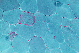
Mitochondrial myopathies are types of myopathies associated with mitochondrial disease. On biopsy, the muscle tissue of patients with these diseases usually demonstrate "ragged red" muscle fibers. These ragged-red fibers contain mild accumulations of glycogen and neutral lipids, and may show an increased reactivity for succinate dehydrogenase and a decreased reactivity for cytochrome c oxidase. Inheritance was believed to be maternal. It is now known that certain nuclear DNA deletions can also cause mitochondrial myopathy such as the OPA1 gene deletion. There are several subcategories of mitochondrial myopathies.

Kearns–Sayre syndrome (KSS), oculocraniosomatic disorder or oculocranionsomatic neuromuscular disorder with ragged red fibers is a mitochondrial myopathy with a typical onset before 20 years of age. KSS is a more severe syndromic variant of chronic progressive external ophthalmoplegia, a syndrome that is characterized by isolated involvement of the muscles controlling movement of the eyelid and eye. This results in ptosis and ophthalmoplegia respectively. KSS involves a combination of the already described CPEO as well as pigmentary retinopathy in both eyes and cardiac conduction abnormalities. Other symptoms may include cerebellar ataxia, proximal muscle weakness, deafness, diabetes mellitus, growth hormone deficiency, hypoparathyroidism, and other endocrinopathies. In both of these diseases, muscle involvement may begin unilaterally but always develops into a bilateral deficit, and the course is progressive. This discussion is limited specifically to the more severe and systemically involved variant.

Ocular myasthenia gravis (MG) is a disease of the neuromuscular junction resulting in hallmark variability in muscle weakness and fatigability. MG is an autoimmune disease where anomalous antibodies are produced against the naturally occurring acetylcholine receptors in voluntary muscles. MG may be limited to the muscles of the eye, leading to abrupt onset of weakness/fatigability of the eyelids or eye movement. MG may also involve other muscle groups.

<span class="mw-page-title-main">MELAS syndrome</span> Medical condition

Mitochondrial encephalopathy, lactic acidosis, and stroke-like episodes (MELAS) is one of the family of mitochondrial diseases, which also include MIDD, MERRF syndrome, and Leber's hereditary optic neuropathy. It was first characterized under this name in 1984. A feature of these diseases is that they are caused by defects in the mitochondrial genome which is inherited purely from the female parent. The most common MELAS mutation is mitochondrial mutation, mtDNA, referred to as m.3243A>G.

<span class="mw-page-title-main">MERRF syndrome</span> Medical condition

MERRF syndrome is a mitochondrial disease. It is extremely rare, and has varying degrees of expressivity owing to heteroplasmy. MERRF syndrome affects different parts of the body, particularly the muscles and nervous system. The signs and symptoms of this disorder appear at an early age, generally childhood or adolescence. The causes of MERRF syndrome are difficult to determine, but because it is a mitochondrial disorder, it can be caused by the mutation of nuclear DNA or mitochondrial DNA. The classification of this disease varies from patient to patient, since many individuals do not fall into one specific disease category. The primary features displayed on a person with MERRF include myoclonus, seizures, cerebellar ataxia, myopathy, and ragged red fibers (RRF) on muscle biopsy, leading to the disease's name. Secondary features include dementia, optic atrophy, bilateral deafness, peripheral neuropathy, spasticity, or multiple lipomata. Mitochondrial disorders, including MERRFS, may present at any age.

<span class="mw-page-title-main">Ptosis (eyelid)</span> Drooping of the upper eyelid over the eye

Ptosis, also known as blepharoptosis, is a drooping or falling of the upper eyelid. This condition is sometimes called "lazy eye," but that term normally refers to the condition amblyopia. If severe enough and left untreated, the drooping eyelid can cause other conditions, such as amblyopia or astigmatism, so it is especially important to treat the disorder in children before it can interfere with vision development.

<span class="mw-page-title-main">Neuropathy, ataxia, and retinitis pigmentosa</span> Medical condition

Neuropathy, ataxia, and retinitis pigmentosa, also known as NARP syndrome, is a rare disease with mitochondrial inheritance that causes a variety of signs and symptoms chiefly affecting the nervous system Beginning in childhood or early adulthood, most people with NARP experience numbness, tingling, or pain in the arms and legs ; muscle weakness; and problems with balance and coordination (ataxia). Many affected individuals also have vision loss caused by changes in the light-sensitive tissue that lines the back of the eye. In some cases, the vision loss results from a condition called retinitis pigmentosa. This eye disease causes the light-sensing cells of the retina gradually to deteriorate.

<span class="mw-page-title-main">Mitochondrial neurogastrointestinal encephalopathy syndrome</span> Medical condition

Mitochondrial neurogastrointestinal encephalopathy syndrome (MNGIE) is a rare autosomal recessive mitochondrial disease. It has been previously referred to as polyneuropathy, ophthalmoplegia, leukoencephalopathy, and POLIP syndrome. The disease presents in childhood, but often goes unnoticed for decades. Unlike typical mitochondrial diseases caused by mitochondrial DNA (mtDNA) mutations, MNGIE is caused by mutations in the TYMP gene, which encodes the enzyme thymidine phosphorylase. Mutations in this gene result in impaired mitochondrial function, leading to intestinal symptoms as well as neuro-ophthalmologic abnormalities. A secondary form of MNGIE, called MNGIE without leukoencephalopathy, can be caused by mutations in the POLG gene.

Pearson syndrome is a mitochondrial disease characterized by sideroblastic anemia and exocrine pancreas dysfunction. Other clinical features are failure to thrive, pancreatic fibrosis with insulin-dependent diabetes and exocrine pancreatic deficiency, muscle and neurologic impairment, and, frequently, early death. It is usually fatal in infancy. The few patients who survive into adulthood often develop symptoms of Kearns–Sayre syndrome. It is caused by a deletion in mitochondrial DNA. Pearson syndrome is very rare, less than a hundred cases have been reported in medical literature worldwide.

<span class="mw-page-title-main">POLG</span> Protein-coding gene in the species Homo sapiens

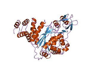
DNA polymerase subunit gamma is an enzyme that in humans is encoded by the POLG gene. Mitochondrial DNA polymerase is heterotrimeric, consisting of a homodimer of accessory subunits plus a catalytic subunit. The protein encoded by this gene is the catalytic subunit of mitochondrial DNA polymerase. Defects in this gene are a cause of progressive external ophthalmoplegia with mitochondrial DNA deletions 1 (PEOA1), sensory ataxic neuropathy dysarthria and ophthalmoparesis (SANDO), Alpers-Huttenlocher syndrome (AHS), and mitochondrial neurogastrointestinal encephalopathy syndrome (MNGIE).

<span class="mw-page-title-main">Twinkle (protein)</span> Mammalian protein found in Homo sapiens

Twinkle protein also known as twinkle mtDNA helicase is a mitochondrial protein that in humans is encoded by the TWNK gene located in the long arm of chromosome 10 (10q24.31).

<span class="mw-page-title-main">POLG2</span> Protein-coding gene in the species Homo sapiens

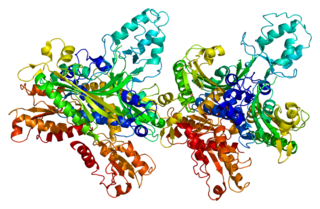
DNA polymerase subunit gamma-2, mitochondrial is a protein that in humans is encoded by the POLG2 gene. The POLG2 gene encodes a 55 kDa accessory subunit protein that imparts high processivity and salt tolerance to the catalytic subunit of DNA polymerase gamma, encoded by the POLG gene. Mutations in this gene result in autosomal dominant progressive external ophthalmoplegia with mitochondrial DNA deletions.

Mitochondrially encoded tRNA lysine also known as MT-TK is a transfer RNA which in humans is encoded by the mitochondrial MT-TK gene.

Mitochondrially encoded tRNA asparagine also known as MT-TN is a transfer RNA which in humans is encoded by the mitochondrial MT-TN gene.

Mitochondrially encoded tRNA tyrosine, also known as MT-TY, is a transfer RNA which in humans is encoded by the mitochondrial MT-TY gene.

Multi/minicore myopathy is a congenital myopathy usually caused by mutations in either the SEPN1 and RYR1 genes. It is characterised the presence of multifocal, well-circumscribed areas with reduction of oxidative staining and low myofibrillar ATPase on muscle biopsy. It is also known as Minicore myopathy, Multicore myopathy, Multiminicore myopathy, Minicore myopathy with external ophthalmoplegia, Multicore myopathy with external ophthalmoplegia and Multiminicore disease with external ophthalmoplegia.

<span class="mw-page-title-main">Mitochondrial DNA depletion syndrome</span> Medical condition

Mitochondrial DNA depletion syndrome, or Alper's disease, is any of a group of autosomal recessive disorders that cause a significant drop in mitochondrial DNA in affected tissues. Symptoms can be any combination of myopathic, hepatopathic, or encephalomyopathic. These syndromes affect tissue in the muscle, liver, or both the muscle and brain, respectively. The condition is typically fatal in infancy and early childhood, though some have survived to their teenage years with the myopathic variant and some have survived into adulthood with the SUCLA2 encephalomyopathic variant. There is currently no curative treatment for any form of MDDS, though some preliminary treatments have shown a reduction in symptoms.

<span class="mw-page-title-main">Sengers syndrome</span> Medical condition

Sengers syndrome is a rare autosomal recessive condition characterised by congenital cataract, hypertrophic cardiomyopathy, muscle weakness and lactic acidosis after exercise. In some cases, they are inherited, in which case they would be called congenital. In addition, heart disease and muscle disease are prevalent, and life expectancy is short for many patients.

Sensory ataxic neuropathy, dysarthria, and ophthalmoparesis, also known as SANDO syndrome, is a very rare genetic disorder which is characterized by ocular and nerve anomalies.

References

  1. 1 2 3 John P.Whitcher; Paul Riordan-Eva (2007-10-18). Vaughan & Asbury's general ophthalmology (17th ed.). McGraw-Hill Medical. p. 293. ISBN   978-0071443142.
  2. Birtel, Johannes; von Landenberg, Christina; et al. (2022). "Mitochondrial Retinopathy". Ophthalmology Retina. 6 (1): 65–79. doi: 10.1016/j.oret.2021.02.017 . PMID   34257060. S2CID   235822866.
  3. Yu Wai Man CY; et al. (2006). "Assessment of visual function in chronic progressive external ophthalmoplegia". Eye. 20 (5): 564–568. doi: 10.1038/sj.eye.6701924 . PMID   15920569.
  4. Millar, N; Newman N (1999). Walsh & Hoyt's Clinical Neuro-Ophthalmology, The Essentials (5th ed.).
  5. Houshmand M, Panahi MS, Hosseini BN, Dorraj GH, Tabassi AR (2006). "Investigation on mtDNA deletions and twinkle gene mutation (G1423C) in Iranian patients with chronic progressive external opthalmoplagia". Neurol India. 54 (2): 182–5. PMID   16804265.
  6. Zeviani M, Di Donauto S (2004). "Mitochondrial disorders". Brain. 127 (10): 2153–2172. doi: 10.1093/brain/awh259 . PMID   15358637.
  7. Copeland WC (2008). "Inherited Mitochondrial Diseases of DNA Replication". Annual Review of Medicine. 59: 131–146. doi:10.1146/annurev.med.59.053006.104646. PMC   2271032 . PMID   17892433.
  8. Van Goethem, Gert; Dermaut, Bart; Löfgren, Ann; Martin, Jean-Jacques; Van Broeckhoven, Christine (2001). "Mutation of POLG is associated with progressive external ophthalmoplegia characterized by mtDNA deletions". Nature Genetics. 28 (3): 211–212. doi:10.1038/90034. ISSN   1061-4036. PMID   11431686. S2CID   35417835.
  9. Behbehani R, Sharfuddin K, Anim JT (2007). "Mitochondrial ophthalmoplegia with fatigable weakness and elevated acetylcholine receptor antibody". J Neuroophthalmol. 27 (1): 41–4. doi: 10.1097/WNO.0b013e31803312fa . PMID   17414872.
  10. Ortube, MC; Bhola, R; Demer, JL (October 2006). "Orbital magnetic resonance imaging of extraocular muscles in chronic progressive external ophthalmoplegia: specific diagnostic findings". Journal of AAPOS. 10 (5): 414–8. doi:10.1016/j.jaapos.2006.04.012. PMC   1850670 . PMID   17070475.
  11. Omar A, Johnson LN (2007). "Tetracycline delays ocular motility decline in chronic progressive external ophthalmoplegia". Neurology. 68 (14): 1159–60. doi:10.1212/01.wnl.0000258659.21421.b0. PMID   17404203. S2CID   26226120.
  12. Rodriguez, MC; MacDonald, JR; Mahoney, DJ; Parise, G; Beal, MF; Tarnopolsky, MA (February 2007). "Beneficial effects of creatine, CoQ10, and lipoic acid in mitochondrial disorders". Muscle & Nerve. 35 (2): 235–42. doi:10.1002/mus.20688. PMID   17080429. S2CID   28962906.
  13. Ahn J, Kim NJ, Choung HK, Hwang SW, Sung M, Lee MJ, Khwarg SI (2008). "Frontalis sling operation using silicone rod for the correction of ptosis in chronic progressive external ophthalmoplegia". Br J Ophthalmol. 92 (12): 1685–8. doi:10.1136/bjo.2008.144816. hdl: 10371/62025 . PMID   18786957. S2CID   21058553.
  14. Tinley, C; Dawson, E; Lee, J (June 2010). "The management of strabismus in patients with chronic progressive external ophthalmoplegia". Strabismus. 18 (2): 41–7. doi:10.3109/09273971003758388. PMID   20521878. S2CID   20074185.

Further reading