Diabetes and deafness

Last updated
Diabetes and deafness (DAD)
Other namesDiabetes mellitus and deafness, maternally inherited, (MIDD); Diabetes-deafness syndrome, maternally transmitted; Ballinger-Wallace syndrome; Noninsulin-dependent diabetes mellitus with deafness; Diabetes mellitus, type II, with deafness
Mitochondrial inheritance.svg
This condition is inherited via a mitochondrial inheritance manner
Symptoms Noninsulin-dependent diabetes, deafness, may also have systemic symptoms including eye, muscle, brain, kidney, heart, and gastrointestinal abnormalities, rarely endocrine abnormalities and osteoporosis
CausesMutation in either MT-TL1, MT-TE, or MT-TK
Differential diagnosis Mitochondrial disease

Diabetes and deafness (DAD) or maternally inherited diabetes and deafness (MIDD) or mitochondrial diabetes is a subtype of diabetes which is caused from a mutation in mitochondrial DNA, which consists of a circular genome. It is associated with the genes MT-TL1, MT-TE, and MT-TK. [1] The point mutation at position 3243A>G, in gene MT-TL1 encoding tRNA leucine 1, is most common. [1] [2] [3] Because mitochondrial DNA is contributed to the embryo by the oocyte and not by spermatozoa, this disease is inherited from maternal family members only. [2] As indicated by the name, MIDD is characterized by diabetes and sensorineural hearing loss. [2] Some individuals also experience more systemic symptoms including eye, muscle, brain, kidney, heart, and gastrointestinal abnormalities, similar to other mitochondrial diseases. [4] [5] [6]

Contents

Signs and symptoms

As suggested by the name, patients with MIDD are subject to sensorineural hearing loss. [2] This begins with a reduction in the perception of frequencies above approximately 5 kHz which progressively declines, over the years, to severe hearing loss at all frequencies. [2] The diabetes that accompanies the hearing loss can be similar to Type 1 diabetes or Type 2 diabetes; however, Type 1-like diabetes is the more common form of the two. MIDD has also been associated with a number of other issues including kidney dysfunction, gastrointestinal problems, and cardiomyopathy. [4]

In the eye, MIDD is characterized by progressive atrophy of the retinal pigment epithelium. Initially, the fovea is spared. Thus, patients often have good visual acuity. However, over time the areas of atrophy expand with eventual loss of central vision. [7]

Table 1: Metabolically active organs that can be affected by the mitochondrial point mutation and the associated complication: [1] [2] [5] [6]

Organ affectedAssociated complication
Ear (cochlea) Sensorineural hearing loss
Brain (Hypothalamus)Short stature
Brain (general)Strokes, seizures, atrophy of cerebellum or cerebrum, ataxia, central nervous system disease, encephalatrophy, basal ganglia calcification, migraine, cerebral infarction, dysarthria
EyeMacular pattern dystrophy, macular degeneration, proliferated retinopathy, external ophthalmoplegia, ptosis
Heart Congestive heart failure, ventricular hypertrophy, arrhythmia
Kidney Focal segmental glomerulosclerosis, nephropathy
IntestineMalabsorption or constipation
Muscle Mitochondrial myopathy, peripheral neuropathy
Testes Hypogonadism
Adrenal glands Hypoaldosteronism
Bones Osteoporosis

Genetics

Penetrance and age of onset

MIDD represents 1% of people who have diabetes. Over 85% of people that carry the mutation in mitochondrial DNA at position 3243 present symptoms of diabetes. The average age at which people who have MIDD are typically diagnosed is 37 years old but has been seen to range anywhere between 11 years to 68 years old. Of these people with diabetes carrying the mitochondrial DNA mutation at position 3243, 75% experience sensorineural hearing loss. [2] In these cases, hearing loss normally appears before the onset of diabetes and is marked by a decrease in perception of high tone frequencies. [4] The associated hearing loss with diabetes is typically more common and more quickly declining in men than in women. [8]

Effect of mutation on tRNALeu(UUR)

Mitochondria have their own circular genome which contains 37 genes, of which 22 code for tRNAs. [9] These tRNAs play an essential role in protein synthesis by transporting amino acids to the ribosome. [2] MIDD is caused by an A to G substitution in the mitochondrial DNA at position 3243, which encodes tRNALeu(UUR). [2] This mutation is typically in heteroplasmic form. A mutation in this gene (A3243G) causes the native conformation to be destabilized, as well as dimerization in the tRNALeu(UUR). The uridine at the anticodon first position of the tRNALeu(UUR) is normally post- transcriptionally modified to ensure correct codon recognition. Such modification is known as taurine modification, which is decreased as a result of the improper structure of the tRNALeu(UUR). [10] Incorrect tRNALeu(UUR) structure also results in decreased aminoacylation. [9] The mutation has also been shown to result in decreased function of the tRNA and thus protein synthesis. [11]

Diabetes characteristics

The A3243G mutation in mitochondrial DNA can be present in any tissue, however, it is more commonly present in tissues with lower replication rates such as muscle. [4] The presence of this mutation can lead to decreased oxygen consumption as a result of reduced function of the respiratory chain and a decrease in oxidative phosphorylation. [12] In some people, this reduction in function of the respiratory chain is suggested to be caused by unbalanced amounts of proteins that are encoded by mitochondrial DNA, due to the presence of the A3243G mutation. [4] However, in other people, the same amount of mitochondrial proteins are generated, but their stability is compromised due to the improper incorporation of amino acids at the UUR codons of the mitochondrial mRNAs. This is a result of the mutated tRNALeu(UUR) with its decreased function in protein synthesis. [12] A decrease in function of the respiratory chain as a result of a mitochondrial DNA mutation could result in a decrease of ATP production. This decrease in ATP could have detrimental effects on other processes in the body. One such process is insulin secretion by pancreatic Beta-cells. [4] In pancreatic Beta-cells, precise levels of ATP/ADP regulate the opening and closing of the KATP channel, which controls the secretion of insulin. When mutations in the mitochondria disrupt the ATP/ADP ratio, this channel cannot function properly and this can result in a person being deficient in insulin. [4] Since the age of onset is later in a person's life, it has been suggested that age plays a role in contributing, along with the reduced ATP/ADP ratio, to the slow deterioration of the function of B-cells. [4]

Deafness characteristics

Hearing loss, as caused by the 3243 mitochondrial DNA mutation, is seen in the form of progressive cochlear dysfunction. Although the mechanism by which the mutation in the tRNALeu(UUR) causes this dysfunction of the cochlea is still under investigation, it has been hypothesized that it involves the ion pumps required for sound transduction. [13] As the mutation in the tRNALeu(UUR) leads to unbalanced amounts or unstable respiratory chain enzymes, respiration and oxidative phosphorylation are reduced, leading to lower levels of ATP. [4] [12] Naturally, the most metabolically active organs in a person will be affected by this ATP deficiency. Included in these metabolically active organs is the cochlear stria vascularis. [2] The stria vascularis and the hair cells, both essential to sound transduction, make use of ion pumps to regulate the concentration of ions including K+, Na+, and Ca2+ using ATP. Without sufficient levels of ATP, these concentration gradients are not maintained and this can lead to cell death in both the stria vascularis and the hair cells, causing hearing loss. [13]

Diagnosis

Physical exams, blood tests, family history, biopsy, DNA testing. [6] Mutations in mitochondrial genes MT-TE, MT-TL1, and MT-TK have been associated with MIDD. [1] The most common mutation is the 3243A>G transition in the mitochondrial tRNA-leucine 1 gene (MT-TL1). [1]

Treatment

Initial

Initially, the person is treated by dietary changes and hypoglycaemic agents. This does not last long before the person has to be started on insulin (within 2 years of diagnosis). [14]

See also

Related Research Articles

<span class="mw-page-title-main">Mitochondrial DNA</span> DNA located in mitochondria

Mitochondrial DNA is the DNA located in mitochondria, cellular organelles within eukaryotic cells that convert chemical energy from food into a form that cells can use, such as adenosine triphosphate (ATP). Mitochondrial DNA is only a small portion of the DNA in a eukaryotic cell; most of the DNA can be found in the cell nucleus and, in plants and algae, also in plastids such as chloroplasts.

<span class="mw-page-title-main">Mitochondrial disease</span> Spontaneously occurring or inherited disorder that involves mitochondrial dysfunction

Mitochondrial disease is a group of disorders caused by mitochondrial dysfunction. Mitochondria are the organelles that generate energy for the cell and are found in every cell of the human body except red blood cells. They convert the energy of food molecules into the ATP that powers most cell functions.

<span class="mw-page-title-main">Wolfram syndrome</span> Human disease

Wolfram syndrome, also called DIDMOAD, is a rare autosomal-recessive genetic disorder that causes childhood-onset diabetes mellitus, optic atrophy, and deafness as well as various other possible disorders including neurodegeneration.

<span class="mw-page-title-main">Leigh syndrome</span> Mitochondrial metabolism disease characterized by progressive loss of mental and movement abilities

Leigh syndrome is an inherited neurometabolic disorder that affects the central nervous system. It is named after Archibald Denis Leigh, a British neuropsychiatrist who first described the condition in 1951. Normal levels of thiamine, thiamine monophosphate, and thiamine diphosphate are commonly found, but there is a reduced or absent level of thiamine triphosphate. This is thought to be caused by a blockage in the enzyme thiamine-diphosphate kinase, and therefore treatment in some patients would be to take thiamine triphosphate daily. While the majority of patients typically exhibit symptoms between the ages of 3 and 12 months, instances of adult onset have also been documented.

<span class="mw-page-title-main">Human mitochondrial genetics</span> Study of the human mitochondrial genome

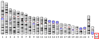
Human mitochondrial genetics is the study of the genetics of human mitochondrial DNA. The human mitochondrial genome is the entirety of hereditary information contained in human mitochondria. Mitochondria are small structures in cells that generate energy for the cell to use, and are hence referred to as the "powerhouses" of the cell.

<span class="mw-page-title-main">Mitochondrial myopathy</span> Medical condition

Mitochondrial myopathies are types of myopathies associated with mitochondrial disease. Adenosine triphosphate (ATP), the chemical used to provide energy for the cell, cannot be produced sufficiently by oxidative phosphorylation when the mitochondrion is either damaged or missing necessary enzymes or transport proteins. With ATP production deficient in mitochondria, there is an over-reliance on anaerobic glycolysis which leads to lactic acidosis either at rest or exercise-induced.

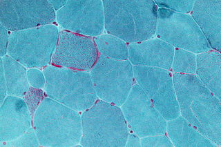
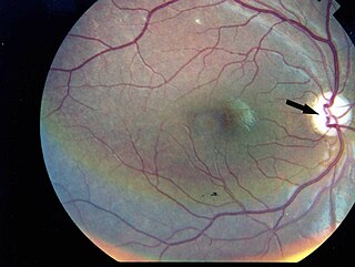
Kearns–Sayre syndrome (KSS), oculocraniosomatic disorder or oculocranionsomatic neuromuscular disorder with ragged red fibers is a mitochondrial myopathy with a typical onset before 20 years of age. KSS is a more severe syndromic variant of chronic progressive external ophthalmoplegia, a syndrome that is characterized by isolated involvement of the muscles controlling movement of the eyelid and eye. This results in ptosis and ophthalmoplegia respectively. KSS involves a combination of the already described CPEO as well as pigmentary retinopathy in both eyes and cardiac conduction abnormalities. Other symptoms may include cerebellar ataxia, proximal muscle weakness, deafness, diabetes mellitus, growth hormone deficiency, hypoparathyroidism, and other endocrinopathies. In both of these diseases, muscle involvement may begin unilaterally but always develops into a bilateral deficit, and the course is progressive. This discussion is limited specifically to the more severe and systemically involved variant.

<span class="mw-page-title-main">MELAS syndrome</span> Medical condition

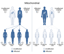
Mitochondrial encephalopathy, lactic acidosis, and stroke-like episodes (MELAS) is one of the family of mitochondrial diseases, which also include MIDD, MERRF syndrome, and Leber's hereditary optic neuropathy. It was first characterized under this name in 1984. A feature of these diseases is that they are caused by defects in the mitochondrial genome which is inherited purely from the female parent. The most common MELAS mutation is mitochondrial mutation, mtDNA, referred to as m.3243A>G.

<span class="mw-page-title-main">MERRF syndrome</span> Medical condition

MERRF syndrome is a mitochondrial disease. It is extremely rare, and has varying degrees of expressivity owing to heteroplasmy. MERRF syndrome affects different parts of the body, particularly the muscles and nervous system. The signs and symptoms of this disorder appear at an early age, generally childhood or adolescence. The causes of MERRF syndrome are difficult to determine, but because it is a mitochondrial disorder, it can be caused by the mutation of nuclear DNA or mitochondrial DNA. The classification of this disease varies from patient to patient, since many individuals do not fall into one specific disease category. The primary features displayed on a person with MERRF include myoclonus, seizures, cerebellar ataxia, myopathy, and ragged red fibers (RRF) on muscle biopsy, leading to the disease's name. Secondary features include dementia, optic atrophy, bilateral deafness, peripheral neuropathy, spasticity, or multiple lipomata. Mitochondrial disorders, including MERRFS, may present at any age.

<span class="mw-page-title-main">MT-RNR1</span> SSU rRNA of the mitochondrial ribosome

Mitochondrially encoded 12S ribosomal RNA is the SSU rRNA of the mitochondrial ribosome. In humans, 12S is encoded by the MT-RNR1 gene and is 959 nucleotides long. MT-RNR1 is one of the 37 genes contained in animal mitochondria genomes. Their 2 rRNA, 22 tRNA and 13 mRNA genes are very useful in phylogenetic studies, in particular the 12S and 16S rRNAs. The 12S rRNA is the mitochondrial homologue of the prokaryotic 16S and eukaryotic nuclear 18S ribosomal RNAs. Mutations in the MT-RNR1 gene may be associated with hearing loss. The rRNA gene also encodes a peptide MOTS-c, also known as Mitochondrial-derived peptide MOTS-c or Mitochondrial open reading frame of the 12S rRNA-c.

<span class="mw-page-title-main">MT-ND2</span> Mitochondrial gene coding for a protein involved in the respiratory chain

MT-ND2 is a gene of the mitochondrial genome coding for the NADH dehydrogenase 2 (ND2) protein. The ND2 protein is a subunit of NADH dehydrogenase (ubiquinone), which is located in the mitochondrial inner membrane and is the largest of the five complexes of the electron transport chain. Variants of human MT-ND2 are associated with mitochondrial encephalomyopathy, lactic acidosis, and stroke-like episodes (MELAS), Leigh's syndrome (LS), Leber's hereditary optic neuropathy (LHON) and increases in adult BMI.

<span class="mw-page-title-main">MT-ATP6</span> Mitochondrial protein-coding gene whose product is involved in ATP synthesis

MT-ATP6 is a mitochondrial gene with the full name 'mitochondrially encoded ATP synthase membrane subunit 6' that encodes the ATP synthase Fo subunit 6. This subunit belongs to the Fo complex of the large, transmembrane F-type ATP synthase. This enzyme, which is also known as complex V, is responsible for the final step of oxidative phosphorylation in the electron transport chain. Specifically, one segment of ATP synthase allows positively charged ions, called protons, to flow across a specialized membrane inside mitochondria. Another segment of the enzyme uses the energy created by this proton flow to convert a molecule called adenosine diphosphate (ADP) to ATP. Mutations in the MT-ATP6 gene have been found in approximately 10 to 20 percent of people with Leigh syndrome.

Mitochondrially encoded tRNA leucine 1 (UUA/G) also known as MT-TL1 is a transfer RNA which in humans is encoded by the mitochondrial MT-TL1 gene.

Mitochondrially encoded tRNA histidine, also known as MT-TH, is a transfer RNA which, in humans, is encoded by the mitochondrial MT-TH gene.

<span class="mw-page-title-main">MT-ND1</span> Mitochondrial gene coding for a protein involved in the respiratory chain

MT-ND1 is a gene of the mitochondrial genome coding for the NADH-ubiquinone oxidoreductase chain 1 (ND1) protein. The ND1 protein is a subunit of NADH dehydrogenase, which is located in the mitochondrial inner membrane and is the largest of the five complexes of the electron transport chain. Variants of the human MT-ND1 gene are associated with mitochondrial encephalomyopathy, lactic acidosis, and stroke-like episodes (MELAS), Leigh's syndrome (LS), Leber's hereditary optic neuropathy (LHON) and increases in adult BMI.

Mitochondrially encoded tRNA glutamic acid also known as MT-TE is a transfer RNA which in humans is encoded by the mitochondrial MT-TE gene. MT-TE is a small 69 nucleotide RNA that transfers the amino acid glutamic acid to a growing polypeptide chain at the ribosome site of protein synthesis during translation.

Mitochondrially encoded tRNA lysine also known as MT-TK is a transfer RNA which in humans is encoded by the mitochondrial MT-TK gene.

Mitochondrially encoded tRNA asparagine also known as MT-TN is a transfer RNA which in humans is encoded by the mitochondrial MT-TN gene.

Mitochondrially encoded tRNA arginine also known as MT-TR is a transfer RNA which in humans is encoded by the mitochondrial MT-TR gene.

<span class="mw-page-title-main">Mitochondrial ribosome</span> Protein complex

The mitochondrial ribosome, or mitoribosome, is a protein complex that is active in mitochondria and functions as a riboprotein for translating mitochondrial mRNAs encoded in mtDNA. The mitoribosome is attached to the inner mitochondrial membrane. Mitoribosomes, like cytoplasmic ribosomes, consist of two subunits — large (mt-LSU) and small (mt-SSU). Mitoribosomes consist of several specific proteins and fewer rRNAs. While mitochondrial rRNAs are encoded in the mitochondrial genome, the proteins that make up mitoribosomes are encoded in the nucleus and assembled by cytoplasmic ribosomes before being implanted into the mitochondria.

References

  1. 1 2 3 4 5 "Diabetes and Deafness, Maternally Inherited; MIDD". Online Mendelian Inheritance in Man (OMIM). Retrieved 2024-03-02.
  2. 1 2 3 4 5 6 7 8 9 10 Murphy R, Turnbull DM, Walker M, Hattersley AT (April 2008). "Clinical features, diagnosis and management of maternally inherited diabetes and deafness (MIDD) associated with the 3243A>G mitochondrial point mutation". Diabetic Medicine. 25 (4): 383–99. doi:10.1111/j.1464-5491.2008.02359.x. PMID   18294221. S2CID   205548877.
  3. de Andrade PB, Rubi B, Frigerio F, van den Ouweland JM, Maassen JA, Maechler P (August 2006). "Diabetes-associated mitochondrial DNA mutation A3243G impairs cellular metabolic pathways necessary for beta cell function". Diabetologia. 49 (8): 1816–1826. doi:10.1007/s00125-006-0301-9. PMID   16736129.
  4. 1 2 3 4 5 6 7 8 9 Maassen JA, 'T Hart LM, Van Essen E, Heine RJ, Nijpels G, Jahangir Tafrechi RS, et al. (February 2004). "Mitochondrial diabetes: molecular mechanisms and clinical presentation". Diabetes. 53 (Suppl 1): S103–S109. doi:10.2337/diabetes.53.2007.S103. PMID   14749274.
  5. 1 2 Finsterer J, Frank M (September 2018). "The Tip of the Iceberg in Maternally Inherited Diabetes and Deafness". Oman Medical Journal. 33 (5): 437–440. doi:10.5001/omj.2018.80. PMC   6131922 . PMID   30210725.
  6. 1 2 3 Yang M, Xu L, Xu C, Cui Y, Jiang S, Dong J, et al. (November 2021). "The Mutations and Clinical Variability in Maternally Inherited Diabetes and Deafness: An Analysis of 161 Patients". Frontiers in Endocrinology. 12: 728043. doi: 10.3389/fendo.2021.728043 . PMC   8654930 . PMID   34899594.
  7. Müller PL, Treis T, Pfau M, Esposti SD, Alsaedi A, Maloca P, et al. (May 2020). "Progression of Retinopathy Secondary to Maternally Inherited Diabetes and Deafness - Evaluation of Predicting Parameters". American Journal of Ophthalmology. 213: 134–144. doi:10.1016/j.ajo.2020.01.013. PMID   31987901. S2CID   210935778.
  8. Uimonen S, Moilanen JS, Sorri M, Hassinen IE, Majamaa K (April 2001). "Hearing impairment in patients with 3243A-->G mtDNA mutation: phenotype and rate of progression". Human Genetics. 108 (4): 284–289. doi:10.1007/s004390100475. PMID   11379873. S2CID   20513165.
  9. 1 2 Wittenhagen LM, Kelley SO (August 2002). "Dimerization of a pathogenic human mitochondrial tRNA". Nature Structural Biology. 9 (8): 586–590. PMID   12101407.
  10. Suzuki T, Suzuki T, Wada T, Saigo K, Watanabe K (December 2002). "Taurine as a constituent of mitochondrial tRNAs: new insights into the functions of taurine and human mitochondrial diseases". The EMBO Journal. 21 (23): 6581–6589. doi:10.1093/emboj/cdf656. PMC   136959 . PMID   12456664.
  11. Flierl A, Reichmann H, Seibel P (October 1997). "Pathophysiology of the MELAS 3243 transition mutation". The Journal of Biological Chemistry. 272 (43): 27189–27196. doi: 10.1074/jbc.272.43.27189 . PMID   9341162.
  12. 1 2 3 Janssen GM, Maassen JA, van Den Ouweland JM (October 1999). "The diabetes-associated 3243 mutation in the mitochondrial tRNA(Leu(UUR)) gene causes severe mitochondrial dysfunction without a strong decrease in protein synthesis rate". The Journal of Biological Chemistry. 274 (42): 29744–29748. doi: 10.1074/jbc.274.42.29744 . PMID   10514449.
  13. 1 2 Yamasoba T, Oka Y, Tsukuda K, Nakamura M, Kaga K (January 1996). "Auditory findings in patients with maternally inherited diabetes and deafness harboring a point mutation in the mitochondrial transfer RNA(Leu) (UUR) gene". The Laryngoscope. 106 (1 Pt 1): 49–53. doi:10.1097/00005537-199601000-00010. PMID   8544627. S2CID   44601058.
  14. "Mitochondrial diabetes - Other types of diabetes mellitus". Diapedia, The Living Textbook of Diabetes. Retrieved 2018-02-06.