Retinopathy

Last updated
Retinopathy
Fundus retinopathy EDA03.JPG
Retinopathy in fundus of eye
Specialty Ophthalmology   OOjs UI icon edit-ltr-progressive.svg

Retinopathy is any damage to the retina of the eyes, which may cause vision impairment. [1] Retinopathy often refers to retinal vascular disease, or damage to the retina caused by abnormal blood flow. [2] Age-related macular degeneration is technically included under the umbrella term retinopathy but is often discussed as a separate entity. Retinopathy, or retinal vascular disease, can be broadly categorized into proliferative and non-proliferative types. Frequently, retinopathy is an ocular manifestation of systemic disease as seen in diabetes or hypertension. [3] Diabetes is the most common cause of retinopathy in the U.S. as of 2008. [4] Diabetic retinopathy is the leading cause of blindness in working-aged people. [5] It accounts for about 5% of blindness worldwide and is designated a priority eye disease by the World Health Organization. [6]

Contents

Signs and symptoms

Many people often do not have symptoms until very late in their disease course. Patients often become symptomatic when there is irreversible damage. [7] Symptoms are usually not painful and can include:

Pathophysiology

The development of retinopathy can be broken down into proliferative and non-proliferative types. Both types cause disease by altering the normal blood flow to the retina through different mechanisms. The retina is supplied by small vessel branches from the central retinal artery. [8] Proliferative retinopathy refers to damage caused by abnormal blood vessel growth. [9] Normally, angiogenesis is a natural part of tissue growth and formation. When there is an unusually high or fast rate of angiogenesis, there is an overgrowth of blood vessels called neovascularization. In the non-proliferative type, abnormal blood flow to the retina occurs due to direct damage or compromise of the blood vessels themselves. Many causes of retinopathy may cause both proliferative and non-proliferative types, though some causes are more associated one type.

Non-proliferative retinopathy

Non-proliferative retinopathy is often caused by direct damage or remodeling of the small blood vessels supplying the retina. [8] Many common causes of non-proliferative damage include hypertensive retinopathy, retinopathy of prematurity, radiation retinopathy, solar retinopathy, sickle cell retinopathy, and anemic retinopathy [10] (including secondary to vitamin B12 deficiency [11] ).

There are three main mechanisms of damage in non-proliferative retinopathy: blood vessel damage or remodeling, direct retinal damage, or occlusion of the blood vessels. The first mechanism is indirect damage by altering the blood vessels that supply the retina. In the case of hypertension, high pressures in the system cause the walls of the artery to thicken, which effectively reduces the amount of blood flow to the retina. [8] This reduction in flow causes tissue ischemia leading to damage. Atherosclerosis, or hardening and narrowing of blood vessels, also reduces flow to the retina. The second mechanism is direct damage to the retina usually caused by free radicals that causes oxidative damage to the retina itself. [12] Radiation, solar retinopathy, and retinopathy of prematurity fall under this category. The third common mechanism is occlusion of blood flow. This can be caused by either physically blocking the vessels of the retinal artery branches or causing the arteries to narrow. [2] Again, the result is reduced blood flow to the retina causing tissue damage. Sickle cell disease compromises blood flow by causing blood to sludge, or thicken and flow slowly, through the retinal arteries. Other disorders that cause hyperviscosity syndrome may also cause blood sludging. Lastly, clots or central artery thrombosis directly blocks flow to the retina causing the cells to die.

Proliferative retinopathy

Proliferative retinopathy during exam Fundus Proliferative retinopathy EDA01.JPG
Proliferative retinopathy during exam

Proliferative retinopathy is the result of aberrant blood flow to the retina due to blood vessel overgrowth, or neovascularization. These pathologically overgrown blood vessels are often fragile, weak, and ineffective at perfusing the retinal tissues. [13] These weak, fragile vessels are also often leaky, allowing fluids, protein, and other debris to leach out into the retina. They are also prone to hemorrhage due to their poor strength. This makes proliferative types of retinopathy more risky since vessel hemorrhaging often leads to vision loss and blindness. [14] Many of the causes mentioned in non-proliferative retinopathy may also cause proliferative retinopathy at later stages. Angiogenesis and neovascularization tend to be a later manifestation of non-proliferative retinopathy. Many types of non-proliferative retinopathies result in tissue ischemia or direct retinal damage. The body responds by trying to increase blood flow to damaged retinal tissues. [15] Diabetes mellitus, which causes diabetic retinopathy, is the most common cause of proliferative retinopathy in the world. [16]

Other causes

Genetic mutations are rare causes of certain retinopathies and are usually X-linked including NDP family of genes causing Norrie disease, FEVR, and Coats disease among others. There is emerging evidence that there may be a genetic predisposition in patients who develop retinopathy of prematurity and diabetic retinopathy. [17] [18] Trauma, especially to the head, and several diseases may cause Purtscher's retinopathy. Physical exertion like weight lifting and aerobic exercise, coughing, sneezing, straining at stool, vomiting, sexual intercourse, blowing up balloons, blowing musical instruments, cardiopulmonary resuscitation or compression injuries may cause valsalva retinopathy. [19]

Diagnosis

Retinopathy is diagnosed by an ophthalmologist or an optometrist during eye examination. The clinician will need to examine the retina, at the back of the eye, to make this diagnosis. There are several ways to examine the retina. The clinician can directly view the retina by looking through the pupil with a light. In most cases, the clinician will dilate the pupil to make for better visualization. [20] Stereoscopic fundus photography is the gold standard for the diagnosis of retinopathy.

Access to care

Telemedicine programs are available that allow primary care clinics to take images using specially designed retinal imaging equipment which can then be shared electronically with specialists at other locations for review. [21] In 2009, Community Health Center, Inc. implemented a telemedicine retinal screening program for low-income patients with diabetes as part of those patients' annual visits at the Federally Qualified Health Center. [22]

Prevention

Retinopathy is often secondary to diseases such as diabetes or hypertension. Controlling blood sugar levels and blood pressure have been shown to help decrease incidence of retinopathy.

Blood sugar control: If someone has diabetes, or is at high risk for diabetes, it is important for them to have their blood sugar levels checked. The gold standard blood sugar test is the A1C test. Many studies have suggested that lowering A1C levels in someone with elevated A1C levels can lower the incidence and progression of retinopathy. Fortunately, blood sugar control can have benefits beyond just the eye. A primary care physician can help with blood sugar control strategies.

Blood pressure control: Controlling blood pressure can also lower the incidence and progression of retinopathy. A primary care physician can help with blood pressure control strategies.

Other: Besides blood sugar and blood pressure control, there are other modifications that can help. Regular exercise may help lower the incidence and progression of retinopathy. If someone has sleep apnea, treatment of sleep apnea may help as well.

Treatment

Treatment is based on the cause of the retinopathy and may include laser therapy to the retina. Laser photocoagulation therapy has been the standard treatment for many types of retinopathy. Evidence shows that laser therapy is generally safe and improves visual symptoms in sickle cell and diabetic retinopathy. [23] [24] In recent years targeting the pathway controlling vessel growth or angiogenesis has been promising. Vascular endothelial growth factor (VEGF) seems to play a vital role in promoting neovascularization. Using anti-VEGF drugs (antibodies to sequester the growth factor), research have shown significant reduction in the extent of vessel outgrowth. Low quality evidence supports the use of anti-VEGF antibodies, such as bevacizumab or pegaptanib which seems to improve outcomes when used in conjunction with laser therapy to treat retinopathy of prematurity, longer term systemic effects are not known however. [25] The evidence is poorer for treatment of diabetic retinopathy. Use of anti-VEGF drugs did not appear to improve outcomes in a clinically significant way when compared to standard laser therapy for diabetic retinopathy. [26]

Epidemiology

The two most common causes of retinopathy include diabetic retinopathy and retinopathy of prematurity. Diabetic retinopathy affects about 5 million people and retinopathy of prematurity affect about 50,000 premature infants each year worldwide. [6] [27] Hypertensive retinopathy is the next most common cause affecting anywhere from 3 to 14% of all non-diabetic adults. [28]

See also

Related Research Articles

<span class="mw-page-title-main">Diabetic retinopathy</span> Medical condition

Diabetic retinopathy, is a medical condition in which damage occurs to the retina due to diabetes. It is a leading cause of blindness in developed countries.

The National Eye Institute (NEI) is part of the U.S. National Institutes of Health (NIH), an agency of the U.S. Department of Health and Human Services. The mission of NEI is "to eliminate vision loss and improve quality of life through vision research." NEI consists of two major branches for research: an extramural branch that funds studies outside NIH and an intramural branch that funds research on the NIH campus in Bethesda, Maryland. Most of the NEI budget funds extramural research.

Retinopathy of prematurity (ROP), also called retrolental fibroplasia (RLF) and Terry syndrome, is a disease of the eye affecting prematurely born babies generally having received neonatal intensive care, in which oxygen therapy is used because of the premature development of their lungs. It is thought to be caused by disorganized growth of retinal blood vessels and may result in scarring and retinal detachment. ROP can be mild and may resolve spontaneously, but it may lead to blindness in serious cases. Thus, all preterm babies are at risk for ROP, and very low birth-weight is an additional risk factor. Both oxygen toxicity and relative hypoxia can contribute to the development of ROP.

<span class="mw-page-title-main">Macular edema</span> Medical condition

Macular edema occurs when fluid and protein deposits collect on or under the macula of the eye and causes it to thicken and swell (edema). The swelling may distort a person's central vision, because the macula holds tightly packed cones that provide sharp, clear, central vision to enable a person to see detail, form, and color that is directly in the centre of the field of view.

<span class="mw-page-title-main">Retinal detachment</span> Medical condition of the eye

Retinal detachment is a disorder of the eye in which the retina peels away from its underlying layer of support tissue. Initial detachment may be localized, but without rapid treatment the entire retina may detach, leading to vision loss and blindness. It is a surgical emergency.

Retinal hemorrhage is a disorder of the eye in which bleeding occurs in the retina, the light sensitive tissue, located on the back wall of the eye. There are photoreceptor cells in the retina called rods and cones, which transduce light energy into nerve signals that can be processed by the brain to form visual images. Retinal hemorrhage is strongly associated with child abuse in infants and young children and often leaves such abused infants permanently blind. In older children and adults, retinal hemorrhage can be caused by several medical conditions such as hypertension, retinal vein occlusion, anemia, leukemia or diabetes.

Neovascularization is the natural formation of new blood vessels, usually in the form of functional microvascular networks, capable of perfusion by red blood cells, that form to serve as collateral circulation in response to local poor perfusion or ischemia.

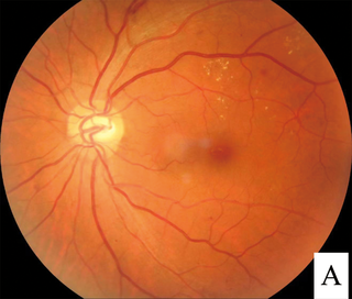
Eales disease is a type of obliterative vasculopathy, also known as angiopathia retinae juvenilis, periphlebitis retinae or primary perivasculitis of the retina. It was first described by the British ophthalmologist Henry Eales (1852–1913) in 1880 and is a rare ocular disease characterized by inflammation and possible blockage of retinal blood vessels, abnormal growth of new blood vessels (neovascularization), and recurrent retinal and vitreal hemorrhages.

Rubeosis iridis is a medical condition of the iris of the eye in which new abnormal blood vessels are found on the surface of the iris.

<span class="mw-page-title-main">Dilated fundus examination</span>

Dilated fundus examination (DFE) is a diagnostic procedure that uses mydriatic eye drops to dilate or enlarge the pupil in order to obtain a better view of the fundus of the eye. Once the pupil is dilated, examiners use ophthalmoscopy to view the eye's interior, which makes it easier to assess the retina, optic nerve head, blood vessels, and other important features. DFE has been found to be a more effective method for evaluating eye health when compared to non-dilated examination, and is the best method of evaluating structures behind the iris. It is frequently performed by ophthalmologists and optometrists as part of an eye examination.

Ocular ischemic syndrome is the constellation of ocular signs and symptoms secondary to severe, chronic arterial hypoperfusion to the eye. Amaurosis fugax is a form of acute vision loss caused by reduced blood flow to the eye; it may be a warning sign of an impending stroke, as both stroke and retinal artery occlusion can be caused by thromboembolism due to atherosclerosis elsewhere in the body. Consequently, those with transient blurring of vision are advised to urgently seek medical attention for a thorough evaluation of the carotid artery. Anterior segment ischemic syndrome is a similar ischemic condition of anterior segment usually seen in post-surgical cases. Retinal artery occlusion leads to rapid death of retinal cells, thereby resulting in severe loss of vision.

<span class="mw-page-title-main">Cotton wool spots</span> Medical condition of the eye

Cotton wool spots are opaque fluffy white patches on the retina of the eye that are considered an abnormal finding during a funduscopic exam. Cotton wool spots are typically a sign of another disease state, most common of which is diabetic retinopathy. The irregularly shaped white patches are a result of ischemia, or reduced blood flow and oxygen, in the retinal nerve fiber layer, which is located in the distribution of the capillaries of the superficial layer of the retina. These areas with reduced blood flow reflect the obstruction of axoplasmic flow due to mechanical or vascular causes and the consequential accumulation as a result of decreased axonal transport. This reduced axonal transport can then cause swelling or bulging on the surface layer of the retina, increasing the potential for nerve fiber damage.

<span class="mw-page-title-main">Macular telangiectasia</span> Disease of the retina affecting central vision

Macular telangiectasia is a condition of the retina, the light-sensing tissue at the back of the eye that causes gradual deterioration of central vision, interfering with tasks such as reading and driving.

<span class="mw-page-title-main">Laser coagulation</span> Procedure widely used in eye surgery

Laser coagulation or laser photocoagulation surgery is used to treat a number of eye diseases and has become widely used in recent decades. During the procedure, a laser is used to finely cauterize ocular blood vessels to attempt to bring about various therapeutic benefits.

<span class="mw-page-title-main">Intraretinal microvascular abnormalities</span> Medical condition

Intraretinal microvascular abnormalities (IRMA) are abnormalities of the blood vessels that supply the retina of the eye, a sign of diabetic retinopathy. IRMA can be difficult to distinguish from and is likely a precursor to retinal neovascularization. One way to distinguish IRMA from retinal neovascularization is to perform fluorescein angiography. Since IRMA blood vessels are patent, unlike neovascular vessels, they do not leak, and therefore exhibit hyperfluorescence on fluorescein angiography.

<span class="mw-page-title-main">Familial exudative vitreoretinopathy</span> Retinal vascular disease

Familial exudative vitreoretinopathy is a genetic disorder affecting the growth and development of blood vessels in the retina of the eye. This disease can lead to visual impairment and sometimes complete blindness in one or both eyes. FEVR is characterized by incomplete vascularization of the peripheral retina. This can lead to the growth of new blood vessels which are prone to leakage and hemorrhage and can cause retinal folds, tears, and detachments. Treatment involves laser photocoagulation of the avascular portions of the retina to reduce new blood vessel growth and risk of complications including leakage of retinal blood vessels and retinal detachments.

<span class="mw-page-title-main">Radiation retinopathy</span> Medical condition

Radiation retinopathy is damage to retina due to exposure to ionizing radiation. Radiation retinopathy has a delayed onset, typically after months or years of radiation, and is slowly progressive. In general, radiation retinopathy is seen around 18 months after treatment with external-beam radiation and with brachytherapy. The time of onset of radiation retinopathy is between 6 months to 3 years.

Joan Whitten Miller is a Canadian-American ophthalmologist and scientist who has made notable contributions to the treatment and understanding of eye disorders. She is credited for developing photodynamic therapy (PDT) with verteporfin (Visudyne), the first pharmacologic therapy for retinal disease. She also co-discovered the role of vascular endothelial growth factor (VEGF) in eye disease and demonstrated the therapeutic potential of VEGF inhibitors, forming the scientific basis of anti-VEGF therapy for age-related macular degeneration (AMD), diabetic retinopathy, and related conditions.

Sickle cell retinopathy can be defined as retinal changes due to blood vessel damage in the eye of a person with a background of sickle cell disease. It can likely progress to loss of vision in late stages due to vitreous hemorrhage or retinal detachment. Sickle cell disease is a structural red blood cell disorder leading to consequences in multiple systems. It is characterized by chronic red blood cell destruction, vascular injury, and tissue ischemia causing damage to the brain, eyes, heart, lungs, kidneys, spleen, and musculoskeletal system.

Drug abuse retinopathy is damage to the retina of the eyes caused by chronic drug abuse. Types of retinopathy caused by drug abuse include maculopathy, Saturday night retinopathy, and talc retinopathy. Common symptoms include temporary and permanent vision loss, blurred vision, and night blindness. Substances commonly associated with this condition include poppers, heroin, cocaine, methamphetamine, tobacco, and alcohol.

References

  1. "Definition of RETINOPATHY". www.merriam-webster.com. Retrieved 2017-03-01.
  2. 1 2 Robbins SL, Kumar V, Cotran RS (2010). Robbins and Cotran Pathologic Basis of Disease: expertconsult (8th ed.). Philadelphia, Pa: Elsevier, Saunders. pp. 1616–1617. ISBN   978-1-4160-3121-5.
  3. "Retina and vitreous". Ophthalmology (2011–2012 ed.). American Academy of Ophthalmology. 2012. p. 271. ISBN   9781615251193.
  4. "Diabetic Retinopathy" (PDF). Center for Disease Control. Retrieved March 1, 2017.
  5. Cheung N, Mitchell P, Wong TY (July 2010). "Diabetic retinopathy". Lancet. 376 (9735): 124–136. doi:10.1016/S0140-6736(09)62124-3. PMID   20580421. S2CID   25347463.
  6. 1 2 "WHO | Priority eye diseases". www.who.int. Archived from the original on March 22, 2006. Retrieved 2017-03-03.
  7. "Diabetic retinopathy: Classification and clinical features". www.uptodate.com. Retrieved 2017-03-09.
  8. 1 2 3 Porth C, Matfin G, eds. (2009-01-01). Pathophysiology : concepts of altered health states. Wolters Kluwer Health/Lippincott Williams & Wilkins. p. 756. ISBN   9780781766166. OCLC   181600926.
  9. "Chapter 10. Retina | Vaughan & Asbury's General Ophthalmology, 18e | AccessMedicine | McGraw-Hill Medical". accessmedicine.mhmedical.com. Retrieved 2017-03-03.
  10. Carraro MC, Rossetti L, Gerli GC (October 2001). "Prevalence of retinopathy in patients with anemia or thrombocytopenia". European Journal of Haematology. 67 (4): 238–244. doi:10.1034/j.1600-0609.2001.00539.x. PMID   11860445. S2CID   20875243.
  11. Jojo V, Singh P (August 2020). "The eye: A lifesaver! An unusual case of Anemic Retinopathy secondary to Malnutrition and its recovery". Journal of Family Medicine and Primary Care. 9 (8): 4421–4424. doi: 10.4103/jfmpc.jfmpc_577_20 . PMC   7586514 . PMID   33110874.
  12. Youseff PN, Sheibani N, Albert DM. Retinal light toxicity. Eye (Lond). 2011; 25(1):1–14.
  13. "Use of the Hand-Held Ophthalmoscope | Harrison's Principles of Internal Medicine, 19e | AccessMedicine | McGraw-Hill Medical". accessmedicine.mhmedical.com. Retrieved 2017-03-06.
  14. "Chapter 52. Atherothrombosis: Disease Burden, Activity, and Vulnerability | Hurst's The Heart, 13e | AccessMedicine | McGraw-Hill Medical". accessmedicine.mhmedical.com. Retrieved 2017-03-06.
  15. "Chapter 10. Retina | Vaughan & Asbury's General Ophthalmology, 18e | AccessMedicine | McGraw-Hill Medical". accessmedicine.mhmedical.com. Retrieved 2017-03-07.
  16. "Diabetic Retinopathy | National Eye Institute". nei.nih.gov. Retrieved 2017-03-07.
  17. Hartnett ME, Cotten CM (December 2015). "Genomics in the neonatal nursery: Focus on ROP". Seminars in Perinatology. 39 (8): 604–610. doi:10.1053/j.semperi.2015.09.007. PMC   4644692 . PMID   26477493.
  18. Zhang Y, Xia W, Lu P, Yuan H (2016-11-06). "The Association between VDR Gene Polymorphisms and Diabetic Retinopathy Susceptibility: A Systematic Review and Meta-Analysis". BioMed Research International. 2016: 5305282. doi: 10.1155/2016/5305282 . PMC   5116338 . PMID   27891515.
  19. "Valsalva Retinopathy - EyeWiki". eyewiki.aao.org. Retrieved 2022-04-24.
  20. "Diabetic retinopathy: Screening". www.uptodate.com. Retrieved 2017-03-07.
  21. "Remote Retinal Screening Facilitates Diagnosis and Treatment of Retinopathy for Poor and/or Uninsured Patients With Diabetes in Rural California". Agency for Healthcare Research and Quality. 2013-10-03. Retrieved 2013-08-01.
  22. "Telemedicine-Based Eye Examinations Enhance Access, Reduce Costs, and Increase Satisfaction for Low-Income and Minority Patients with Diabetes". Agency for Healthcare Research and Quality. 2013-07-17. Retrieved 2013-08-01.
  23. Evans JR, Michelessi M, Virgili G (November 2014). "Laser photocoagulation for proliferative diabetic retinopathy". The Cochrane Database of Systematic Reviews. 2014 (11): CD011234. doi:10.1002/14651858.CD011234.pub2. PMC   6823265 . PMID   25420029.
  24. Myint KT, Sahoo S, Thein AW, Moe S, Ni H (December 2022). "Laser therapy for retinopathy in sickle cell disease". The Cochrane Database of Systematic Reviews. 2022 (12): CD010790. doi:10.1002/14651858.CD010790.pub3. PMC   9744465 . PMID   36508693.
  25. Sankar MJ, Sankar J, Chandra P (January 2018). "Anti-vascular endothelial growth factor (VEGF) drugs for treatment of retinopathy of prematurity". The Cochrane Database of Systematic Reviews. 1 (1): CD009734. doi:10.1002/14651858.CD009734.pub3. PMC   6491066 . PMID   29308602.
  26. Martinez-Zapata MJ, Salvador I, Martí-Carvajal AJ, Pijoan JI, Cordero JA, Ponomarev D, et al. (March 2023). "Anti-vascular endothelial growth factor for proliferative diabetic retinopathy". The Cochrane Database of Systematic Reviews. 2023 (3): CD008721. doi:10.1002/14651858.CD008721.pub3. PMC   10026605 . PMID   36939655.
  27. "Retinopathy of Prematurity – Europe". American Academy of Ophthalmology. 2013-11-07. Retrieved 2017-03-03.
  28. "Retinal Physician -". www.retinalphysician.com. Retrieved 2017-03-03.