Laser coagulation

Last updated
Laser coagulation
Transpupilliarnaia lazernaia koaguliatsiia setchatki.jpg
Other namesLaser photocoagulation
MeSH D017075

Laser coagulation or laser photocoagulation surgery is used to treat a number of eye diseases and has become widely used in recent decades. During the procedure, a laser is used to finely cauterize ocular blood vessels to attempt to bring about various therapeutic benefits.

Contents

The procedure is used mostly to close blood vessels in the eye, in certain kinds of diabetic retinopathy; it is no longer used in age-related macular degeneration in favor of anti-VEGF drugs.

Medical uses

Diabetic retinopathy

The American Academy of Ophthalmology practice guidelines recommend laser coagulation for people who have both mild to moderate nonproliferative diabetic retinopathy (NPDR) and clinically significant macular edema outside the fovea; treatment with anti-VEGF drugs is better than laser coagulation for clinically significant macular edema in the fovea. [1] For people with severe NPDR and no macular edema, the AAO recommends laser photocoagulation for the whole retina; when there is macular edema, the laser coagulation focused on major lesions is recommended. [1] While there is evidence that anti-VEGF drugs is useful for proliferative diabetic retinopathy, laser coagulation across the whole retina is still preferred in the AAO recommendations, as there is long-term follow up data for laser treatment but not for drug treatment. [1]

Diabetic macular edema

Anti-VEGF drugs are potentially superior to laser coagulation for diabetic macular edema; some specialists are using the drugs over laser coagulation. [2]

Macular degeneration

The American Academy of Ophthalmology practice guidelines do not recommend laser coagulation therapy for macular degeneration, but said that it may be useful in people with new blood vessels in the choroid outside of the fovea who do not respond to treatment with anti-VEGF drugs. [3] [4]

Argon, krypton, dye and diode lasers have been used with varying levels of energy to try to prevent age-related macular degeneration by eliminating drusen. A Cochrane review published in 2015 found that while laser treatment reduces drusen, there is no difference from placebo at 2 years with respect to preventing vision loss. [5]

A 2007 Cochrane review found that laser photocoagulation of new blood vessels in the choroid outside of the fovea using blue-green argon, green argon, red krypton, or near-infrared diode is effective and economical method, but that the benefits are limited for vessels next to or below the fovea. [6]

Retinal tears

The laser is used to create a row of microscopic burns in the target tissue to cause scarring which will prevent the edges of the tear from detaching from the layer below. Laser photocoagulation can help prevent the deterioration of some retinal disorders and reduce the risk of future vision loss, but it cannot restore vision once it has been lost. The procedure is safe and effective for treating indicated retinal disorders, such as tears and glaucoma. It is typically an outpatient procedure lasting 15 to 20 minutes. The procedure is not entirely without risk. Damage will occur to light sensitive cells of the retina cauterised by the laser which will result in some loss of vision. [7] [8] Light from the laser is absorbed by the retinal pigment epithelium and by the underlying choroid, which raises the temperature by 20 to 30 degrees Celsius. These thermal burns denature tissue protein, causing death of the affected retinal cells and coagulative necrosis. [9]

Retinopathy caused by sickle cell disease

Laser coagulation has been used in people with sickle cell retinopathy. A 2015 Cochrane review found two clinical trials conducted in the 1980s using three approaches - one single-center trial employed sectoral scatter laser photocoagulation using an argon laser; and in the second, two-center trial focused on feeder vessel coagulation, one center used an argon laser and the other used a xenon arc laser. Based on weak evidence, it appears that laser coagulation may be effective in preventing visual loss and vitreous haemorrhage in this condition but that it does not have an effect on regression of proliferative sickle retinopathy or preventing the development of new vessel growth. [10]

Radiation proctitis

When radiation therapy is administered to treat cancers like cervical cancer, prostate cancer, and colorectal cancer, radiation proctitis can occur, which involves chronic bleeding in the rectum. Treatment with Nd:YAG lasers and with Nd:YAG laser passed through a Potassium titanyl phosphate crystal, and with an argon laser has been studied in small clinical trials. [11] Nd:YAG laser has been abandoned due to risks of damaging the colon wall, fibrosis, stricture formation, and recto-vaginal fistula, and severe damage in case of accidents, as well as the cost. [11] The other two modalities were largely replaced by argon plasma coagulation by 2011, which is safer and less expensive. [11]

Complications

In the eye, side effects and complications of laser photocoagulation are not infrequent[ clarification needed ] and include loss of vision, worsening visual acuity, reduced night vision, and hemorrhaging in the eye. [6] In about 8% of cases can cause scarring which in turn can lead to permanent central vision loss. [1] [12]

History

German ophthalmologist, Gerhard Meyer-Schwickerath is widely credited with developing the predecessor of laser coagulation, photocoagulation. In 1946, he started conducted the first experiments on light coagulation. In 1949, he performed the first successful treatment of a retinal detachment with a light beam (light coagulation) using a self-constructed device on the roof of the ophthalmic clinic at the University of Hamburg-Eppendorf. [13] [14] Results of using laser coagulation to treat diabetic retinopathy were first published in 1954. [2] [15] Conventional macular focal and grid laser photocoagulation were established as the treatment of choice for diabetic macular edema in the Early Treatment Diabetic Retinopathy Study (ETDRS), which was published in 1985. [2] [16]

See also

Related Research Articles

<span class="mw-page-title-main">Retinopathy</span> Medical condition

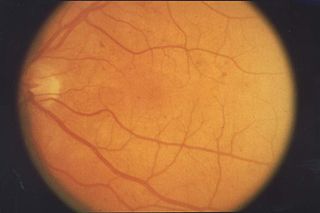
Retinopathy is any damage to the retina of the eyes, which may cause vision impairment. Retinopathy often refers to retinal vascular disease, or damage to the retina caused by abnormal blood flow. Age-related macular degeneration is technically included under the umbrella term retinopathy but is often discussed as a separate entity. Retinopathy, or retinal vascular disease, can be broadly categorized into proliferative and non-proliferative types. Frequently, retinopathy is an ocular manifestation of systemic disease as seen in diabetes or hypertension. Diabetes is the most common cause of retinopathy in the U.S. as of 2008. Diabetic retinopathy is the leading cause of blindness in working-aged people. It accounts for about 5% of blindness worldwide and is designated a priority eye disease by the World Health Organization.

<span class="mw-page-title-main">Diabetic retinopathy</span> Medical condition

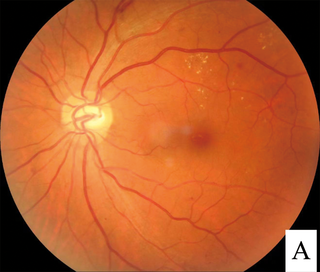
Diabetic retinopathy, is a medical condition in which damage occurs to the retina due to diabetes. It is a leading cause of blindness in developed countries.

<span class="mw-page-title-main">Vitrectomy</span> Surgery to remove vitreous humor from the eye

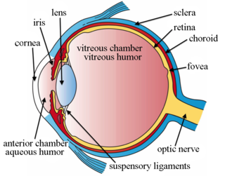
Vitrectomy is a surgery to remove some or all of the vitreous humor from the eye.

The National Eye Institute (NEI) is part of the U.S. National Institutes of Health (NIH), an agency of the U.S. Department of Health and Human Services. The mission of NEI is "to eliminate vision loss and improve quality of life through vision research." NEI consists of two major branches for research: an extramural branch that funds studies outside NIH and an intramural branch that funds research on the NIH campus in Bethesda, Maryland. Most of the NEI budget funds extramural research.

<span class="mw-page-title-main">Macular edema</span> Medical condition

Macular edema occurs when fluid and protein deposits collect on or under the macula of the eye and causes it to thicken and swell (edema). The swelling may distort a person's central vision, because the macula holds tightly packed cones that provide sharp, clear, central vision to enable a person to see detail, form, and color that is directly in the centre of the field of view.

<span class="mw-page-title-main">Macular degeneration</span> Medical condition associated with vision loss

Macular degeneration, also known as age-related macular degeneration, is a medical condition which may result in blurred or no vision in the center of the visual field. Early on there are often no symptoms. Over time, however, some people experience a gradual worsening of vision that may affect one or both eyes. While it does not result in complete blindness, loss of central vision can make it hard to recognize faces, drive, read, or perform other activities of daily life. Visual hallucinations may also occur.

<span class="mw-page-title-main">Central serous chorioretinopathy</span> Eye disease characterized by leakage of fluid under the retina

Central serous chorioretinopathy, also known as central serous retinopathy (CSR), is an eye disease that causes visual impairment, often temporary, usually in one eye. When the disorder is active it is characterized by leakage of fluid under the retina that has a propensity to accumulate under the central macula. This results in blurred or distorted vision (metamorphopsia). A blurred or gray spot in the central visual field is common when the retina is detached. Reduced visual acuity may persist after the fluid has disappeared.

Ranibizumab, sold under the brand name Lucentis among others, is a monoclonal antibody fragment (Fab) created from the same parent mouse antibody as bevacizumab. It is an anti-angiogenic that is approved to treat the "wet" type of age-related macular degeneration, diabetic retinopathy, and macular edema due to branch retinal vein occlusion or central retinal vein occlusion.

<span class="mw-page-title-main">Angioid streaks</span> Medical condition

Angioid streaks, also called Knapp streaks or Knapp striae, are small breaks in Bruch's membrane, an elastic tissue containing membrane of the retina that may become calcified and crack. Up to 50% of angioid streak cases are idiopathic. It may occur secondary to blunt trauma, or it may be associated with many systemic diseases. The condition is usually asymptomatic, but decrease in vision may occur due to choroidal neovascularization.

<span class="mw-page-title-main">Choroidal neovascularization</span> Creation of new blood vessels in the choroid layer of the eye

Choroidal neovascularization (CNV) is the creation of new blood vessels in the choroid layer of the eye. Choroidal neovascularization is a common cause of neovascular degenerative maculopathy commonly exacerbated by extreme myopia, malignant myopic degeneration, or age-related developments.

Aflibercept, sold under the brand names Eylea among others, is a medication used to treat wet macular degeneration and metastatic colorectal cancer. It was developed by Regeneron Pharmaceuticals and is approved in the United States and the European Union.

<span class="mw-page-title-main">Macular telangiectasia</span> Disease of the retina affecting central vision

Macular telangiectasia is a condition of the retina, the light-sensing tissue at the back of the eye that causes gradual deterioration of central vision, interfering with tasks such as reading and driving.

<span class="mw-page-title-main">Branch retinal vein occlusion</span> Medical condition

Branch retinal vein occlusion is a common retinal vascular disease of the elderly. It is caused by the occlusion of one of the branches of central retinal vein.

<span class="mw-page-title-main">Radiation retinopathy</span> Medical condition

Radiation retinopathy is damage to retina due to exposure to ionizing radiation. Radiation retinopathy has a delayed onset, typically after months or years of radiation, and is slowly progressive. In general, radiation retinopathy is seen around 18 months after treatment with external-beam radiation and with brachytherapy. The time of onset of radiation retinopathy is between 6 months to 3 years.

Joan Whitten Miller is a Canadian-American ophthalmologist and scientist who has made notable contributions to the treatment and understanding of eye disorders. She is credited for developing photodynamic therapy (PDT) with verteporfin (Visudyne), the first pharmacologic therapy for retinal disease. She also co-discovered the role of vascular endothelial growth factor (VEGF) in eye disease and demonstrated the therapeutic potential of VEGF inhibitors, forming the scientific basis of anti-VEGF therapy for age-related macular degeneration (AMD), diabetic retinopathy, and related conditions.

Anti–vascular endothelial growth factor therapy, also known as anti-VEGF therapy or medication, is the use of medications that block vascular endothelial growth factor. This is done in the treatment of certain cancers and in age-related macular degeneration. They can involve monoclonal antibodies such as bevacizumab, antibody derivatives such as ranibizumab (Lucentis), or orally-available small molecules that inhibit the tyrosine kinases stimulated by VEGF: sunitinib, sorafenib, axitinib, and pazopanib.

<span class="mw-page-title-main">Faricimab</span> Medication for macular degeneration

Faricimab, sold under the brand name Vabysmo, is a monoclonal antibody used for the treatment of neovascular age-related macular degeneration (nAMD) and diabetic macular edema (DME). Faricimab is the first bispecific monoclonal antibody to target both vascular endothelial growth factor (VEGF) and angiopoietin 2 (Ang-2). By targeting these pathways, faricimab stabilizes blood vessels in the retina. It is given by intravitreal injection by an ophthalmologist.

<span class="mw-page-title-main">Intravitreal injection</span> Method of administration of drugs into the eye by injection with a fine needle

Intravitreal injection is the method of administration of drugs into the eye by injection with a fine needle. The medication will be directly applied into the vitreous humor. It is used to treat various eye diseases, such as age-related macular degeneration (AMD), diabetic retinopathy, and infections inside the eye such as endophthalmitis. As compared to topical administration, this method is beneficial for a more localized delivery of medications to the targeted site, as the needle can directly pass through the anatomical eye barrier and dynamic barrier. It could also minimize adverse drug effects on other body tissues via the systemic circulation, which could be a possible risk for intravenous injection of medications. Although there are risks of infections or other complications, with suitable precautions throughout the injection process, chances for these complications could be lowered.

Sickle cell retinopathy can be defined as retinal changes due to blood vessel damage in the eye of a person with a background of sickle cell disease. It can likely progress to loss of vision in late stages due to vitreous hemorrhage or retinal detachment. Sickle cell disease is a structural red blood cell disorder leading to consequences in multiple systems. It is characterized by chronic red blood cell destruction, vascular injury, and tissue ischemia causing damage to the brain, eyes, heart, lungs, kidneys, spleen, and musculoskeletal system.

Conbercept, sold under the commercial name Lumitin, is a novel vascular endothelial growth factor (VEGF) inhibitor used to treat neovascular age-related macular degeneration (AMD) and diabetic macular edema (DME). The anti-VEGF was approved for the treatment of neovascular AMD by the China State FDA (CFDA) in December 2013. As of December 2020, conbercept is undergoing phase III clinical trials through the U.S. Food and Drug Administration’s PANDA-1 and PANDA-2 development programs.

References

  1. 1 2 3 4 "Diabetic Retinopathy PPP - Updated 2019". American Academy of Ophthalmology Preferred Practice Pattern. 24 October 2016. Retrieved 11 December 2021.
  2. 1 2 3 Chen, X; Modjtahedi, BS; Young, LH (2015). "Management of Diabetic Macular Edema: Is It Time to Say Goodbye to Macular Laser?". International Ophthalmology Clinics. 55 (4): 113–22. doi:10.1097/iio.0000000000000091. PMID   26322431. S2CID   12297465.
  3. "Age-Related Macular Degeneration PPP - Updated 2015". American Academy of Ophthalmology Preferred Practice Pattern. 29 January 2015. Archived from the original on 21 October 2016. Retrieved 22 October 2016.
  4. Lindsley, K; Li, T; Ssemanda, E; Virgili, G; Dickersin, K (April 2016). "Interventions for Age-Related Macular Degeneration: Are Practice Guidelines Based on Systematic Reviews?". Ophthalmology. 123 (4): 884–97. doi:10.1016/j.ophtha.2015.12.004. PMC   4808456 . PMID   26804762.
  5. Virgili, G; Michelessi, M; Parodi, MB; Bacherini, D; Evans, JR (23 October 2015). "Laser treatment of drusen to prevent progression to advanced age-related macular degeneration". The Cochrane Database of Systematic Reviews. 2015 (10): CD006537. doi:10.1002/14651858.CD006537.pub3. PMC   4733883 . PMID   26493180.
  6. 1 2 Virgili, G; Bini, A (18 July 2007). "Laser photocoagulation for neovascular age-related macular degeneration". The Cochrane Database of Systematic Reviews (3): CD004763. doi:10.1002/14651858.CD004763.pub2. PMID   17636773.
  7. "Laser photocoagulation - eye". medlineplus.gov. Retrieved 8 March 2023.
  8. NORLASE (24 January 2022). "Laser Photocoagulation: Concept, Procedure, and Efficiency of Green Laser for Eye Conditions" . Retrieved 8 March 2023.
  9. Weng, Christina Y.; Bhagat, Neelakshi; Lim, Jennifer I; Karth, Peter A. (17 December 2022). Weng, Christina Y. (ed.). "Panretinal Photocoagulation".
  10. Myint, Kay Thi; Sahoo, Soumendra; Thein, Aung Win; Moe, Soe; Ni, Han (2022-12-12). "Laser therapy for retinopathy in sickle cell disease". The Cochrane Database of Systematic Reviews. 2022 (12): CD010790. doi:10.1002/14651858.CD010790.pub3. ISSN   1469-493X. PMC   9744465 . PMID   36508693.
  11. 1 2 3 Rustagi, T; Mashimo, H (7 November 2011). "Endoscopic management of chronic radiation proctitis". World Journal of Gastroenterology. 17 (41): 4554–62. doi: 10.3748/wjg.v17.i41.4554 . PMC   3225092 . PMID   22147960.
  12. Laser Eye Surgery
  13. Goes, Frank (2013). Eye in history. New Delhi: Jaypee Brothers. ISBN   9789350902745. OCLC   813930522.
  14. Boyd, Benjamin (2010). Modern ophthalmology : the highlights : the account of a master wintnessing a 60 year epoch of evolution and progress (1950-2010). Panama: Jaypee-Highlights Medical Publishers. ISBN   9789962678168. OCLC   720191230.
  15. Meyer-Schwickerath, G (1954). "[Light coagulation; a method for treatment and prevention of the retinal detachment]". Albrecht von Graefe's Archiv für Ophthalmologie (in German). 156 (1): 2–34. doi:10.1007/BF00703328. PMID   14349833. S2CID   131234164.
  16. "Photocoagulation for diabetic macular edema. Early Treatment Diabetic Retinopathy Study report number 1. Early Treatment Diabetic Retinopathy Study research group". Archives of Ophthalmology. 103 (12): 1796–806. December 1985. doi:10.1001/archopht.1985.01050120030015. PMID   2866759.