Thyrotoxic periodic paralysis

Last updated

Thyrotoxic periodic paralysis
Illu08 thyroid.jpg
Thyrotoxic periodic paralysis occurs when the thyroid gland releases excessive amounts of thyroxine (thyroid hormone).
Specialty Endocrinology

Thyrotoxic periodic paralysis (TPP) is a rare condition featuring attacks of muscle weakness in the presence of hyperthyroidism (overactivity of the thyroid gland). Hypokalemia (a decreased potassium level in the blood) is usually present during attacks. The condition may be life-threatening if weakness of the breathing muscles leads to respiratory failure, or if the low potassium levels lead to abnormal heart rhythms. [1] [2] If untreated, it is typically recurrent in nature. [1]

Contents

The condition has been linked with genetic mutations in genes that code for certain ion channels that transport electrolytes (sodium and potassium) across cell membranes. The main ones are the L-type calcium channel α1-subunit [1] and potassium inward rectifier 2.6; [3] it is therefore classified as a channelopathy. [3] The abnormality in the channel is thought to lead to shifts of potassium into cells, under conditions of high thyroxine (thyroid hormone) levels, usually with an additional precipitant.

Treatment of the low levels of potassium in the blood, followed by correction of the hyperthyroidism, leads to complete resolution of the attacks. It occurs predominantly in males of Chinese, Japanese, Vietnamese, Filipino, and Korean descent. [1] TPP is one of several conditions that can cause periodic paralysis. [4]

Signs and symptoms

An attack often begins with muscle pain, cramping, and stiffness. [5] This is followed by weakness or paralysis that tends to develop rapidly, usually in late evening or the early hours of the morning. The weakness is usually symmetrical; [5] the limb muscles closer to the trunk (proximal) are predominantly affected, and weakness tends to start in the legs and spread to the arms. Muscles of the mouth and throat, eyes, and breathing are usually not affected, but occasionally weakness of the respiratory muscles can cause life-threatening respiratory failure. Attacks typically resolve within several hours to several days, even in the absence of treatment. [1] [2] [5] On neurological examination during an attack, flaccid weakness of the limbs is noted; reflexes are usually diminished, but the sensory system is unaffected. [1] [5] Mental status is not affected. [5]

Attacks may be brought on by physical exertion, drinking alcohol, or eating food high in carbohydrates or salt. This may explain why attacks are more common in summer when more people drink sugary drinks and engage in exercise. Exercise-related attacks tend to occur during a period of rest immediately after exercise; exercise may, therefore, be recommended to abort an attack. [1]

There may be symptoms of thyroid overactivity, such as weight loss, a fast or irregular heart rate, tremor, and perspiration; [1] [2] but such symptoms occur in only half of all cases. [5] The most common type of hyperthyroidism, Graves' disease, may additionally cause eye problems (Graves' ophthalmopathy) and skin changes of the legs (pretibial myxedema). [6] Thyroid disease may also cause muscle weakness in the form of thyrotoxic myopathy, but this is constant rather than episodic. [5]

Causes

Genetics

Genetic mutations in the L-type calcium channel α1-subunit (Cav1.1) have been described in Southern Chinese with TPP. The mutations are located in a different part of the gene from those described in the related condition familial periodic paralysis. In TPP, the mutations described are single-nucleotide polymorphisms located in the hormone response element responsive to thyroid hormone, implying that transcription of the gene and production of ion channels may be altered by increased thyroid hormone levels. Furthermore, mutations have been reported in the genes coding for potassium voltage-gated channel, Shaw-related subfamily, member 4 (Kv3.4) and sodium channel protein type 4 subunit alpha (Na41.4). [1]

Of people with TPP, 33% from various populations were demonstrated to have mutations in KCNJ18, the gene coding for Kir2.6, an inward-rectifier potassium ion channel. This gene, too, harbors a thyroid response element. [3]

Certain forms of human leukocyte antigen (HLA)especially B46, DR9, DQB1*0303, A2, Bw22, AW19, B17, and DRW8are more common in TPP. Linkage to particular forms of HLA, which plays a central role in the immune response, might imply an immune system cause, but it is uncertain whether this directly causes TPP or whether it increases the susceptibility to Graves' disease, a known autoimmune disease. [1]

Thyroid disease

The most common underlying form of thyroid disease associated with TPP is Graves' disease, a syndrome due to an autoimmune reaction that leads to overproduction of thyroid hormone. [6] TPP has also been described in people with other thyroid problems such as thyroiditis, toxic nodular goiter, toxic adenoma, TSH-producing pituitary adenoma, excessive ingestion of thyroxine or iodine, [1] and amiodarone-induced hyperthyroidism. [2]

Mechanism

Na /K -ATPase maintains the normal gradients of sodium and potassium between cells and extracellular fluid, expending the cellular fuel ATP in doing so. Scheme sodium-potassium pump-en.svg
Na /K -ATPase maintains the normal gradients of sodium and potassium between cells and extracellular fluid, expending the cellular fuel ATP in doing so.

The muscle weakness and increased risk of irregular heart beat in TPP result from markedly reduced levels of potassium in the bloodstream. Potassium is not in fact lost from the body, but increased Na+/K+-ATPase activity (the enzyme that moves potassium into cells and keeps sodium in the blood) leads to shift of potassium into tissues, and depletes the circulation. In other types of potassium derangement, the acid-base balance is usually disturbed, with metabolic alkalosis and metabolic acidosis often being present. In TPP, these disturbances are generally absent. Hypokalemia leads to hyperpolarization of muscle cells, making the neuromuscular junction less responsive to normal nerve impulses and leading to decreased contractility of the muscles. [1]

It is not clear how the described genetic defects increase the Na+/K+-ATPase activity, but it is suspected that the enzyme becomes more active due to increased thyroid hormone levels. Hyperthyroidism increases the levels of catecholamines (such as adrenaline) in the blood, increasing Na+/K+-ATPase activity. [5] The enzyme activity is then increased further by the precipitating causes. For instance, increased carbohydrate intake leads to increased insulin levels; this is known to activate Na+/K+-ATPase. Once the precipitant is removed, the enzyme activity returns to normal levels. [1] It has been postulated that male hormones increase Na+/K+-ATPase activity, and that this explains why males are at a higher risk of TPP despite thyroid disease being more common in females. [2]

TPP is regarded as a model for related conditions, known as "channelopathies", which have been linked with mutations in ion channels; the majority of these conditions occurs episodically. [3]

Diagnosis

Hypokalemia (low blood potassium levels) commonly occurs during attacks; levels below 3.0 mmol/L are typically encountered. Magnesium and phosphate levels are often found to be decreased. Creatine kinase levels are elevated in two thirds of cases, usually due to a degree of muscle injury; severe elevations suggestive of rhabdomyolysis (muscle tissue destruction) are rare. [1] [2] Electrocardiography (ECG/EKG) may show tachycardia (a fast heart rate) due to the thyroid disease, abnormalities due to cardiac arrhythmia (atrial fibrillation, ventricular tachycardia), and conduction changes associated with hypokalemia (U waves, QRS widening, QT prolongation, and T wave flattening). [2] Electromyography shows changes similar to those encountered in myopathies (muscle diseases), with a reduced amplitude of the compound muscle action potentials (CMAPs); [4] they resolve when treatment has commenced. [1]

TPP is distinguished from other forms of periodic paralysis (especially hypokalemic periodic paralysis) with thyroid function tests on the blood. These are normal in the other forms, and in thyrotoxicosis the levels of thyroxine and triiodothyronine are elevated, with resultant suppression of TSH production by the pituitary gland. [1] [6] Various other investigations are usually performed to separate the different causes of hyperthyroidism. [6]

Treatment

The non-selective beta blocker propranolol can rapidly improve the symptoms of hyperthyroidism, including attacks of TPP. Propranolol.svg
The non-selective beta blocker propranolol can rapidly improve the symptoms of hyperthyroidism, including attacks of TPP.

In the acute phase of an attack, administration of potassium will quickly restore muscle strength and prevent complications. However, caution is advised as the total amount of potassium in the body is not decreased, and it is possible for potassium levels to overshoot ("rebound hyperkalemia"); slow infusions of potassium chloride are therefore recommended while other treatment is commenced. [1]

The effects of excess thyroid hormone typically respond to the administration of a non-selective beta blocker, such as propranolol (as most of the symptoms are driven by increased levels of adrenaline and its effect on the β-adrenergic receptors). Subsequent attacks may be prevented by avoiding known precipitants, such as high salt or carbohydrate intake, until the thyroid disease has been adequately treated. [1]

Treatment of the thyroid disease usually leads to resolution of the paralytic attacks. Depending on the nature of the disease, the treatment may consist of thyrostatics (drugs that reduce production of thyroid hormone), radioiodine, or occasionally thyroid surgery. [1] [2]

Epidemiology

TPP occurs predominantly in males of Chinese, Japanese, Vietnamese, Filipino, and Korean descent, [1] as well as Thais, [3] with much lower rates in people of other ethnicities. [1] In Chinese and Japanese people with hyperthyroidism, 1.8–1.9% experience TPP. This is in contrast to North America, where studies report a rate of 0.1–0.2%. [1] [2] Native Americans, who share a genetic background with East Asians, are at an increased risk. [1]

The typical age of onset is 20–40. It is unknown why males are predominantly affected, with rates in males being 17- to 70-fold those in females, despite thyroid overactivity being much more common in women. [1] [2]

History

Karl Friedrich Otto Westphal Westphal.jpg
Karl Friedrich Otto Westphal

After several case reports in the 18th and 19th centuries, periodic paralysis was first described in full by the German neurologist Karl Friedrich Otto Westphal (1833–1890) in 1885. [7] [8] In 1926 the Japanese physician Tetsushiro Shinosaki, from Fukuoka, observed the high rate of thyroid disease in Japanese people with periodic paralysis. [9] [10] The first English-language report, in 1931, originated from Dunlap and Kepler, physicians at the Mayo Clinic; they described the condition in a patient with features of Graves' disease. [2] [10] In 1937 periodic paralysis was linked with hypokalemia, as well as precipitation of attacks with glucose and insulin. [11] [12] This phenomenon has been used as a diagnostic test. [12]

In 1974 it was discovered that propranolol could prevent attacks. [13] The concept of channelopathies and the link with specific ion channel mutations emerged at the end of the 20th century. [1] [3] [4]

Related Research Articles

<span class="mw-page-title-main">Hyperthyroidism</span> Thyroid gland disease that involves an overproduction of thyroid hormone

Hyperthyroidism is the condition that occurs due to excessive production of thyroid hormones by the thyroid gland. Thyrotoxicosis is the condition that occurs due to excessive thyroid hormone of any cause and therefore includes hyperthyroidism. Some, however, use the terms interchangeably. Signs and symptoms vary between people and may include irritability, muscle weakness, sleeping problems, a fast heartbeat, heat intolerance, diarrhea, enlargement of the thyroid, hand tremor, and weight loss. Symptoms are typically less severe in the elderly and during pregnancy. An uncommon but life-threatening complication is thyroid storm in which an event such as an infection results in worsening symptoms such as confusion and a high temperature; this often results in death. The opposite is hypothyroidism, when the thyroid gland does not make enough thyroid hormone.

<span class="mw-page-title-main">Thyroid</span> Endocrine gland in the neck; secretes hormones that influence metabolism

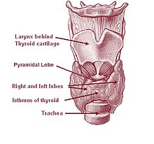
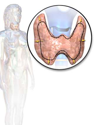
The thyroid, or thyroid gland, is an endocrine gland in vertebrates. In humans, it is in the neck and consists of two connected lobes. The lower two thirds of the lobes are connected by a thin band of tissue called the isthmus (pl.: isthmi). The thyroid gland is a butterfly-shaped gland located in the neck below the Adam's apple. Microscopically, the functional unit of the thyroid gland is the spherical thyroid follicle, lined with follicular cells (thyrocytes), and occasional parafollicular cells that surround a lumen containing colloid. The thyroid gland secretes three hormones: the two thyroid hormones – triiodothyronine (T3) and thyroxine (T4) – and a peptide hormone, calcitonin. The thyroid hormones influence the metabolic rate and protein synthesis and growth and development in children. Calcitonin plays a role in calcium homeostasis. Secretion of the two thyroid hormones is regulated by thyroid-stimulating hormone (TSH), which is secreted from the anterior pituitary gland. TSH is regulated by thyrotropin-releasing hormone (TRH), which is produced by the hypothalamus.

Myotonia is a symptom of a small handful of certain neuromuscular disorders characterized by delayed relaxation of the skeletal muscles after voluntary contraction or electrical stimulation, and the muscle shows an abnormal EMG.

<span class="mw-page-title-main">Primary aldosteronism</span> Medical condition

Primary aldosteronism (PA), also known as primary hyperaldosteronism or Conn's syndrome, refers to the excess production of the hormone aldosterone from the adrenal glands, resulting in low renin levels and high blood pressure. This abnormality is caused by hyperplasia or tumors. Many experience fatigue, potassium deficiency and high blood pressure which may cause poor vision, confusion or headaches. Symptoms may also include: muscular aches and weakness, muscle spasms, low back and flank pain from the kidneys, trembling, tingling sensations, dizziness/vertigo, nocturia and excessive urination. Complications include cardiovascular disease such as stroke, myocardial infarction, kidney failure and abnormal heart rhythms.

<span class="mw-page-title-main">Hypokalemia</span> Medical condition with insufficient potassium

Hypokalemia is a low level of potassium (K+) in the blood serum. Mild low potassium does not typically cause symptoms. Symptoms may include feeling tired, leg cramps, weakness, and constipation. Low potassium also increases the risk of an abnormal heart rhythm, which is often too slow and can cause cardiac arrest.

Hyperkalemic periodic paralysis is an inherited autosomal dominant disorder that affects sodium channels in muscle cells and the ability to regulate potassium levels in the blood. It is characterized by muscle hyperexcitability or weakness which, exacerbated by potassium, heat or cold, can lead to uncontrolled shaking followed by paralysis. Onset usually occurs in early childhood, but it still occurs with adults.

<span class="mw-page-title-main">Andersen–Tawil syndrome</span> Rare autosomal dominant genetic disorder

Andersen–Tawil syndrome, also called Andersen syndrome and long QT syndrome 7, is a rare genetic disorder affecting several parts of the body. The three predominant features of Andersen–Tawil syndrome include disturbances of the electrical function of the heart characterised by an abnormality seen on an electrocardiogram and a tendency to abnormal heart rhythms, physical characteristics including low-set ears and a small lower jaw, and intermittent periods of muscle weakness known as hypokalaemic periodic paralysis.

<span class="mw-page-title-main">Channelopathy</span> Diseases caused by disturbed function of ion channel subunits or the proteins that regulate them

Channelopathies are a group of diseases caused by the dysfunction of ion channel subunits or their interacting proteins. These diseases can be inherited or acquired by other disorders, drugs, or toxins. Mutations in genes encoding ion channels, which impair channel function, are the most common cause of channelopathies. There are more than 400 genes that encode ion channels, found in all human cell types and are involved in almost all physiological processes. Each type of channel is a multimeric complex of subunits encoded by a number of genes. Depending where the mutation occurs it may affect the gating, conductance, ion selectivity, or signal transduction of the channel.

Myotonia congenita is a congenital neuromuscular channelopathy that affects skeletal muscles. It is a genetic disorder. The hallmark of the disease is the failure of initiated contraction to terminate, often referred to as delayed relaxation of the muscles (myotonia) and rigidity. Symptoms include delayed relaxation of the muscles after voluntary contraction (myotonia), and may also include stiffness, hypertrophy (enlargement), transient weakness in some forms of the disorder, severe masseter spasm, and cramping. The condition is sometimes referred to as fainting goat syndrome, as it is responsible for the eponymous 'fainting' seen in fainting goats when presented with a sudden stimulus. Of note, myotonia congenita has no association with malignant hyperthermia (MH).

Periodic paralysis is a group of rare genetic diseases that lead to weakness or paralysis from common triggers such as cold, heat, high carbohydrate meals, not eating, stress or excitement and physical activity of any kind. The underlying mechanism of these diseases are malfunctions in the ion channels in skeletal muscle cell membranes that allow electrically charged ions to leak in or out of the muscle cell, causing the cell to depolarize and become unable to move.

<span class="mw-page-title-main">Thyroid disease</span> Medical condition

Thyroid disease is a medical condition that affects the function of the thyroid gland. The thyroid gland is located at the front of the neck and produces thyroid hormones that travel through the blood to help regulate many other organs, meaning that it is an endocrine organ. These hormones normally act in the body to regulate energy use, infant development, and childhood development.

<span class="mw-page-title-main">Hypokalemic periodic paralysis</span> Medical condition

Hypokalemic periodic paralysis (hypoKPP), also known as familial hypokalemic periodic paralysis (FHPP), is a rare, autosomal dominant channelopathy characterized by muscle weakness or paralysis when there is a fall in potassium levels in the blood. In individuals with this mutation, attacks sometimes begin in adolescence and most commonly occur with individual triggers such as rest after strenuous exercise, high carbohydrate meals, meals with high sodium content, sudden changes in temperature, and even excitement, noise, flashing lights, cold temperatures and stress. Weakness may be mild and limited to certain muscle groups, or more severe full-body paralysis. During an attack, reflexes may be decreased or absent. Attacks may last for a few hours or persist for several days. Recovery is usually sudden when it occurs, due to release of potassium from swollen muscles as they recover. Some patients may fall into an abortive attack or develop chronic muscle weakness later in life.

<i>Paramyotonia congenita</i> Medical condition

Paramyotonia congenita (PC) is a rare congenital autosomal dominant neuromuscular disorder characterized by "paradoxical" myotonia. This type of myotonia has been termed paradoxical because it becomes worse with exercise whereas classical myotonia, as seen in myotonia congenita, is alleviated by exercise. PC is also distinguished as it can be induced by cold temperatures. Although more typical of the periodic paralytic disorders, patients with PC may also have potassium-provoked paralysis. PC typically presents within the first decade of life and has 100% penetrance. Patients with this disorder commonly present with myotonia in the face or upper extremities. The lower extremities are generally less affected. While some other related disorders result in muscle atrophy, this is not normally the case with PC. This disease can also present as hyperkalemic periodic paralysis and there is debate as to whether the two disorders are actually distinct.

Na<sub>v</sub>1.4 Protein-coding gene in the species Homo sapiens

Sodium channel protein type 4 subunit alpha is a protein that in humans is encoded by the SCN4A gene.

Thyrotoxic myopathy (TM) is a neuromuscular disorder that develops due to the overproduction of the thyroid hormone thyroxine. Also known as hyperthyroid myopathy, TM is one of many myopathies that lead to muscle weakness and muscle tissue breakdown. Evidence indicates the onset may be caused by hyperthyroidism. Physical symptoms of TM may include muscle weakness, the breakdown of muscle tissue, fatigue, and heat intolerance. Physical acts such as lifting objects and climbing stairs may become increasingly difficult. If untreated, TM can be an extremely debilitating disorder that can, in extreme rare cases, lead to death. If diagnosed and treated properly the effects can be controlled and in most cases reversed leaving no lasting effects.

Ca<sub>v</sub>1.1 Mammalian protein found in Homo sapiens

Cav1.1 also known as the calcium channel, voltage-dependent, L type, alpha 1S subunit, (CACNA1S), is a protein which in humans is encoded by the CACNA1S gene. It is also known as CACNL1A3 and the dihydropyridine receptor.

<span class="mw-page-title-main">Thyroid hormones</span> Hormones produced by the thyroid gland

Thyroid hormones are any hormones produced and released by the thyroid gland, namely triiodothyronine (T3) and thyroxine (T4). They are tyrosine-based hormones that are primarily responsible for regulation of metabolism. T3 and T4 are partially composed of iodine, derived from food. A deficiency of iodine leads to decreased production of T3 and T4, enlarges the thyroid tissue and will cause the disease known as simple goitre.

The Kir2.6 also known as inward rectifier potassium channel 18 is a protein that in humans is encoded by the KCNJ18 gene. Kir2.6 is an inward-rectifier potassium ion channel.

Louis Ptáček is an American neurologist and professor who contributed greatly to the field of genetics and neuroscience. He was also an HHMI investigator from 1997 to 2018. His chief areas of research include the understanding of inherited Mendelian disorders and circadian rhythm genes. Currently, Ptáček is a neurology professor and a director of the Division of Neurogenetics in University of California, San Francisco, School of Medicine. His current investigations primarily focus on extensive clinical studies in families with hereditary disorders, which include identifying and characterizing the genes responsible for neurological variations.

Hyperkalemic periodic paralysis is a genetic disorder that occurs in horses. It is also known as Impressive syndrome, after an index case in a horse named Impressive. It is an inherited autosomal dominant disorder that affects sodium channels in muscle cells and the ability to regulate potassium levels in the blood. It is characterized by muscle hyperexcitability or weakness which, exacerbated by potassium, heat or cold, can lead to uncontrolled shaking followed by paralysis.

References

  1. 1 2 3 4 5 6 7 8 9 10 11 12 13 14 15 16 17 18 19 20 21 22 23 24 25 Kung AW (July 2006). "Clinical review: Thyrotoxic periodic paralysis: a diagnostic challenge". The Journal of Clinical Endocrinology and Metabolism. 91 (7): 2490–5. doi: 10.1210/jc.2006-0356 . PMID   16608889.
  2. 1 2 3 4 5 6 7 8 9 10 11 Pothiwala P, Levine SN (2010). "Analytic review: thyrotoxic periodic paralysis: a review". Journal of Intensive Care Medicine. 25 (2): 71–7. doi:10.1177/0885066609358849. PMID   20089526. S2CID   24394963.
  3. 1 2 3 4 5 6 Ryan DP, Ptácek LJ (October 2010). "Episodic neurological channelopathies". Neuron. 68 (2): 282–92. doi: 10.1016/j.neuron.2010.10.008 . PMID   20955935. S2CID   16230992.
  4. 1 2 3 Fontaine B (2008). "Periodic paralysis". Advances in Genetics. 63: 3–23. doi:10.1016/S0065-2660(08)01001-8. ISBN   978-0-12-374527-9. PMID   19185183.
  5. 1 2 3 4 5 6 7 8 Lin SH (January 2005). "Thyrotoxic periodic paralysis" (PDF). Mayo Clinic Proceedings. 80 (1): 99–105. doi: 10.4065/80.1.99 . PMID   15667036.
  6. 1 2 3 4 Weetman AP (October 2000). "Graves' disease". The New England Journal of Medicine. 343 (17): 1236–48. doi:10.1056/NEJM200010263431707. PMID   11071676.
  7. Westphal CF (1885). "Über einen merkwürdigen Fall von periodischer Lähmung aller vier Extremitäten mit gleichzeitigem Erlöschen der elektrischen Erregbarkeit während der Lähmung". Berl. Klin. Wochenschr. (in German). 22: 489–91 and 509–11.
  8. Weber F, Lehmann-Horn F (28 April 2009). Adam MP, Ardinger HH, Pagon RA, Wallace SE, Bean LJ, Stephens K, Amemiya A (eds.). "Hypokalemic Periodic Paralysis". GeneReviews. PMID   20301512.
  9. Shinosaki T (1926). "Klinische Studien über die periodische Extremitätenlähmung". Zeitschrift für die gesamte Neurologie und Psychiatrie (in German). 100 (1): 564–611. doi:10.1007/BF02970940. S2CID   76554730.
  10. 1 2 Dunlap H, Kepler K (1931). "A syndrome resembling familial periodic paralysis occurring in the course of exophthalmic goiter". Endocrinology. 15 (6): 541–6. doi:10.1210/endo-15-6-541.
  11. Aitken RS, Allott EN, Castleden LI, Walker M (1937). "Observations on a case of familial periodic paralysis". Clin. Sci. 3: 47–57.
  12. 1 2 McFadzean AJ, Yeung R (February 1967). "Periodic paralysis complicating thyrotoxicosis in Chinese". British Medical Journal. 1 (5538): 451–5. doi:10.1136/bmj.1.5538.451. PMC   1840834 . PMID   6017520.
  13. Yeung RT, Tse TF (October 1974). "Thyrotoxic periodic paralysis. Effect of propranolol". The American Journal of Medicine. 57 (4): 584–90. doi:10.1016/0002-9343(74)90010-2. PMID   4432863.