Neuromuscular junction disease

Last updated
Neuromuscular junction disease
Specialty Neurology

Neuromuscular junction disease is a medical condition where the normal conduction through the neuromuscular junction fails to function correctly. [1]

Contents

Autoimmune

In diseases such as myasthenia gravis, the end plate potential (EPP) fails to effectively activate the muscle fiber due to an autoimmune reaction against acetylcholine receptors, resulting in muscle weakness and fatigue. [2]

Myasthenia gravis is caused most commonly by auto-antibodies against the acetylcholine receptor. It has recently been realized that a second category of gravis is due to auto-antibodies against MuSK. A different condition, Lambert–Eaton myasthenic syndrome, is usually associated with presynaptic antibodies to the voltage-dependent calcium channel. It is possible for these conditions to coexist. [3]

Neuromuscular junction

The neuromuscular junction is a specialized synapse between a neuron and the muscle it innervates. It allows efferent signals from the nervous system to contract muscle fibers causing them to contract. In vertebrates, the neuromuscular junction is always excitatory, therefore to stop contraction of the muscle, inhibition must occur at the level of the efferent motor neuron. In other words, the inhibition must occur at the level of the spinal cord.[ citation needed ]

Release of acetylcholine vesicles from the presynaptic terminal occurs only after adequate depolarization of the efferent nerve. Once a motor nerve action potential reaches the presynaptic nerve terminal it causes an increase in intracellular calcium concentration by causing an increase in ion conductance through voltage gated calcium channels. This increase in calcium concentration allows the acetylcholine vesicles to fuse with the plasma membrane at the presynaptic membrane, in a process called exocytosis, thus releasing acetylcholine into the synapse. Once acetylcholine is present in the synapse it is able to bind to nicotinic acetylcholine receptors increasing conductance of certain cations, sodium and potassium in the postsynaptic membrane and producing an excitatory end т и ироооurrent. As cations flow into the postsynaptic cell, this causes a depolarization, as the membrane voltage increases above normal resting potential. If the signal is of sufficient magnitude, than an action potential will be generated post synaptically. The action potential will propagate through the sarcolemma to the interior of the muscle fibers eventually leading to an increase in intracellular calcium levels and subsequently initiating the process of Excitation–contraction coupling. Once coupling begins it allows the sarcomeres of the muscles to shorten, thus leading to the contraction of the muscle.[ citation needed ]

Neuromuscular junction diseases are a result of a malfunction in one or more steps of the above pathway. As a result, normal functioning can be completely or partially inhibited, with the symptoms largely presenting themselves as problems in mobility and muscle contraction as expected from disorders in motor end plates. Neuromuscular junction diseases can also be referred to as end plate diseases or disorders.[ citation needed ]

Among neuromuscular diseases some can be autoimmune disease, or hereditary disorders. They can affect either presynaptic mechanisms or postsynaptic mechanisms, preventing the junction from functioning normally. The most studied diseases affecting the human acetylcholine receptor are myasthenia gravis and some forms of congenital myasthenic syndrome. Other diseases include the Lambert–Eaton syndrome and botulism.[ citation needed ]

Classification

There are two ways to classify neuromuscular diseases. The first relies on its mechanism of action, or how the action of the diseases affects normal functioning (whether it is through mutations in genes or more direct pathways such as poisoning). This category divides neuromuscular diseases into three broad categories: immune-mediated disease, toxic/metabolic and congenital syndromes.[ citation needed ]

The second classification method divides the diseases according to the location of their disruption. In the neuromuscular junction, the diseases will either act on the presynaptic membrane of the motor neuron, the synapse separating the motor neuron from the muscle fiber, or the postsynaptic membrane (the muscle fiber).

Immune-mediated

Immune-mediated diseases include a variety of diseases not only affecting the neuromuscular junction. Immune-mediated disorders range from simple and common problems such as allergies to disorders such as HIV/AIDS. Within this classification, autoimmune disorders are considered to be a subset of immune-mediated syndromes. Autoimmune diseases occur when the body's immune system begins to target its own cells, often causing harmful effects.[ citation needed ]

The neuromuscular junction diseases present within this subset are myasthenia gravis, and Lambert-Eaton syndrome.(reference 26) In each of these diseases, a receptor or other protein essential to normal function of the junction is targeted by antibodies in an autoimmune attack by the body.[ citation needed ]

Toxic/metabolic

Metabolic diseases are usually a result of abnormal functioning of one of the metabolic processes required for regular production and utilization of energy in a cell. This can occur by damaging or disabling an important enzyme, or when a feedback system is abnormally functioning. Toxic diseases are a result of a form of poison that effects neuromuscular junction functioning. Most commonly animal venom or poison, or other toxic substances are the origin of the problem.[ citation needed ]

Neuromuscular junction diseases in this category include snake venom poisoning, botulism, arthropod poisoning, organophosphates and hypermagnesemia.(reference 13) Organophosphates are present in many insecticides and herbicides. They are also the basis of many nerve gases.(reference 27) Hypermagnesmia is a condition where the balance of magnesium in the body is unstable and concentrations are higher than normal baseline values.(reference 28)

Congenital

Congenital syndromes affecting the neuromuscular junction are considered a very rare form of disease, occurring in 1 out of 200,000 in the United Kingdom.(reference 29) These are genetically inherited disorders. Symptoms are seen early since the affected individuals carry the mutation from birth. Congenital syndromes are usually classified by the location of the affected gene products. Congenital syndromes can have multiple targets affecting either the presynaptic, synaptic or postsynaptic parts of the neuromuscular junction.(reference 30) For example, if the malfunctioning or inactive protein is acetylcholinesterase, this would be classified as a synapse congenital syndrome.(reference 29)

Presynaptic

The diseases that act on the presynaptic membrane are autoimmune neuromyotonia, Lambert–Eaton syndrome, congenital myasthenia gravis and botulism.(reference 5) All of these disorders negatively affect the presynaptic membrane in some way. Neuromyotonia causes antibodies to damage the normal function of potassium rectifier channels, while Lambert–Eaton syndrome causes antibodies to attack presynaptic calcium channels.(reference 7) Congenital myasthenia gravis is a large group of diseases, since the genetic defects can affect any point in the chain of events leading to successful transmission across the junction. One discovered type of congenital myasthenia gravis can affect the junction presynaptically by a mutation in the gene encoding choline acetyl transferase.(reference 29) This protein is an enzyme that is responsible for catalyzing the reaction that combines acetyl-coenzyime A with choline, yielding acetylcholine.(reference 31)

There are many mechanisms through which presynaptic function can be impaired. Most often this causes a decrease in the release of acetylcholine. It can also impair vesicle exocytosis by interfering with the complex guiding vesicle fusion and release of contents. Mechanism of action can also impair the calcium channels that induce exocytosis of the vesicles. Other ion channels can also be disrupted, such as the potassium channels causing inefficient repolarization at the presynaptic membrane as in neuromyotonia.(reference 5)

Synapse

At the synaptic cleft, the neurotransmitter normally diffuses across the synapse to eventually contact postsynaptic receptors. However, after exiting the presynaptic membrane, the neurotransmitters can be hindered by a subset of diseases that interfere with the transmission of the neurotransmitter across the synapse. The mechanism currently known that operates via the synaptic cleft causing impairment of normal functioning is another congenital myasthenia gravis.(reference 7) This mechanism is the only currently known disease that acts on the synapse.(reference 12) It acts by impairing the function of the enzyme that breaks down acetylcholine causing it to become very hypertonic at the synapse.(reference 12) This increase in acetylcholine in the synapse disrupts normal functioning of the junction,.(reference 32)(reference 33)

Postsynaptic

The highest number of diseases affect the neuromuscular junction postsynaptically. In other words, it is the most susceptible to negative intervention.(reference 7) The targets of these postsynaptic diseases can be multiple different proteins. Immune-mediated myasthenia gravis being the most common, effecting the acetylcholine receptors at the post synaptic membrane.(reference 35) All the diseases that affect the postsynaptic membrane are forms of myasthenia gravis.(reference 5) Here is a list of the diseases: myasthenia gravis, neonatal myasthenia gravis, drug-induced myasthenia gravis and several types of Congenital myasthenia where the product of the mutated gene is a postsynaptic protein (reference renamed from 5)

Most common diseases

Myasthenia gravis

Myasthenia gravis is the most common neuromuscular disease affecting function of the end plate in patients. It is present in 1 person out of 10,000 in the population, and its onset is usually in either younger or older individuals. (reference 14)

Acquired myasthenia gravis is the most common neuromuscular junction disease.(reference 7) Important observations were made by Patrick and Lindstrom in 1973 when they found that antibodies attacking the acetylcholine receptors were present in around 85% of cases of myasthenia gravis.(reference renamed form 13)(reference 36) The remaining diseases were also a result of antibody attacks on vital proteins, but instead of the acetylcholine receptor, the culprits were MuSK, a muscle-specific serum kinase, and lipoprotein receptor-related protein.(reference 36) So these mechanisms describe myasthenia gravis that is acquired, and not congenital, affecting these vital proteins by an immunological response against self-antigens. The cases not caused by antibodies against the acetylcholine receptors became by convention called seronegative myasthenia gravis.(reference 37) The term seronegative came about because scientists would be testing for acetylcholine receptor antibodies in patients that had myasthenia gravis resulting in negative tests in the serum. This does not imply that there are no antibodies present, but this terminology only became present because scientists were testing for the wrong antigen.(reference 36)(reference 38)

Neonatal myasthenia gravis is a very rare condition in which a mother with myasthenia gravis passes down her antibodies to her infant through the placenta, causing it to be born with antibodies that will attach self-antigens.(reference 12)

Drug-induced myasthenia gravis is also a very rare condition in which pharmacological drugs cause a blockade or disruption of the NMJ machinery.(reference 12) Robert W. Barrons summarizes the possible causes of drug-induced myasthenia gravis: "Prednisone was most commonly implicated as aggravating myasthenia gravis, and D-penicillamine was most commonly associated with myasthenic syndrome. The greatest frequency of drug-induced neuromuscular blockade was seen with aminoglycoside-induced postoperative respiratory depression. However, drugs most likely to impact myasthenic patients negatively are those used in the treatment of the disease. These include overuse of anticholinesterase drugs, high-dose prednisone, and anesthesia and neuromuscular blockers for thymectomy."(reference 39)

Lambert-Eaton myasthenic syndrome (LEMS)

Lambert-Eaton myasthenic syndrome (LEMS) is similar to myasthenia gravis in that it is an immune-mediated response acting against a specific protein in the neuromuscular junction. [4] The difference is that LEMS is a result of an autoimmune response on the voltage-gated calcium channels of the presynaptic membrane.(reference 14) The antibodies attack the voltage-gated calcium channels of the P/Q type.(reference 35) Abnormal activity of this ion channel, which usually initiates the process of acetylcholine vesicles from the presynaptic membrane once the membrane is sufficiently depolarized, causes less acetylcholine to be released into the synapse.(reference 12) LEMS is about 20 times more rare than myasthenia gravis.(reference 40)

LEMS also differs from myasthenia gravis in that it is usually associated with small-cell lung cancer, which is present in 60% percent of patients.(reference 40) It seems that as cancer develops, the body will begin to develop antibodies against the cancer, and in some cases the antibodies can also attack the calcium channels present at the presynaptic membrane.(reference 12) In the cases where no cancer is present in the patient, there is usually an underlying different autoimmune disease that causes the immune system to become hyperactive attacking its own antigens.(reference 40)

Other diseases

Neuromyotonia

Neuromyotonia is classified into three types.(reference 14) The most common form of this disease is acquired neuromyotonia, which is the result of an autoimmune attack on rectifier voltage-gated potassium channels.(reference 12) This causes the presynaptic membrane to remain hyperpolarized, making it difficult for adequate depolarizations to occur.(reference 5)

Congenital myasthenia

This is the most complex and diverse congenital myasthenic syndrome.(reference 29) Since this is a genetic disorder, there are infinite possibilities of genes that could be mutated in different ways that could disrupt normal functioning of the neuromuscular junction. Around 11 gene targets have been specified.(reference 3) Its prevalence in the population is very difficult to measure since it is a rare genetic disorder that presents itself as a neuromuscular junction disorder, but in the United Kingdom, estimates are 1 in 200,000 of the population.(reference 29) The major signs that indicate a congenital syndrome are symptoms present at birth, such as weakness and a depressive response to repetitive nerve stimulation.(reference 29)

Since the disease is genetic in nature and is not immune-mediated, any serum test will show up negative since congenital myasthenia is not a result of antibodies attacking the vital proteins of the NMJ.(reference 7) Knowledge of this disease is very plastic as new genes that could be "effected" (affected? effective?) could be discovered as we gain more insight into the different types.

Botulism

Neurotoxin may act on the neuromuscular junction either post synaptically or presynaptically as there are several different forms of toxins that the NMJ is sensitive to.(reference 14) Common mechanisms of action include blockage of acetylcholine release at the synapse via inhibition of the SNARE protein, thus causing the NMJ to become abnormal in function.(reference 12)

Diagnosis

Tests

Treatment

Symptomatic treatment

Cholinesterase inhibitors at AChR

Immunosuppressive treatment

Related Research Articles

Angela Vincent is Emeritus professor at the University of Oxford and a Fellow of Somerville College, Oxford.

<span class="mw-page-title-main">Lambert–Eaton myasthenic syndrome</span> Medical condition

Lambert–Eaton myasthenic syndrome (LEMS) is a rare autoimmune disorder characterized by muscle weakness of the limbs.

<span class="mw-page-title-main">Myasthenia gravis</span> Autoimmune disease resulting in skeletal muscle weakness

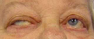
Myasthenia gravis (MG) is a long-term neuromuscular junction disease that leads to varying degrees of skeletal muscle weakness. The most commonly affected muscles are those of the eyes, face, and swallowing. It can result in double vision, drooping eyelids, and difficulties in talking and walking. Onset can be sudden. Those affected often have a large thymus or develop a thymoma.

Neuromyotonia (NMT) is a form of peripheral nerve hyperexcitability that causes spontaneous muscular activity resulting from repetitive motor unit action potentials of peripheral origin. NMT along with Morvan's syndrome are the most severe types in the Peripheral Nerve Hyperexciteability spectrum. Example of two more common and less severe syndromes in the spectrum are Cramp Fasciculation Syndrome and Benign Fasciculation Syndrome. NMT can have both hereditary and acquired forms. The prevalence of NMT is unknown.

<span class="mw-page-title-main">Acetylcholine</span> Organic chemical and neurotransmitter

Acetylcholine (ACh) is an organic compound that functions in the brain and body of many types of animals as a neurotransmitter. Its name is derived from its chemical structure: it is an ester of acetic acid and choline. Parts in the body that use or are affected by acetylcholine are referred to as cholinergic.

<span class="mw-page-title-main">Acetylcholine receptor</span> Integral membrane protein

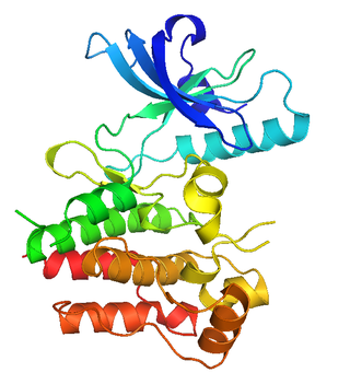
An acetylcholine receptor is an integral membrane protein that responds to the binding of acetylcholine, a neurotransmitter.

<span class="mw-page-title-main">Excitatory synapse</span> Sort of synapse

An excitatory synapse is a synapse in which an action potential in a presynaptic neuron increases the probability of an action potential occurring in a postsynaptic cell. Neurons form networks through which nerve impulses travels, each neuron often making numerous connections with other cells of neurons. These electrical signals may be excitatory or inhibitory, and, if the total of excitatory influences exceeds that of the inhibitory influences, the neuron will generate a new action potential at its axon hillock, thus transmitting the information to yet another cell.

<span class="mw-page-title-main">Edrophonium</span>

Edrophonium is a readily reversible acetylcholinesterase inhibitor. It prevents breakdown of the neurotransmitter acetylcholine and acts by competitively inhibiting the enzyme acetylcholinesterase, mainly at the neuromuscular junction. It is sold under the trade names Tensilon and Enlon.

<span class="mw-page-title-main">Neuromuscular junction</span> Junction between the axon of a motor neuron and a muscle fiber

A neuromuscular junction is a chemical synapse between a motor neuron and a muscle fiber.

<span class="mw-page-title-main">End-plate potential</span>

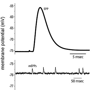
End plate potentials (EPPs) are the voltages which cause depolarization of skeletal muscle fibers caused by neurotransmitters binding to the postsynaptic membrane in the neuromuscular junction. They are called "end plates" because the postsynaptic terminals of muscle fibers have a large, saucer-like appearance. When an action potential reaches the axon terminal of a motor neuron, vesicles carrying neurotransmitters are exocytosed and the contents are released into the neuromuscular junction. These neurotransmitters bind to receptors on the postsynaptic membrane and lead to its depolarization. In the absence of an action potential, acetylcholine vesicles spontaneously leak into the neuromuscular junction and cause very small depolarizations in the postsynaptic membrane. This small response (~0.4mV) is called a miniature end plate potential (MEPP) and is generated by one acetylcholine-containing vesicle. It represents the smallest possible depolarization which can be induced in a muscle.

<span class="mw-page-title-main">MuSK protein</span> Mammalian protein found in Homo sapiens

MuSK is a receptor tyrosine kinase required for the formation and maintenance of the neuromuscular junction. It is activated by a nerve-derived proteoglycan called agrin, which is similarly also required for neuromuscular junction formation.

<span class="mw-page-title-main">Neuromuscular disease</span> Medical condition

A neuromuscular disease is any disease affecting the peripheral nervous system (PNS), the neuromuscular junctions, or skeletal muscles, all of which are components of the motor unit. Damage to any of these structures can cause muscle atrophy and weakness. Issues with sensation can also occur.

<span class="mw-page-title-main">Agrin</span> Mammalian protein found in Homo sapiens

Agrin is a large proteoglycan whose best-characterised role is in the development of the neuromuscular junction during embryogenesis. Agrin is named based on its involvement in the aggregation of acetylcholine receptors during synaptogenesis. In humans, this protein is encoded by the AGRN gene.

Dok-7 is a non-catalytic cytoplasmic adaptor protein that is expressed specifically in muscle and is essential for the formation of neuromuscular synapses. Further, Dok-7 contains pleckstrin homology (PH) and phosphotyrosine-binding (PTB) domains that are critical for Dok-7 function. Finally, mutations in Dok-7 are commonly found in patients with limb-girdle congenital myasthenia.

Congenital myasthenic syndrome (CMS) is an inherited neuromuscular disorder caused by defects of several types at the neuromuscular junction. The effects of the disease are similar to Lambert-Eaton Syndrome and myasthenia gravis, the difference being that CMS is not an autoimmune disorder. There are only 600 known family cases of this disorder and it is estimated that its overall frequency in the human population is 1 in 200,000.

<span class="mw-page-title-main">RAPSN</span> Protein-coding gene in the species Homo sapiens

43 kDa receptor-associated protein of the synapse (rapsyn) is a protein that in humans is encoded by the RAPSN gene.

<span class="mw-page-title-main">CHRNE</span> Protein-coding gene

Acetylcholine receptor subunit epsilon is a protein that in humans is encoded by the CHRNE gene.

<span class="mw-page-title-main">CHRNB1</span> Protein-coding gene in the species Homo sapiens

Acetylcholine receptor subunit beta is a protein that in humans is encoded by the CHRNB1 gene.

Repetitive nerve stimulation is a variant of the nerve conduction study where electrical stimulation is delivered to a motor nerve repeatedly several times per second. By observing the change in the muscle electrical response (CMAP) after several stimulations, a physician can assess for the presence of a neuromuscular junction disease, and differentiate between presynaptic and postsynaptic conditions. The test was first described by German neurologist Friedrich Jolly in 1895, and is also known as Jolly's test.

<span class="mw-page-title-main">Active zone</span>

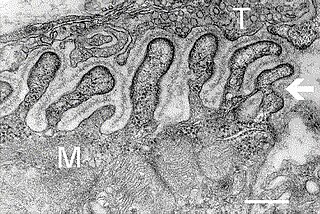
The active zone or synaptic active zone is a term first used by Couteaux and Pecot-Dechavassinein in 1970 to define the site of neurotransmitter release. Two neurons make near contact through structures called synapses allowing them to communicate with each other. As shown in the adjacent diagram, a synapse consists of the presynaptic bouton of one neuron which stores vesicles containing neurotransmitter, and a second, postsynaptic neuron which bears receptors for the neurotransmitter, together with a gap between the two called the synaptic cleft. When an action potential reaches the presynaptic bouton, the contents of the vesicles are released into the synaptic cleft and the released neurotransmitter travels across the cleft to the postsynaptic neuron and activates the receptors on the postsynaptic membrane.

References

  1. Levitan, Irwin; Kaczmarek, Leonard (August 19, 2015). "Intercellular communication". The Neuron: Cell and Molecular Biology (4th ed.). New York, NY: Oxford University Press. pp. 153–328. ISBN   978-0199773893.
  2. Hoch W, McConville J, Helms S, Newsom-Davis J, Melms A, Vincent A (2001). "Auto-antibodies to the receptor tyrosine kinase MuSK in patients with myasthenia gravis without acetylcholine receptor antibodies". Nat Med. 7 (3): 365–8. doi:10.1038/85520. PMID   11231638. S2CID   18641849.
  3. Sha SJ, Layzer RB (July 2007). "Myasthenia gravis and Lambert-Eaton myasthenic syndrome in the same patient". Muscle Nerve. 36 (1): 115–7. doi:10.1002/mus.20735. PMID   17206662. S2CID   297071.
  4. "Lambert-Eaton Myasthenic Syndrome". The Lecturio Medical Concept Library. Retrieved 1 August 2021.