Nemaline myopathy

Last updated
Nemaline myopathy
Other namesNemaline rod myopathy
Specialty Neurology   OOjs UI icon edit-ltr-progressive.svg

Nemaline myopathy (also called rod myopathy or nemaline rod myopathy) is a congenital, often hereditary neuromuscular disorder with many symptoms that can occur such as muscle weakness, hypoventilation, swallowing dysfunction, and impaired speech ability. The severity of these symptoms varies and can change throughout one's life to some extent. The prevalence is estimated at 1 in 50,000 live births. [1] It is the most common non-dystrophic myopathy. [2] [3]

Contents

"Myopathy" means muscle disease. Muscle fibers from a person with nemaline myopathy contains thread-like [4] rods, sometimes called nemaline bodies. [5] While the rods are diagnostic of the disorder, they are more likely a byproduct of the disease process rather than causing any dysfunction on their own. People with nemaline myopathy (NM) usually experience delayed motor development, or no motor development in severe cases, and weakness may occur in all of the skeletal muscles, such as muscles in the arms, legs, torso, neck flexors, throat, and face. The weakness tends to be more severe in the proximal muscles rather than the distal muscles. The ocular muscles are normally spared.

The disorder is often clinically categorized into groups with wide ranges of overlapping severity, from the most severe neonatal form which is incompatible with life, to a form so mild that it may not be diagnosed since the person appears to function at the lowest end of normal strength and breathing adequacy. Sporadic late onset nemaline myopathy (SLONM) is not a congenital disorder and is considered a different muscle disease from NM, which has its onset at birth or early childhood. [6] Respiratory problems are usually a primary concern for people with all forms of NM, and respiratory infections are quite common. NM shortens life expectancy, particularly in the more severe forms, but aggressive and proactive care allows most individuals to survive and even lead active lives.

Nemaline myopathy is one of the neuromuscular diseases covered by the Muscular Dystrophy Association in the United States.

Signs and symptoms

Signs vary from person to person. Young children and babies lack movement and have a difficult time eating and breathing. For young children not diagnosed immediately at birth, these are usually the first visible symptoms. One sign is a swollen face in disproportional areas. Other examples in newborns include swaying and a difficulty in moving. Other symptoms include feeble muscles in the neck and upper rib cage area. In adults, the most common symptom is respiratory problems. [7] Other symptoms in adults could range from mild to severe speech impediments. It is common to be diagnosed with scoliosis in relations to nemaline myopathy. [8] As babies that have NM develop and become of age when they should start walking, many take longer than average due to the lack of muscle, or just muscle fatigue. [9]

Since facial muscles are involved in NM takeover, elongated faces and a lower mandible are often observed in people with NM. People affected by NM usually will begin to feel muscle exhaustion between ages 20–50. NM is usually not progressive. Gastroesophageal reflux, although not common, is associated with NM. Heart abnormalities can occur as a result of NM, but the likelihood of that happening are not high. [7]

Mobility and orthopedics

Most children with mild NM eventually walk independently, although often at a later age than their peers. Some use wheelchairs or other devices, such as walkers or braces, to enhance their mobility. Individuals with severe NM generally have limited limb movement and use wheelchairs full-time. Due to weakness in the trunk muscles, people with NM are prone to scoliosis, which usually develops in childhood and worsens during puberty. Many individuals with NM undergo spinal fusion surgery to straighten and stabilize their backs.[ citation needed ]

Although patients early on often have mobility in their joints that is past the normal range, as they age, joint deformities and scoliosis usually occur. [10] If the person with nemaline myopathy keeps an eye on his or her joints early on, the problems with them can be detected when they begin and their progression can be delayed. Treatment of joint problems ranges from stretching exercises with physical therapy to surgical introduction of braces. The benefits of exercise in people with nemaline myopathy are still being studied, however, researchers have seen improvements in muscle function from low-intensity exercise. Vigorous exercise and the use of heavy weights should be avoided.[ citation needed ]

Respiratory involvement

Attention to respiratory issues is critical to the health of many people with NM. Infants with severe NM frequently experience respiratory distress at or soon after birth, although this is only found in the rarest forms. Though respiratory compromise may not be immediately apparent in people with intermediate or mild NM, it often, but not always, exists to some extent. As in many neuromuscular disorders, hypoventilation can begin insidiously, and it may cause serious health problems if not remedied by the use of noninvasive mechanical devices to assist breathing, particularly at night.[ citation needed ]

Communication and eating

Bulbar (throat) muscle weakness is a main feature of nemaline myopathy. Individuals with the most severe forms of NM are unable to swallow and receive their nutrition through feeding tubes. Most people with intermediate and mild NM take some or all of their nutrition orally. Bulbar muscle impairment may also lead to difficulty with communication. People with NM often have hypernasal speech as a result of poor closure of the velopharyngeal port (between the soft palate and the back of the throat). This may be able to be surgically corrected. Communicative skills may be enhanced through speech therapy, oral prosthetic devices, surgery, and augmentative communication devices. NM does not have any impact on cognition or intelligence.[ citation needed ]

Physical characteristics and effects

Physical expression of nemaline myopathy varies greatly, but weakness is usually concentrated in the proximal muscles, particularly respiratory, bulbar and trunk muscles. [11] People with severe NM show obvious symptoms at birth, while those with intermediate or mild NM may initially appear unaffected. Babies with NM are frequently observed to be "floppy" and hypotonic. Children born with NM often gain strength as they grow, though the effect of muscle weakness on body features may become more evident with time. Adults with NM typically have a very slender physique. [11]

Causes

Nemaline myopathy is caused by mutations in one of at least 11 different genes. [2] [12] Nemaline myopathy is a clinically and genetically heterogeneous disorder and both autosomal dominant and autosomal recessive forms can occur. Diagnosis is made based upon clinical signs such as muscle weakness, absent or low deep tendon reflexes (hyporeflexia), and a high-arched palate, along with electron-dense aggregates, called nemaline rods, being observed at the microscopic level within muscle fibers. Genetic confirmation through identification of a known genetic mutation in the patient is also an important component of diagnosis. [13] [14]

The two most common gene mutations causing nemaline myopathy are found on NEB or ACTA1 . [15] Mutations of the NEB gene usually result in symptoms present at birth or beginning in early childhood. This mutation results in about 50% of affected nemaline myopathy patients. The most common inheritance pathway for those with mutations in NEB is autosomal recessive in which each parent carries one mutated copy along with one normal functioning copy of the gene, and they pass the mutated copy to their offspring. In some cases, occasionally with ACTA1 mutations, NM can be caused by an inheritance pattern of autosomal dominance. This mutation results in about 15 to 25 percent of NM cases. [16] One reason why this is lower is because NM is associated with de novo mutations in ACTA1, occurring spontaneously in the egg or sperm. [9] When the condition is heritable, each pregnancy with the same partners has the same risk of passing the mutated genes to offspring. New mutations (de novo) can also occur causing NM and de novo mutations have been most often found to occur in the ACTA1 gene. [16] MYPN is the last found gene related to NM The risk of all cases of nemaline myopathy is the same in males and females. [9]

An example of muscle cells with rods. The rods have been stained red and are located on the inside of the plasma membrane. Biopsy nemaline myopathy gomori.jpg
An example of muscle cells with rods. The rods have been stained red and are located on the inside of the plasma membrane.
Summary of Molecular Genetic Testing Used in Nemaline Myopathy [17]
Gene 1Proportion of Nemaline Myopathy Attributed to Mutation of This GeneTest Method
NEB Up to 50%Sequence analysis
Deletion/duplication analysis & targeted analysis for pathogenic variants
ACTA1 15%-25%Sequence analysis
Deletion/duplication analysis
TPM3 2%-3%Sequence analysis
Deletion/duplication analysis
TPM2 <1%Sequence analysis
Deletion/duplication analysis
TNNT1 Almost exclusively in Old AmishSequence analysis
Deletion/duplication analysis
CFL2 RareSequence analysis
KBTBD13UnknownSequence analysis
KLHL4020%Sequence analysis
KLHL41 UnknownSequence analysis
LMOD3 UnknownSequence analysis
UnknownNANA

The physical capabilities of a given person with NM do not correlate well either with genotype or with muscle pathology as observed in the biopsy. [18]

Mechanism

Muscle cells contract in complex mechanical and chemical processes. If any part of the process or structure is disrupted, dysfunction will likely result, as in the case of those with genetic variations. In those with nemaline myopathy, muscle contraction is adversely affected. At the electron microscopic level, rod-shaped components can often be seen in some of the muscle cells, and when seen, are diagnostic for the condition called nemaline rod myopathy. The presence of these rods is not itself causing muscle weakness; rather they appear as a result of something going wrong within the muscle fiber. There is no connection between the number of rods found in the muscle cells and the amount of weakness a person has. All of the different gene mutations leading to the condition called nemaline myopathy that have been found so far are in genes that encode different components of the sarcomere. [11] In normal muscle cells, the various parts of the muscle fibers that make up the sarcomere are distributed evenly in a pattern for effective muscle contraction. Evidence suggests that some kinds of NM affect the arrangement of these muscle fibers, causing the muscles to be unable to contract as efficiently or effectively.[ citation needed ]

This is a simple example of a healthy sarcomere. Normal Sarcomere.svg
This is a simple example of a healthy sarcomere.
Example of a sarcomere with nemaline rods. Nemaline myopathy sarcomere.svg
Example of a sarcomere with nemaline rods.

Nemaline myopathy is usually genetic and shows traits in the affected individual from birth or an early age. However, there are some cases of symptoms of nemaline myopathy not showing up until adulthood. These cases are usually not genetic. Of the genes that have been linked to nemaline myopathy, most are also involved in encoding proteins in the sarcomeres in the muscle cells. [11] Respiratory muscles are often more affected than other skeletal muscle groups. Cardiac muscle is usually not affected in nemaline myopathy; however, in cases where it does, patients often present with dilated cardiomyopathy. The ocular muscles are usually spared.[ citation needed ]

The different genes whose mutations lead to the different kinds of nemaline myopathies affect the cells and the person's body differently. The first kind of nemaline myopathy identified is due to the Slow α-Tropomyosin Gene TPM3 and varies from case to case with its severity. In this kind of nemaline myopathy, affected people are weaker and more affected in their lower limbs than their upper limbs. [11]

As stated above, the most common genetic form of NM is caused by a mutation in the nebulin gene, called Nebulin, [19] [20] and has a range of severity levels. All published cases up to this point where NM is thought to be caused by a mutation in the NEB gene have been autosomal recessive and are the most common cause of nemaline myopathy. [21] Patients with this kind of NM are more affected in the muscles in their head, rather than their proximal muscles at the core of their body. Consequently, patients with this genetic mutation often cannot lift their heads and speak with a nasal voice. There have been cases that suggest this kind of NM may lead to patients having higher intellect. [11]

A third kind of nemaline myopathy in the Skeletal Muscle α-Actin Gene ACTA1 is due to a recessive null mutation. [11] These patients do not always show the typical nemaline bodies in their muscle cells. The only abnormality they show is an abnormal distribution of muscle fibers.

There are several other identified kinds of mutations that lead to Nemaline Myopathies. One affects slow skeletal muscles, one leads to the formation of both nemaline bodies and other abnormal, core-like, structures forming in the patient's muscles.[ citation needed ]

Diagnosis

Treatment

At present, Nemaline myopathy does not have a cure. Nemaline myopathy is a very rare disease that only affects 1 out of 50,000 on average, although recent studies show that this number is even smaller. There are a number of treatments to minimize the symptoms of the disease. The treatments and procedures to help patients with nemaline myopathy vary depending on the severity of the disease. A possible accommodation could be the use of a stabilizer, such as a brace. Other means include moderate stretching and moderate exercise to help target muscles maintain maximum health. [22] As people with NM grow and develop throughout their lives, it is important for them to see a variety of health professionals regularly, including a neurologist, physical therapist, and others, such as speech therapists and psychologists, to help both the patient and family adjust to everyday life. [10]

Outcome

Although there is no cure for NM, it is possible, and common for many people live healthy active lives even with moderate to severe cases. [23] Research continues to seek ways to ameliorate debilitating symptoms and lengthen the life-span in quality ways for those affected. Some people have seen mild improvements in secretion handling, energy level, and physical functioning with supplemental L-tyrosine, an amino acid that is available through health centers. [24] Some symptoms may worsen as the patient ages. Muscle loss increases with age naturally, but it is even more significant with nemaline myopathy. [25]

Current research

New research resources have become available for the NM community, such as the CMDIR (registry) and the CMD-TR (biorepository). These two resources connect families and individuals interested in participating in research with the scientists that aim to treat or cure NM. Some research on NM seeks to better understand the molecular effects the gene mutations have on muscle cells and the rest of the body [26] and to observe any connections NM may have to other diseases and health complications.

History

"Rod myopathy" was first identified by Douglas Reye, an Australian physician, in 1958. [27] However, Reye's results were never published because another doctor dismissed his finding of rods in the muscle tissue as an artifact of the biopsy. Forty years later, Reye's "rod myopathy" patient was confirmed to have nemaline myopathy. Another group of Australian researchers has since published an article recognizing Reye for his work. [28]

"Nemaline myopathy" was first named in a published paper in 1963 by North American researchers P.E. Cohen and G. M. Shy. Shy and his team discovered rod- like structures in muscle fibers of patients with muscle weakness by performing muscle biopsies on multiple patients. [22] Laboratories performing research on NM are located around the world, notably in the United States, Canada, England, Finland, and Australia.

Society and culture

In 1999, the NM Community [29] and first website (www.nemaline.org [30] ) for nemaline myopathy was launched by David McDougall, and in October 2004, the first Nemaline Myopathy Conference was hosted in Toronto, Canada, by Nemaline Myopathy Support Group. Many more conferences and social events have been held since, and all events organized since 2008 have been co-sponsored by A Foundation Building Strength for Nemaline Myopathy (AFBS), [31] the only foundation focused on supporting treatment development and social events for the NM community. In March 2006, Niki Shisler released a book, Fragile, in which she recounted her experiences surrounding the birth of twin sons with severe NM. In 2014, a team of experts collaborated with affected individuals and families caring for someone with a congenital myopathy to develop the first guidebook on managing life with a congenital myopathy[ citation needed ]

Related Research Articles

Inclusion body myositis (IBM) is the most common inflammatory muscle disease in older adults. The disease is characterized by slowly progressive weakness and wasting of both proximal muscles and distal muscles, most apparent in the finger flexors and knee extensors. IBM is often confused with an entirely different class of diseases, called hereditary inclusion body myopathies (hIBM). The "M" in hIBM is an abbreviation for "myopathy" while the "M" in IBM is for "myositis". In IBM, two processes appear to occur in the muscles in parallel, one autoimmune and the other degenerative. Inflammation is evident from the invasion of muscle fibers by immune cells. Degeneration is characterized by the appearance of holes, deposits of abnormal proteins, and filamentous inclusions in the muscle fibers. sIBM is a rare disease, with a prevalence ranging from 1 to 71 individuals per million.

In medicine, myopathy is a disease of the muscle in which the muscle fibers do not function properly. This results in muscular weakness. Myopathy means muscle disease. This meaning implies that the primary defect is within the muscle, as opposed to the nerves or elsewhere. Muscle cramps, stiffness, and spasm can also be associated with myopathy.

Hereditary inclusion body myopathies (HIBM) are a group of rare genetic disorders which have different symptoms. Generally, they are neuromuscular disorders characterized by muscle weakness developing in young adults. Hereditary inclusion body myopathies comprise both autosomal recessive and autosomal dominant muscle disorders that have a variable expression (phenotype) in individuals, but all share similar structural features in the muscles.

<span class="mw-page-title-main">MELAS syndrome</span> Medical condition

Mitochondrial encephalopathy, lactic acidosis, and stroke-like episodes (MELAS) is one of the family of mitochondrial diseases, which also include MIDD, MERRF syndrome, and Leber's hereditary optic neuropathy. It was first characterized under this name in 1984. A feature of these diseases is that they are caused by defects in the mitochondrial genome which is inherited purely from the female parent. The most common MELAS mutation is mitochondrial mutation, mtDNA, referred to as m.3243A>G.

<span class="mw-page-title-main">Centronuclear myopathy</span> Medical condition

Centronuclear myopathies (CNM) are a group of congenital myopathies where cell nuclei are abnormally located in the center of muscle cells instead of their normal location at the periphery.

<span class="mw-page-title-main">Congenital muscular dystrophy</span> Medical condition

Congenital muscular dystrophies are autosomal recessively-inherited muscle diseases. They are a group of heterogeneous disorders characterized by muscle weakness which is present at birth and the different changes on muscle biopsy that ranges from myopathic to overtly dystrophic due to the age at which the biopsy takes place.

<span class="mw-page-title-main">MERRF syndrome</span> Medical condition

MERRF syndrome is a mitochondrial disease. It is extremely rare, and has varying degrees of expressivity owing to heteroplasmy. MERRF syndrome affects different parts of the body, particularly the muscles and nervous system. The signs and symptoms of this disorder appear at an early age, generally childhood or adolescence. The causes of MERRF syndrome are difficult to determine, but because it is a mitochondrial disorder, it can be caused by the mutation of nuclear DNA or mitochondrial DNA. The classification of this disease varies from patient to patient, since many individuals do not fall into one specific disease category. The primary features displayed on a person with MERRF include myoclonus, seizures, cerebellar ataxia, myopathy, and ragged red fibers (RRF) on muscle biopsy, leading to the disease's name. Secondary features include dementia, optic atrophy, bilateral deafness, peripheral neuropathy, spasticity, or multiple lipomata. Mitochondrial disorders, including MERRFS, may present at any age.

<span class="mw-page-title-main">Myotonic dystrophy</span> Genetic disorder that impairs muscle function

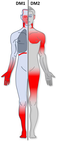
Myotonic dystrophy (DM) is a type of muscular dystrophy, a group of genetic disorders that cause progressive muscle loss and weakness. In DM, muscles are often unable to relax after contraction. Other manifestations may include cataracts, intellectual disability and heart conduction problems. In men, there may be early balding and an inability to father children. While myotonic dystrophy can occur at any age, onset is typically in the 20s and 30s.

Congenital myopathy is a very broad term for any muscle disorder present at birth. This defect primarily affects skeletal muscle fibres and causes muscular weakness and/or hypotonia. Congenital myopathies account for one of the top neuromuscular disorders in the world today, comprising approximately 6 in 100,000 live births every year. As a whole, congenital myopathies can be broadly classified as follows:

<span class="mw-page-title-main">Bethlem myopathy</span> Medical condition

Bethlem myopathy is predominantly an autosomal dominant myopathy, classified as a congenital form of muscular dystrophy. There are two types of Bethlem myopathy, based on which type of collagen is affected.

<span class="mw-page-title-main">Central core disease</span> Autosomal dominant genetic disorder

Central core disease (CCD), also known as central core myopathy, is an autosomal dominant inherited muscle disorder present from birth that negatively affects the skeletal muscles. It was first described by Shy and Magee in 1956. It is characterized by the appearance of the myofibril under the microscope.

Danon disease is a metabolic disorder. Danon disease is an X-linked lysosomal and glycogen storage disorder associated with hypertrophic cardiomyopathy, skeletal muscle weakness, and intellectual disability. It is inherited in an X-linked dominant pattern.

<span class="mw-page-title-main">Neutral lipid storage disease</span> Congenital autosomal recessive disorder

Neutral lipid storage disease is a congenital autosomal recessive disorder characterized by accumulation of triglycerides in the cytoplasm of leukocytes, muscle, liver, fibroblasts, and other tissues. It commonly occurs as one of two subtypes, cardiomyopathic neutral lipid storage disease (NLSD-M), or ichthyotic neutral lipid storage disease (NLSD-I) which is also known as Chanarin–Dorfman syndrome), which are characterized primarily by myopathy and ichthyosis, respectively. Normally, the ichthyosis that is present is typically non-bullous congenital ichthyosiform erythroderma which appears as white scaling.

Mitochondrially encoded tRNA glutamic acid also known as MT-TE is a transfer RNA which in humans is encoded by the mitochondrial MT-TE gene. MT-TE is a small 69 nucleotide RNA that transfers the amino acid glutamic acid to a growing polypeptide chain at the ribosome site of protein synthesis during translation.

Mitochondrially encoded tRNA phenylalanine also known as MT-TF is a transfer RNA which in humans is encoded by the mitochondrial MT-TF gene.

Mitochondrially encoded tRNA lysine also known as MT-TK is a transfer RNA which in humans is encoded by the mitochondrial MT-TK gene.

<span class="mw-page-title-main">X-linked spinal muscular atrophy type 2</span> Medical condition

X-linked spinal muscular atrophy type 2, also known as arthrogryposis multiplex congenita X-linked type 1 (AMCX1), is a rare neurological disorder involving death of motor neurons in the anterior horn of spinal cord resulting in generalised muscle wasting (atrophy). The disease is caused by a mutation in UBA1 gene and is passed in an X-linked recessive manner by carrier mothers to affected sons.

Desmin-related myofibrillar myopathy, is a subgroup of the myofibrillar myopathy diseases and is the result of a mutation in the gene that codes for desmin which prevents it from forming protein filaments, instead forming aggregates of desmin and other proteins throughout the cell.

Sporadic late-onset nemaline myopathy, or SLONM, is a very rare disease, one of the nemaline myopathies, causing loss of muscle bulk and weakness in the legs but sparing the cranial nerves, and beginning its clinical course after age 40. It was first identified in 1966 at the Mayo Clinic, by A.G. Engel, and that same year W.K. Engel and J.S. Resnick noted another case that they elaborated in 1975. The diagnosis of the disease rests on subacutely evolving weakness after age 40, normal to low CK level, a myopathic EMG with fibrillations, and often a monoclonal gammopathy. The diagnosis is confirmed by visualizing rods in cryosections on light and electron microscopy. The associated monoclonal gammopathy has an unfavorable prognosis.

Autophagic vacuolar myopathy (AVM) consists of multiple rare genetic disorders with common histological and pathological features on muscle biopsy. The features highlighted are vacuolar membranes of the autophagic vacuoles having sarcolemmal characteristics and an excess of autophagic vacuoles. There are currently five types of AVM identified. The signs and symptoms become more severe over the course of the disease. It begins with an inability to pick up small objects and progresses to difficulty in walking. The age of onset varies from early childhood to late adulthood, affecting people of all ages.

References

  1. Yuen, Michaela; Sandaradura, Sarah A.; Dowling, James J.; Kostyukova, Alla S.; Moroz, Natalia; Quinlan, Kate G.; Lehtokari, Vilma-Lotta; Ravenscroft, Gianina; Todd, Emily J. (2014-11-03). "Leiomodin-3 dysfunction results in thin filament disorganization and nemaline myopathy". The Journal of Clinical Investigation. 124 (11): 4693–4708. doi:10.1172/JCI75199. ISSN   0021-9738. PMC   4347224 . PMID   25250574.
  2. 1 2 de Winter, Josine Marieke; Buck, Danielle; Hidalgo, Carlos; Jasper, Jeffrey R.; Malik, Fady I.; Clarke, Nigel F.; Stienen, Ger J. M.; Lawlor, Michael W.; Beggs, Alan H. (2013-06-01). "Troponin activator augments muscle force in nemaline myopathy patients with nebulin mutations". Journal of Medical Genetics. 50 (6): 383–392. doi:10.1136/jmedgenet-2012-101470. ISSN   1468-6244. PMC   3865762 . PMID   23572184.
  3. Ottenheijm, Coen A. C.; Lawlor, Michael W.; Stienen, Ger J. M.; Granzier, Henk; Beggs, Alan H. (2011-05-15). "Changes in cross-bridge cycling underlie muscle weakness in patients with tropomyosin 3-based myopathy". Human Molecular Genetics. 20 (10): 2015–2025. doi:10.1093/hmg/ddr084. ISSN   1460-2083. PMC   3080611 . PMID   21357678.
  4. nemaline myopathy
  5. "Nemaline Myopathy - NORD (National Organization for Rare Disorders)". NORD (National Organization for Rare Disorders). Retrieved 2016-04-25.
  6. Chahin, Nizar; Selcen, Duygu; Engel, Andrew G. (2005-10-25). "Sporadic late onset nemaline myopathy". Neurology. 65 (8): 1158–1164. doi:10.1212/01.wnl.0000180362.90078.dc. ISSN   1526-632X. PMID   16148261. S2CID   23334154.
  7. 1 2 3 4 5 "Muscle Disorders: MedlinePlus". www.nlm.nih.gov. Retrieved 2016-04-10.
  8. "Diseases - Inherited / Endocrine Myopathies - Type Of (Nemaline myopathy)". Muscular Dystrophy Association. 2015-12-18. Retrieved 2016-04-10.
  9. 1 2 3 Piteau, SJ; Rossiter, JP; Smith, RG; MacKenzie, JJ (2014). "Congenital Myopathy With Cap-Like Structures and Nemaline Rods: Case Report and Literature Review". Pediatric Neurology. Elsevier. 51 (2): 192–197. doi:10.1016/j.pediatrneurol.2014.04.002. PMID   25079567.
  10. 1 2 Ohlsson, Monica. "Nemaline Myopathy". Socialstyrelsen. The Swedish Information Centre for Rare Diseases. Archived from the original on 2019-04-08. Retrieved 2016-04-12.
  11. 1 2 3 4 5 6 7 Wallgren, Petterson; Sewry, CA; Nowak, KJ; Laing, NG (2011). "Nemaline Myopathies". Semin Pediatr Neurol. 18 (4): 230–8. doi:10.1016/j.spen.2011.10.004. PMID   22172418.
  12. Sandaradura, Sarah A.; North, Kathryn N. (2015-08-26). "LMOD3: the "missing link" in nemaline myopathy?". Oncotarget. 6 (29): 26548–26549. doi:10.18632/oncotarget.5267. ISSN   1949-2553. PMC   4694930 . PMID   26337340.
  13. North, Kathryn N.; Wang, Ching H.; Clarke, Nigel; Jungbluth, Heinz; Vainzof, Mariz; Dowling, James J.; Amburgey, Kimberly; Quijano-Roy, Susana; Beggs, Alan H. (2014-02-01). "Approach to the diagnosis of congenital myopathies". Neuromuscular Disorders. 24 (2): 97–116. doi:10.1016/j.nmd.2013.11.003. ISSN   1873-2364. PMC   5257342 . PMID   24456932.
  14. Romero, Norma B.; Sandaradura, Sarah A.; Clarke, Nigel F. (2013-10-01). "Recent advances in nemaline myopathy". Current Opinion in Neurology. 26 (5): 519–526. doi:10.1097/WCO.0b013e328364d681. ISSN   1473-6551. PMID   23995272. S2CID   12185842.
  15. Wallgren-Pettersson, Carina; Sewry, Caroline A.; Nowak, Kristen J.; Laing, Nigel G. (2011-12-01). "Nemaline myopathies". Seminars in Pediatric Neurology. 18 (4): 230–238. doi:10.1016/j.spen.2011.10.004. ISSN   1558-0776. PMID   22172418.
  16. 1 2 Laing, Nigel G.; Dye, Danielle E.; Wallgren-Pettersson, Carina; Richard, Gabriele; Monnier, Nicole; Lillis, Suzanne; Winder, Thomas L.; Lochmüller, Hanns; Graziano, Claudio (2009-09-01). "Mutations and Polymorphisms of the Skeletal Muscle α-Actin Gene (ACTA1)". Human Mutation. 30 (9): 1267–1277. doi:10.1002/humu.21059. ISSN   1059-7794. PMC   2784950 . PMID   19562689.
  17. North, Kathryn N.; Ryan, Monique M. (2015-06-11). "Table 1. [Summary of Molecular Genetic Testing Used in Nemaline Myopathy]. - GeneReviews® - NCBI Bookshelf" . Retrieved 2016-04-25.
  18. Ryan MM, Ilkovski B, Strickland CD, et al. (February 2003). "Clinical course correlates poorly with muscle pathology in nemaline myopathy". Neurology. 60 (4): 665–73. doi:10.1212/01.WNL.0000046585.81304.BC. PMID   12601110. S2CID   11652174.
  19. "WikiGenes - Collaborative Publishing". WikiGenes - Collaborative Publishing. Retrieved 2016-04-10.
  20. Li, Frank; Buck, Danielle; De Winter, Josine; Kolb, Justin; Meng, Hui; Birch, Camille; Slater, Rebecca; Escobar, Yael Natelie; Smith, John E. (2015-09-15). "Nebulin deficiency in adult muscle causes sarcomere defects and muscle-type-dependent changes in trophicity: novel insights in nemaline myopathy". Human Molecular Genetics. 24 (18): 5219–5233. doi:10.1093/hmg/ddv243. ISSN   1460-2083. PMC   4550825 . PMID   26123491.
  21. Ottenheijm, Lawlor, Steiner, Granzier, Beggs. "Changes in cross-bridge cycling underlie muscle weakness in patients with tropomyosin 3-based myopathy"
  22. 1 2 "Nemaline Myopathy - NORD (National Organization for Rare Disorders)". NORD (National Organization for Rare Disorders). Retrieved 2016-04-10.
  23. "Diseases-Inherited/Endocrine Myopathies". Muscular Dystrophy Association. 2015-12-18. Retrieved 27 April 2016.
  24. Ryan, Monique M.; Sy, Catherine; Rudge, Sian; Ellaway, Carolyn; Ketteridge, David; Roddick, Laurence G.; Iannaccone, Susan T.; Kornberg, Andrew J.; North, Kathryn N. (2008-06-01). "Dietary L-tyrosine supplementation in nemaline myopathy". Journal of Child Neurology. 23 (6): 609–613. doi:10.1177/0883073807309794. ISSN   0883-0738. PMID   18079309. S2CID   23371464.
  25. "Nemaline myopathy". www.socialstyrelsen.se. Archived from the original on 2019-04-08. Retrieved 2016-04-10.
  26. "Nemaline Myopathy". Genetics Home Reference. U.S. National Library of Medicine.
  27. "Congenital myopathies and muscular dystrophies team". Westmead Children's Hospital. 9 August 2005. Retrieved 13 February 2012.
  28. Schnell C, Kan A, North KN (June 2000). "'An artefact gone awry': identification of the first case of nemaline myopathy by Dr R.D.K. Reye". Neuromuscul. Disord. 10 (4–5): 307–12. doi:10.1016/S0960-8966(99)00123-6. PMID   10838259. S2CID   38543084.
  29. "Nemaline Myopathy Support Group: The official community for NM | Facebook". www.facebook.com.
  30. "Nemaline Myopathy Support Group: The original and official community for NM". Nemaline Myopathy Support Group. 1999.
  31. "A Foundation Building Strength for Nemaline Myopathy Research". 14 December 2015.