Feeding tube

Last updated
Feeding tube
Enteral feeding tube stylet retracted.png
ICD-9-CM 96.35
MeSH D004750

A feeding tube is a medical device used to provide nutrition to people who cannot obtain nutrition by mouth, are unable to swallow safely, or need nutritional supplementation. The state of being fed by a feeding tube is called gavage, enteral feeding or tube feeding. Placement may be temporary for the treatment of acute conditions or lifelong in the case of chronic disabilities. A variety of feeding tubes are used in medical practice. They are usually made of polyurethane or silicone. The diameter of a feeding tube is measured in French units (each French unit equals 13 mm). They are classified by the site of insertion and intended use. [1]

Contents

Medical uses

There are dozens of conditions that may require tube feeding (enteral nutrition) to prevent or treat malnutrition. Conditions that necessitate feeding tubes include prematurity, failure to thrive (or malnutrition), neurologic and neuromuscular disorders, inability to swallow, anatomical and post-surgical malformations of the mouth and esophagus, cancer, Sanfilippo syndrome, and digestive disorders. [2]

Children

Feeding tubes are used widely in children with excellent success for a wide variety of conditions. Some children use them temporarily until they are able to eat on their own, while other children require them for a longer time. Some children only use feeding tubes to supplement their oral diet, while others rely on them exclusively. [3] [4]

Dementia

People with advanced dementia who get feeding assistance rather than feeding tubes have better outcomes. [5] Feeding tubes do not increase life expectancy for such people, or protect them from aspiration pneumonia. [5] [6] Feeding tubes can also increase the risk of pressure ulcers, require pharmacological or physical restraints, and lead to distress. [5] [7] [8] [9] In the final stages of dementia, assisted feeding may still be preferred over a feeding tube to bring benefits of palliative care and human interaction even when nutritional goals are not being met.[ citation needed ]

ICU

Feeding tubes are often used in the intensive care unit (ICU) to provide nutrition to people who are critically ill while their medical conditions are addressed; as of 2016 there was no consensus as to whether nasogastric or gastric tubes led to better outcomes. [10]

Mechanical obstruction and dysmotility

There is at least moderate evidence for feeding tubes improving outcomes for chronic malnutrition in people with cancers of the head and neck that obstruct the esophagus and would limit oral intake, [11] [12] people with advanced gastroparesis, [13] and ALS. [14] For long term use, gastric tubes appear to have better outcomes than nasogastric tubes. [15]

GI surgery

People who have surgery on their throat or stomach often have a feeding tube while recovering from surgery; a tube leading through the nose and down to the middle part of the small intestine is used, or a tube is directly placed through the abdomen to the small intestine. There is some evidence to suggest that people with a tube through the nose were able to start eating normally sooner. [16]

Product types

Medical nutrition companies make flavored products for drinking and unflavored for tube feeding. In the USA these are regulated as medical foods, which are defined in section 5(b) of the Orphan Drug Act (21 U.S.C. 360ee (b) (3)) as "a food which is formulated to be consumed or administered enterally under the supervision of a physician and which is intended for the specific dietary management of a disease or condition for which distinctive nutritional requirements, based on recognized scientific principles, are established by medical evaluation." [17] [18]

Types

The most common types of tubes include those placed through the nose, including nasogastric, nasoduodenal, and nasojejunal tubes, and those placed directly into the abdomen, such as a gastrostomy, gastrojejunostomy, or jejunostomy feeding tube. [19] [20]

Nasogastric feeding tube

A nasogastric feeding tube or NG-tube is passed through the nares (nostril), down the esophagus and into the stomach. This type of feeding tube is generally used for short term feeding, usually less than a month, though some infants and children may use an NG-tube longterm. Individuals who need tube feeding for a longer period of time are typically transitioned to a more permanent gastric feeding tube. The primary advantage of the NG-tube is that it is temporary and relatively non-invasive to place, meaning it can be removed or replaced at any time without surgery. NG-tubes can have complications, particularly related to accidental removal of the tube and nasal irritation. [21] More specifically, when nasogastric or nasoenteric tubes are placed incorrectly, they can damage patients' vocal cords, lungs, or trachea, resulting in serious injuries or even death. [22]

In March 2022, Avanos Medical's Cortrak2 EAS recall, for instance, has been classified as a Class I recall by the FDA, following reports of injuries and patient deaths caused by misplaced nasoenteric or nasogastric tubes. [22]

Nasojejunal feeding tube

A nasojejunal or NJ-tube is similar to an NG-tube except that it is threaded through the stomach and into the jejunum, the middle section of the small intestine. In some cases, a nasoduodenal or ND-tube may be placed into the duodenum, the first part of the small intestine. These types of tube are used for individuals who are unable to tolerate feeding into the stomach, due to dysfunction of the stomach, impaired gastric motility, severe reflux or vomiting. These types of tubes must be placed in a hospital setting. [16] [23]

Gastrostomy or gastric feeding tube

A gastric feeding tube (G-tube or "button") is a tube inserted through a small incision in the abdomen into the stomach and is used for long-term enteral nutrition. One type is the percutaneous endoscopic gastrostomy (PEG) tube which is placed endoscopically. The position of the endoscope can be visualized on the outside of the person's abdomen because it contains a powerful light source. A needle is inserted through the abdomen, visualized within the stomach by the endoscope, and a suture passed through the needle is grasped by the endoscope and pulled up through the esophagus. The suture is then tied to the end of the PEG tube that will be external, and pulled back down through the esophagus, stomach, and out through the abdominal wall. The insertion takes about 20 minutes. The tube is kept within the stomach either by a balloon on its tip (which can be deflated) or by a retention dome which is wider than the tract of the tube. G-tubes may also be placed surgically, using either an open or laparoscopic technique. [24]

Gastric feeding tubes are suitable for long-term use, though they sometimes need to be replaced if used long-term. The G-tube can be useful where there is difficulty with swallowing because of neurologic or anatomic disorders (stroke, esophageal atresia, tracheoesophageal fistula, radiotherapy for head and neck cancer), and to decrease the risk of aspiration pneumonia. [25] However, in people with advanced dementia or adult failure to thrive it does not decrease the risk of pneumonia. There is moderate quality evidence suggesting that the risk of aspiration pneumonia may be reduced by inserting the feeding tube into the duodenum or the jejunum (post-pyloric feeding), when compared to inserting the feeding tube into the stomach. [25] People with dementia may attempt to remove the PEG, which causes complications. [26]

Gastric drainage tube

A G-tube may instead be used for gastric drainage as a longer-term solution to the condition where blockage in the proximal small intestine causes bile and acid to accumulate in the stomach, typically leading to periodic vomiting, or if the vagus nerve is damaged. Where such conditions are only short term, as in a hospital setting, a nasal tube connected to suction is usually used. A blockage lower in the intestinal tract may be addressed with a surgical procedure known as a colostomy, and either type of blockage may be corrected with a bowel resection under appropriate circumstances. If such correction is not possible or practical, nutrition may be supplied by parenteral nutrition. [27] [28]

Gastrojejunal feeding tube

A gastrojejunostomy or GJ feeding tube is a combination device that includes access to both the stomach and the jejunum, or middle part of the small intestine. Typical tubes are placed in a G-tube site or stoma, with a narrower long tube continuing through the stomach and into the small intestine. The GJ-tube is used widely in individuals with severely impaired gastric motility, high risk of aspiration, or an inability to feed into the stomach. It allows the stomach to be continually vented or drained while simultaneously feeding into the small intestine. GJ-tubes are typically placed by an interventional radiologist in a hospital setting. The primary complication of a GJ-tube is migration of the long portion of the tube out of the intestine and back into the stomach. [29]

Jejunal feeding tube

A jejunostomy feeding tube (J-tube) is a tube surgically or endoscopically inserted through the abdomen and into the jejunum (the second part of the small intestine). [16]

Complications

Nasogastric and nasojejeunal tubes are meant to convey liquid food to the stomach or intestines. When inserted incorrectly, the tip may rest in the respiratory system instead of the stomach or intestines; in this case, the liquid food will enter the lungs, resulting in pneumonia and can, in rare cases, lead to death. [23] [30] [31]

Complications associated with gastrostomy tubes (inserted through the abdomen and into the stomach or intestines) include leakage of gastric contents (containing hydrochloric acid) around the tube into the abdominal (peritoneal) cavity resulting in peritonitis, a serious complication which will cause death if it is not properly treated. Septic shock is another possible complication. [32] Minor leakage may cause irritation of the skin around the gastrostomy site or stoma. Barrier creams, to protect the skin from the corrosive acid, are used to manage this. [33]

A phenomenon called "tube dependency" has been discussed in the medical literature, in which a child refuses to eat after being on a feeding tube, but it is not recognized as a disorder in the ICD or DSM and its epidemiology is unknown. [34]

Oral and dental complications

Guidelines for dental care for children fed by tube are poorly established. Many dental complications arise due to poor oral health that may result from reluctance or intolerance towards oral hygiene practices by patients and caregivers, abundance of dental plaque and/or tooth decay, and lack of oral simulation. [35] Although many studies on this topic involve a relatively small sample size, the findings are important as they are associated with the development of various oral conditions, dental diseases and even systemic diseases such as aspiration pneumonia. [36] [37]

Calculus

Adults fed by tube have previously shown a significantly higher rate and quantity of calculus deposition than adults fed orally. Even with an intensive oral hygiene program in place, adults fed by tube still demonstrate a greater quantity of supra-gingival calculus accumulation, [35] which can be a risk factor for several oral diseases including periodontal diseases and aspiration pneumonia. Although calculus removal may be difficult for caregivers to perform and provide an unpleasant experience for patients with a feeding tube, the implications of calculus in the initiation of aspiration pneumonia make it clear that it poses a serious health risk. Research suggests that the best course of treatment for patients with a gastric tube is periodic professional cleaning, maintained with routine home use of a non-foaming anti-calculus dentifrice (toothpaste). [36]

Caries

Dental caries is a localized disease in which susceptible tooth structure is broken down by bacteria that are able to ferment carbohydrates into acid. Although it has not been extensively studied, researchers speculate that individuals fed by tube may be less prone to the development of caries as they are not exposed to carbohydrates orally. Examination of dental plaque from tube-fed individuals found that it contained fewer caries-associated microorganisms (lactobacilli and streptococcus) and had reduced ability to produce acids, suggesting an overall weaker ability to cause caries. Further, studies with animal subjects found that tube-feeding was not associated with tooth decay, even when combined with reduced salivation. [35] Thus, tube-feeding alone does not necessarily directly promote the development of caries.

Periodontal diseases

To date, no published studies have been conducted on periodontal disease indicators (including clinical attachment loss, pocket depth, or periodontal indices) among tube-fed individuals. However, since tube feeding is correlated with calculus build-up, which is known to be a risk factor in the development of periodontal diseases, further investigation is critical to determine what role tube-feeding might have in the development of periodontal disease. [35]

Dental erosion

Dental erosion is the dissolution of the tooth's hard structures (enamel, dentin & cementum) by exposure to acids not caused by bacteria. In the case of individuals fed by gastric tube, acid may enter the oral cavity through reflux of gastric contents. Gastroesophageal reflux (GER) affects up to 67% of children and young adults with central nervous system dysfunction, a condition which in itself is normally an indicator for tube feeding. The effects of gastric acid on the teeth may sometimes be masked or minimized by the abundance of calculus. Tube feeding may either resolve, exacerbate or introduce the issue of GER in individuals. [35]

Aspiration pneumonia

Individuals fed by tube are susceptible to aspiration through a multitude of factors. Firstly, undisturbed plaque is known to shift towards the type of bacteria (Gram-negative anaerobic) implicated in aspiration pneumonia. Additionally, tube-fed patients are commonly affected by gastroesophageal reflux and a breakdown in the airway protection reflex (breathing while swallowing). This results in the inadvertent inhalation of bacteria-containing gastric juices as they are re-swallowed, leading to the development of aspiration pneumonia. [35]

Oral hypersensitivity

While a child undergoes a period of tube-feeding, there is a lack of oral stimulation that can lead to the development of oral hypersensitivity. This can complicate the delivery of dental care and serve as a barrier for the child's return to oral feeding. This can also lead to dysphagia (difficulty swallowing), muscle weakness and improper airway protection, resulting in longer periods of tube-feeding and increased risk of dental complications. A dentist may prescribe a "desensitization program", which involves routine stimulation of intra-oral and extra-oral structures, and encourage oral hygiene procedures to be performed at home. [35]

History

While enemas were previously used for supplemental enteral nutrition, the practice of surgically inserting feeding tubes emerged in the mid to late 1800s. Initially, these procedures were largely unsuccessful, but they quickly improved with advancements in technique. [38]

Originally, the nasogastric tube (NGT) was described by John Hunter in the 18th century as a combination of eelskin and whalebone. It was initially utilized to provide liquid nutrition to the ill. [39]

See also

Related Research Articles

<span class="mw-page-title-main">Esophageal atresia</span> Congenital discontinuity of the oesophagus

Esophageal atresia is a congenital medical condition that affects the alimentary tract. It causes the esophagus to end in a blind-ended pouch rather than connecting normally to the stomach. It comprises a variety of congenital anatomic defects that are caused by an abnormal embryological development of the esophagus. It is characterized anatomically by a congenital obstruction of the esophagus with interruption of the continuity of the esophageal wall.

<span class="mw-page-title-main">Dysphagia</span> Difficulty in swallowing

Dysphagia is difficulty in swallowing. Although classified under "symptoms and signs" in ICD-10, in some contexts it is classified as a condition in its own right.

<span class="mw-page-title-main">Nasogastric intubation</span> Feeding tube going into the stomach through the nose and throat

Nasogastric intubation is a medical process involving the insertion of a plastic tube through the nose, down the esophagus, and down into the stomach. Orogastric intubation is a similar process involving the insertion of a plastic tube through the mouth. Abraham Louis Levin invented the NG tube. Nasogastric tube is also known as Ryle's tube in Commonwealth countries, after John Alfred Ryle.

<span class="mw-page-title-main">Pulmonary aspiration</span> Entry of materials into the larynx (voice box) and lower respiratory tract

Pulmonary aspiration is the entry of material such as pharyngeal secretions, food or drink, or stomach contents from the oropharynx or gastrointestinal tract, into the larynx and lower respiratory tract, the portions of the respiratory system from the trachea (windpipe) to the lungs. A person may inhale the material, or it may be delivered into the tracheobronchial tree during positive pressure ventilation. When pulmonary aspiration occurs during eating and drinking, the aspirated material is often colloquially referred to as "going down the wrong pipe".

<span class="mw-page-title-main">Upper gastrointestinal bleeding</span> Medical condition

Upper gastrointestinal bleeding is gastrointestinal bleeding (hemorrhage) in the upper gastrointestinal tract, commonly defined as bleeding arising from the esophagus, stomach, or duodenum. Blood may be observed in vomit or in altered form as black stool. Depending on the amount of the blood loss, symptoms may include shock.

<span class="mw-page-title-main">Esophagogastroduodenoscopy</span> Diagnostic endoscopic procedure

Esophagogastroduodenoscopy (EGD) or oesophagogastroduodenoscopy (OGD), also called by various other names, is a diagnostic endoscopic procedure that visualizes the upper part of the gastrointestinal tract down to the duodenum. It is considered a minimally invasive procedure since it does not require an incision into one of the major body cavities and does not require any significant recovery after the procedure. However, a sore throat is common.

<span class="mw-page-title-main">Percutaneous endoscopic gastrostomy</span> Feeding tube going into the stomach through the abdominal wall

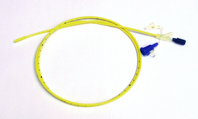
Percutaneous endoscopic gastrostomy (PEG) is an endoscopic medical procedure in which a tube is passed into a patient's stomach through the abdominal wall, most commonly to provide a means of feeding when oral intake is not adequate. This provides enteral nutrition despite bypassing the mouth; enteral nutrition is generally preferable to parenteral nutrition. The PEG procedure is an alternative to open surgical gastrostomy insertion, and does not require a general anesthetic; mild sedation is typically used. PEG tubes may also be extended into the small intestine by passing a jejunal extension tube through the PEG tube and into the jejunum via the pylorus.

<span class="mw-page-title-main">Aspiration pneumonia</span> Medical condition

Aspiration pneumonia is a type of lung infection that is due to a relatively large amount of material from the stomach or mouth entering the lungs. Signs and symptoms often include fever and cough of relatively rapid onset. Complications may include lung abscess, acute respiratory distress syndrome, empyema, and parapneumonic effusion. Some include chemical induced inflammation of the lungs as a subtype, which occurs from acidic but non-infectious stomach contents entering the lungs.

<span class="mw-page-title-main">Gastrostomy</span> Surgical procedure creating opening in stomach

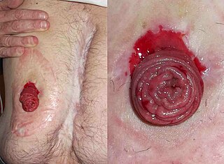
A gastrostomy is the creation of an artificial external opening into the stomach for nutritional support or gastric decompression. Typically this would include an incision in the patient's epigastrium as part of a formal operation. When originally devised over a century ago the procedure was completed through open surgery using the Stamm technique. It can be performed through surgical approach, percutaneous approach by interventional radiology, percutaneous endoscopic gastrostomy (PEG) or percutaneous ultrasound gastrostomy (PUG).

Gastric lavage, also commonly called stomach pumping or gastric irrigation, is the process of cleaning out the contents of the stomach using a tube. Since its first recorded use in early 19th century, it has become one of the most routine means of eliminating poisons from the stomach. Such devices are normally used on a person who has ingested a poison or overdosed on a drug such as ethanol. They may also be used before surgery, to clear the contents of the digestive tract before it is opened.

<span class="mw-page-title-main">Pseudocyst</span> Medical condition

Pseudocysts are like cysts, but lack epithelial or endothelial cells. Initial management consists of general supportive care. Symptoms and complications caused by pseudocysts require surgery. Computed tomography (CT) scans are used for initial imaging of cysts, and endoscopic ultrasounds are used in differentiating between cysts and pseudocysts. Endoscopic drainage is a popular and effective method of treating pseudocysts.

<span class="mw-page-title-main">Oropharyngeal dysphagia</span> Difficulty controlling the mouth or throat for swallowing

Oropharyngeal dysphagia is the inability to empty material from the oropharynx into the esophagus as a result of malfunction near the esophagus. Oropharyngeal dysphagia manifests differently depending on the underlying pathology and the nature of the symptoms. Patients with dysphagia can experience feelings of food sticking to their throats, coughing and choking, weight loss, recurring chest infections, or regurgitation. Depending on the underlying cause, age, and environment, dysphagia prevalence varies. In research including the general population, the estimated frequency of oropharyngeal dysphagia has ranged from 2 to 16 percent.

<span class="mw-page-title-main">Whole bowel irrigation</span> Medical process

Whole bowel irrigation (WBI) is a medical process involving the rapid administration of large volumes of an osmotically balanced macrogol solution, either orally or via a nasogastric tube, to flush out the entire gastrointestinal tract.

<span class="mw-page-title-main">Gastroparesis</span> Medical condition

Gastroparesis, is a medical disorder consisting of weak muscular contractions (peristalsis) of the stomach, resulting in food and liquid remaining in the stomach for a prolonged period of time. Stomach contents thus exit more slowly into the duodenum of the digestive tract. This can result in irregular absorption of nutrients, inadequate nutrition, and poor glycemic control.

Therapeutic endoscopy is the medical term for an endoscopic procedure during which treatment is carried out via the endoscope. This contrasts with diagnostic endoscopy, where the aim of the procedure is purely to visualize a part of the gastrointestinal, respiratory or urinary tract in order to aid diagnosis. In practice, a procedure which starts as a diagnostic endoscopy may become a therapeutic endoscopy depending on the findings, such as in cases of upper gastrointestinal bleeding, or the finding of polyps during colonoscopy.

<span class="mw-page-title-main">Jejunostomy</span>

Jejunostomy is the surgical creation of an opening (stoma) through the skin at the front of the abdomen and the wall of the jejunum. It can be performed either endoscopically, or with open surgery.

Cholecystostomy or (cholecystotomy) is a medical procedure used to drain the gallbladder through either a percutaneous or endoscopic approach. The procedure involves creating a stoma in the gallbladder, which can facilitate placement of a tube or stent for drainage, first performed by American surgeon, Dr. John Stough Bobbs, in 1867. It is sometimes used in cases of cholecystitis or other gallbladder disease where the person is ill, and there is a need to delay or defer cholecystectomy. The first endoscopic cholecystostomy was performed by Drs. Todd Baron and Mark Topazian in 2007 using ultrasound guidance to puncture the stomach wall and place a plastic biliary catheter for gallbladder drainage.

<span class="mw-page-title-main">Assisted feeding</span>

Assisted feeding, also called hand feeding or oral feeding, is the action of a person feeding another person who cannot otherwise feed themselves. The term is used in the context of some medical issue or in response to a disability, such as when a person living with dementia is no longer able to manage eating alone. The person being fed must be able to eat by mouth, but lacks either the cognitive or physical ability to self-feed. Individuals who are born with a disability like cerebral palsy, or arthrogryposis multiplex congenita (AMC) may be unable to feed themselves. Also, those who acquire a disability due to an accident or a disease like amyotrophic lateral sclerosis (ALS) may require hand feeding because they may become unable to pick-up and bring food to their own mouth.

Salamat Ahuoiza Aliu is a Nigerian neurosurgeon.

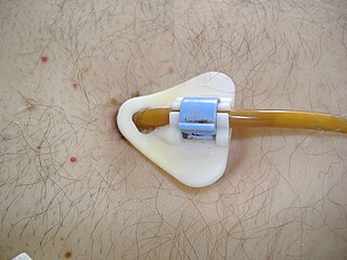
Buried bumper syndrome (BBS) is a condition that affects feeding tubes placed into the stomach through the abdominal wall. Gastrostomy tubes include an internal bumper, which secures the inner portion of the tube inside the stomach, and external bumper, which secures the outer portion of the tube and opposes the abdomen. Buried bumper syndrome occurs when the internal bumper of a gastrostomy tube erodes into the wall of the stomach. The internal bumper may become entirely buried within the fistulous tract. The main causative factor is excessive tightening of the external bumper, leading to increased pressure of the internal bumper on the wall of the stomach. Additional risk factors include: obesity, weight gain, malnutrition, corticosteroid therapy, and poor wound healing.

References

  1. "Tube feeding". 2019-05-09.
  2. Yasuda, Hideto; Kondo, Natsuki; Yamamoto, Ryohei; Asami, Sadaharu; Abe, Takayuki; Tsujimoto, Hiraku; Tsujimoto, Yasushi; Kataoka, Yuki (2021-09-27). "Monitoring of gastric residual volume during enteral nutrition". The Cochrane Database of Systematic Reviews. 2021 (9): CD013335. doi:10.1002/14651858.CD013335.pub2. ISSN   1469-493X. PMC   8498989 . PMID   34596901.
  3. Hannah, E; John, RM (November 2013). "Everything the nurse practitioner should know about pediatric feeding tubes". Journal of the American Association of Nurse Practitioners. 25 (11): 567–77. doi:10.1002/2327-6924.12075. PMID   24170530. S2CID   6405475. Open Access logo PLoS transparent.svg
  4. Craig, GM (December 2013). "Psychosocial aspects of feeding children with neurodisability". European Journal of Clinical Nutrition. 67 (Suppl 2): S17–20. doi: 10.1038/ejcn.2013.226 . PMID   24301004. Open Access logo PLoS transparent.svg
  5. 1 2 3 American Academy of Hospice and Palliative Medicine, "Five Things Physicians and Patients Should Question", Choosing Wisely: an initiative of the ABIM Foundation, American Academy of Hospice and Palliative Medicine, retrieved 1 August 2013
  6. Murray, A; Mulkerrin, S; O'Keeffe, ST (2019). "The perils of 'risk feeding'". Age and Ageing. 48 (4): 478–481. doi: 10.1093/ageing/afz027 . PMID   30939597.
  7. Hanson, L. C.; Ersek, M.; Gilliam, R.; Carey, T. S. (2011). "Oral Feeding Options for People with Dementia: A Systematic Review". Journal of the American Geriatrics Society. 59 (3): 463–472. doi:10.1111/j.1532-5415.2011.03320.x. PMC   3164780 . PMID   21391936.
  8. Kuo, S.; Rhodes, R. L.; Mitchell, S. L.; Mor, V.; Teno, J. M. (2009). "Natural History of Feeding-Tube Use in Nursing Home Residents with Advanced Dementia". Journal of the American Medical Directors Association. 10 (4): 264–270. doi:10.1016/j.jamda.2008.10.010. PMC   2733212 . PMID   19426943.
  9. Schlein, K (August 2016). "Gastric Versus Small Bowel Feeding in Critically Ill Adults". Nutrition in Clinical Practice. 31 (4): 514–22. doi:10.1177/0884533616629633. PMID   26920643.
  10. Bishop, S; Reed, WM (December 2015). "The provision of enteral nutritional support during definitive chemoradiotherapy in head and neck cancer patients". Journal of Medical Radiation Sciences. 62 (4): 267–76. doi:10.1002/jmrs.132. PMC   4968562 . PMID   27512573.
  11. Canadian Agency for Drugs and Technologies in Health (13 August 2014). "Nasogastric Feeding Tubes versus Percutaneous Endoscopic Gastrostomy for Patients with Head or Neck Cancer: A Review of Clinical Effectiveness and Guidelines". Rapid Response Reports. CADTH Rapid Response Reports. PMID   25411678.
  12. Sarosiek, I; Davis, B; Eichler, E; McCallum, RW (March 2015). "Surgical approaches to treatment of gastroparesis: gastric electrical stimulation, pyloroplasty, total gastrectomy and enteral feeding tubes". Gastroenterology Clinics of North America. 44 (1): 151–67. doi:10.1016/j.gtc.2014.11.012. PMID   25667030.
  13. Greenwood, DI (June 2013). "Nutrition management of amyotrophic lateral sclerosis". Nutrition in Clinical Practice. 28 (3): 392–9. doi:10.1177/0884533613476554. PMID   23466470.
  14. Lai, L; Ali, SF (September 2015). "Percutaneous Endoscopic Gastrostomy and Open Gastrostomy". Atlas of the Oral and Maxillofacial Surgery Clinics of North America. 23 (2): 165–8. doi:10.1016/j.cxom.2015.05.001. PMID   26333904.
  15. 1 2 3 Wang, L; Tian, Z; Liu, Y (January 2017). "Nasoenteric tube versus jejunostomy for enteral nutrition feeding following major upper gastrointestinal operations: a meta-analysis" (PDF). Asia Pacific Journal of Clinical Nutrition. 26 (1): 20–26. doi:10.6133/apjcn.112015.05. PMID   28049257.
  16. Affairs, Office of Regulatory (2019-02-09). "Medical Foods Guidance Documents & Regulatory Information". Food and Drug Administration .
  17. Nutrition, Center for Food Safety and Applied. "Medical Foods - Guidance for Industry: Frequently Asked Questions About Medical Foods; Second Edition". Food and Drug Administration .
  18. Guenter, Peggi (2001). "Enteral Feeding Access Devices". In Guenter, Peggi; Silkroski, Marcia (eds.). Tube Feeding: Practical Guidelines and Nursing Protocols. Gaithersburg: Aspen Publishers. pp. 51–67. ISBN   978-0-8342-1939-7.
  19. "Nutrition support for adults: oral nutrition support, enteral tube feeding and parenteral nutrition". NICE. August 2017. Retrieved 30 January 2018.
  20. "Dysphagia Treatment". NHS Choices. 19 January 2015.
  21. 1 2 "Avanos Medical Recalls Cortrak*2 Enteral Access System for Risk of Misplaced Enteral Tubes Could Cause Patient Harm". fda.gov. 16 May 2022. Retrieved 23 October 2022.
  22. 1 2 Tiancha, H; Jiyong, J; Min, Y (July 2015). "How to Promote Bedside Placement of the Postpyloric Feeding Tube: A Network Meta-Analysis of Randomized Controlled Trials". Journal of Parenteral and Enteral Nutrition. 39 (5): 521–30. doi:10.1177/0148607114546166. PMID   25146431.
  23. Thaker, AM; Sedarat, A (September 2016). "Laparoscopic-Assisted Percutaneous Endoscopic Gastrostomy". Current Gastroenterology Reports. 18 (9): 46. doi:10.1007/s11894-016-0520-2. PMID   27422123. S2CID   19146165.
  24. 1 2 Alkhawaja, Sana; Martin, Claudio; Butler, Ronald J.; Gwadry-Sridhar, Femida (2015-08-04). "Post-pyloric versus gastric tube feeding for preventing pneumonia and improving nutritional outcomes in critically ill adults". The Cochrane Database of Systematic Reviews. 2018 (8): CD008875. doi:10.1002/14651858.CD008875.pub2. ISSN   1469-493X. PMC   6516803 . PMID   26241698.
  25. Siamak Milanchi; Matthew T Wilson (January–March 2008). "Malposition of percutaneous endoscopic-guided gastrostomy: Guideline and management". J Minim Access Surg. 4 (1): 1–4. doi: 10.4103/0972-9941.40989 . PMC   2699054 . PMID   19547728.
  26. Costa, G; Ruscelli, P; Balducci, G; Buccoliero, F; Lorenzon, L; Frezza, B; Chirletti, P; Stagnitti, F; Miniello, S; Stella, F (2016). "Clinical strategies for the management of intestinal obstruction and pseudo-obstruction. A Delphi Consensus study of SICUT (Società Italiana di Chirurgia d'Urgenza e del Trauma)". Annali Italiani di Chirurgia. 87: 105–17. PMID   27179226.
  27. Mulholland, Michael W.; Doherty, Gerard M. (2011). Complications in Surgery. Lippincott Williams & Wilkins. p. 270. ISBN   978-1-60547-530-1.
  28. Gershman, George; Ament, Marvin (2008). Practical Pediatric Gastrointestinal Endoscopy. John Wiley & Sons. p. 126. ISBN   978-0-470-98731-5.
  29. Santos, SC; Woith, W; Freitas, MI; Zeferino, EB (September 2016). "Methods to determine the internal length of nasogastric feeding tubes: An integrative review". International Journal of Nursing Studies. 61: 95–103. doi:10.1016/j.ijnurstu.2016.06.004. PMID   27328376.
  30. Metheny, NA; Meert, KL (May 2014). "Effectiveness of an electromagnetic feeding tube placement device in detecting inadvertent respiratory placement". American Journal of Critical Care. 23 (3): 240–7, quiz 248. doi: 10.4037/ajcc2014954 . PMID   24786813. S2CID   40766751. Open Access logo PLoS transparent.svg
  31. Melis M, Fichera A, Ferguson MK (July 2006). "Bowel necrosis associated with early jejunal tube feeding: A complication of postoperative enteral nutrition". Arch Surg. 141 (7): 701–4. doi: 10.1001/archsurg.141.7.701 . PMID   16847244.
  32. Townley, A; Wincentak, J; Krog, K; Schippke, J; Kingsnorth, S (2018). "Pediatric gastrostomy stoma complications and treatments: A rapid scoping review". Journal of Clinical Nursing. 27 (7–8): 1369–1380. doi:10.1111/jocn.14233. PMID   29266535. S2CID   3914260.
  33. Krom, H; de Winter, JP; Kindermann, A (June 2017). "Development, prevention, and treatment of feeding tube dependency". European Journal of Pediatrics. 176 (6): 683–688. doi:10.1007/s00431-017-2908-x. PMC   5432583 . PMID   28409284.
  34. 1 2 3 4 5 6 7 Dyment, Heather A.; Casas, Michael J. (1999). "Dental care for children fed by tube: a critical review". Special Care in Dentistry. 19 (5): 220–224. doi:10.1111/j.1754-4505.1999.tb01389.x. ISSN   1754-4505. PMID   10765889.
  35. 1 2 Brown, Laurie M.; Casamassimo, Paul S.; Griffen, Ann; Tatakis, Dimitris (Sep–Oct 2006). "Supragingival calculus in children with gastrostomy feeding: significant reduction with a caregiver-applied tartar-control dentifrice". Pediatric Dentistry. 28 (5): 410–414. ISSN   0164-1263. PMID   17036705.
  36. Cardona‐Soria, Sandra; Cahuana‐Cárdenas, Abel; Rivera‐Baró, Alejandro; Miranda‐Rius, Jaume; Martín de Carpi, Javier; Brunet‐Llobet, Lluís (2019-11-07). "Oral health status in pediatric patients with cerebral palsy fed by oral versus enteral route". Special Care in Dentistry. 40 (1): 35–40. doi:10.1111/scd.12429. ISSN   0275-1879. PMID   31697430. S2CID   207956249.
  37. Minard, Gayle (December 2006). "The history of surgically placed feeding tubes". Nutrition in Clinical Practice. 21 (6): 626–633. doi:10.1177/0115426506021006626. ISSN   0884-5336. PMID   17119170.
  38. "Nasogastric Tube". ScienceDirect. Retrieved 2023-02-15.