Percutaneous endoscopic gastrostomy

Last updated
Percutaneous endoscopic gastrostomy
Percutaneous endoscopic gastrostomy-tube.jpg
Percutaneous endoscopic gastrostomy
Other namesPEG tube
Specialty Gastroenterology
ComplicationsInfection, Hemorrhage, Gastrointestinal perforation, Gastrocolic fistula, Buried bumper syndrome
ICD-9-CM 43.11
OPS-301 code sec

Percutaneous endoscopic gastrostomy (PEG) is an endoscopic medical procedure in which a tube (PEG tube) is passed into a patient's stomach through the abdominal wall, most commonly to provide a means of feeding when oral intake is not adequate (for example, because of dysphagia or sedation). This provides enteral nutrition (making use of the natural digestion process of the gastrointestinal tract) despite bypassing the mouth; enteral nutrition is generally preferable to parenteral nutrition (which is only used when the GI tract must be avoided). The PEG procedure is an alternative to open surgical gastrostomy insertion, and does not require a general anesthetic; mild sedation is typically used. PEG tubes may also be extended into the small intestine by passing a jejunal extension tube (PEG-J tube) through the PEG tube and into the jejunum via the pylorus. [1]

Contents

PEG administration of enteral feeds is the most commonly used method of nutritional support for patients in the community. Many stroke patients, for example, are at risk of aspiration pneumonia due to poor control over the swallowing muscles; some will benefit from a PEG performed to maintain nutrition. PEGs may also be inserted to decompress the stomach in cases of gastric volvulus. [2]

Indications

Gastrostomy may be indicated in numerous situations, usually those in which normal (or nasogastric) feeding is impossible. The causes for these situations may be neurological (e.g. stroke), anatomical (e.g. cleft lip and palate during the process of correction) or other (e.g. radiation therapy for tumors in head & neck region).[ citation needed ]

In certain situations where normal or nasogastric feeding is not possible, gastrostomy may be of no clinical benefit. In advanced dementia, studies show that PEG placement does not in fact prolong life. [3] Instead, oral assisted feeding is preferable. [4] Quality improvement protocols have been developed with the aim of reducing the number of non-beneficial gastrostomies in patients with dementia. [5]

A gastrostomy can be placed to decompress the stomach contents in a patient with a malignant bowel obstruction. This is referred to as a "venting PEG" and is placed to prevent and manage nausea and vomiting.

A gastrostomy can also be used to treat volvulus of the stomach, where the stomach twists along one of its axes. The tube (or multiple tubes) is used for gastropexy, or adhering the stomach to the abdominal wall, preventing twisting of the stomach. [2]

A PEG tube can be used in providing gastric or post-surgical drainage. [6]

Techniques

PEG tube, cannula and guidewire (Pull Technique) PEG tube kit.jpg
PEG tube, cannula and guidewire (Pull Technique)

Two major techniques for placing PEGs have been described in the literature.

The Gauderer-Ponsky technique involves performing a gastroscopy to evaluate the anatomy of the stomach. The anterior stomach wall is identified and techniques are used to ensure that there is no organ between the wall and the skin:

An angiocath is used to puncture the abdominal wall through a small incision, and a soft guidewire is inserted through this and pulled out of the mouth. The feeding tube is attached to the guidewire and pulled through the mouth, esophagus, stomach, and out of the incision. [2]

In the Russell introducer technique, the Seldinger technique is used to place a wire into the stomach, and a series of dilators are used to increase the size of the gastrostomy. The tube is then pushed in over the wire. [7]

Anesthetic management

There are several techniques such as moderate sedation with left transversus abdominis plane block, and moderate sedation with local anesthetic infiltration at feeding tube site. [8]

Contraindications

As with other types of feeding tubes, care must be made to place PEGs into an appropriate population. The following are contraindications to PEG use: [9]

Absolute contraindications

Relative contraindications

In advanced dementia

The American Medical Directors Association, the American Geriatrics Society and the American Academy of Hospice and Palliative Medicine recommend against inserting percutaneous feeding tubes in individuals with advanced dementia and, instead, recommend oral assisted feedings. Artificial nutrition neither prolongs life nor improves its quality in patients with advanced dementia. It may increase the risk of the patient inhaling food, it does not reduce suffering, it may cause fluid overload, diarrhea, abdominal pain and local complications, and it can reduce the amount of human interaction the patient experiences. [10]

Complications

Removal of PEG tubes

Endoscopic removal of PEG tube PEG removal.jpg
Endoscopic removal of PEG tube

Indications

Techniques

PEG tubes with rigid, fixed "bumpers" are removed endoscopically. The PEG tube is pushed into the stomach so that part of the tube is visible behind the bumper. An endoscopy snare is then passed through the endoscope, and passed over the bumper so that the tube adjacent to the bumper is grasped. The external part of the tube is then cut, and the tube is withdrawn into the stomach, and then pulled up into the esophagus and removed through the mouth. The PEG site heals without intervention.[ citation needed ]

PEG tubes with a collapsible or deflatable bumper can be removed using traction (simply by pulling the PEG tube out through the abdominal wall).

History

The first percutaneous endoscopic gastrostomy performed on a child was on June 12, 1979, at the Rainbow Babies & Children's Hospital, University Hospitals of Cleveland. Dr. Michael W.L. Gauderer, pediatric surgeon, Dr. Jeffrey Ponsky, endoscopist, and Dr. James Bekeny, surgical resident, performed the procedure on a 4+12-month-old child with inadequate oral intake. [15] The authors of the technique, Dr. Michael W.L. Gauderer and Dr. Jeffrey Ponsky, first published the technique in 1980. [15] In 2001, the details of the development of the procedure were published, the first author being the originator of the technique itself. [2]

See also

Related Research Articles

<span class="mw-page-title-main">Gastroenterology</span> Branch of medicine focused on the digestive system and its disorders

Gastroenterology is the branch of medicine focused on the digestive system and its disorders. The digestive system consists of the gastrointestinal tract, sometimes referred to as the GI tract, which includes the esophagus, stomach, small intestine and large intestine as well as the accessory organs of digestion which include the pancreas, gallbladder, and liver. The digestive system functions to move material through the GI tract via peristalsis, break down that material via digestion, absorb nutrients for use throughout the body, and remove waste from the body via defecation. Physicians who specialize in the medical specialty of gastroenterology are called gastroenterologists or sometimes GI doctors. Some of the most common conditions managed by gastroenterologists include gastroesophageal reflux disease, gastrointestinal bleeding, irritable bowel syndrome, inflammatory bowel disease (IBD) which includes Crohn's disease and ulcerative colitis, peptic ulcer disease, gallbladder and biliary tract disease, hepatitis, pancreatitis, colitis, colon polyps and cancer, nutritional problems, and many more.

<span class="mw-page-title-main">Laparoscopy</span> Minimally invasive operations within the abdominal or pelvic cavities

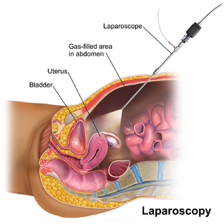
Laparoscopy is an operation performed in the abdomen or pelvis using small incisions with the aid of a camera. The laparoscope aids diagnosis or therapeutic interventions with a few small cuts in the abdomen.

<span class="mw-page-title-main">Esophageal atresia</span> Congenital discontinuity of the oesophagus

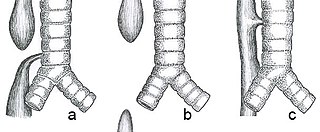
Esophageal atresia is a congenital medical condition that affects the alimentary tract. It causes the esophagus to end in a blind-ended pouch rather than connecting normally to the stomach. It comprises a variety of congenital anatomic defects that are caused by an abnormal embryological development of the esophagus. It is characterized anatomically by a congenital obstruction of the esophagus with interruption of the continuity of the esophageal wall.

<span class="mw-page-title-main">Nasogastric intubation</span> Feeding tube going into the stomach through the nose and throat

Nasogastric intubation is a medical process involving the insertion of a plastic tube through the nose, down the esophagus, and down into the stomach. Orogastric intubation is a similar process involving the insertion of a plastic tube through the mouth. Abraham Louis Levin invented the NG tube. Nasogastric tube is also known as Ryle's tube in Commonwealth countries, after John Alfred Ryle.

<span class="mw-page-title-main">Cholecystectomy</span> Surgical removal of the gallbladder

Cholecystectomy is the surgical removal of the gallbladder. Cholecystectomy is a common treatment of symptomatic gallstones and other gallbladder conditions. In 2011, cholecystectomy was the eighth most common operating room procedure performed in hospitals in the United States. Cholecystectomy can be performed either laparoscopically, or via an open surgical technique.

<span class="mw-page-title-main">Feeding tube</span> Medical device used to provide nutrition to people

A feeding tube is a medical device used to provide nutrition to people who cannot obtain nutrition by mouth, are unable to swallow safely, or need nutritional supplementation. The state of being fed by a feeding tube is called gavage, enteral feeding or tube feeding. Placement may be temporary for the treatment of acute conditions or lifelong in the case of chronic disabilities. A variety of feeding tubes are used in medical practice. They are usually made of polyurethane or silicone. The diameter of a feeding tube is measured in French units. They are classified by the site of insertion and intended use.

<span class="mw-page-title-main">Esophagogastroduodenoscopy</span> Diagnostic endoscopic procedure

Esophagogastroduodenoscopy (EGD) or oesophagogastroduodenoscopy (OGD), also called by various other names, is a diagnostic endoscopic procedure that visualizes the upper part of the gastrointestinal tract down to the duodenum. It is considered a minimally invasive procedure since it does not require an incision into one of the major body cavities and does not require any significant recovery after the procedure. However, a sore throat is common.

<span class="mw-page-title-main">Gastric bypass surgery</span> Type of bariatric surgery

Gastric bypass surgery refers to a technique in which the stomach is divided into a small upper pouch and a much larger lower "remnant" pouch and then the small intestine is rearranged to connect to both. Surgeons have developed several different ways to reconnect the intestine, thus leading to several different gastric bypass procedures (GBP). Any GBP leads to a marked reduction in the functional volume of the stomach, accompanied by an altered physiological and physical response to food.

<span class="mw-page-title-main">Gastrostomy</span> Surgical procedure creating opening in stomach

A Gastrostomy is the creation of an artificial external opening into the stomach for nutritional support or gastric decompression. Typically this would include an incision in the patient's epigastrium as part of a formal operation. When originally devised over a century ago the procedure was completed through open surgery using the Stamm technique. It can be performed through surgical approach, percutaneous approach by interventional radiology, percutaneous endoscopic gastrostomy (PEG) or percutaneous ultrasound gastrostomy (PUG).

<span class="mw-page-title-main">Pseudocyst</span> Medical condition

Pseudocysts are like cysts, but lack epithelial or endothelial cells. Initial management consists of general supportive care. Symptoms and complications caused by pseudocysts require surgery. Computed tomography (CT) scans are used for initial imaging of cysts, and endoscopic ultrasounds are used in differentiating between cysts and pseudocysts. Endoscopic drainage is a popular and effective method of treating pseudocysts.

<span class="mw-page-title-main">Ascending cholangitis</span> Medical condition

Ascending cholangitis, also known as acute cholangitis or simply cholangitis, is inflammation of the bile duct, usually caused by bacteria ascending from its junction with the duodenum. It tends to occur if the bile duct is already partially obstructed by gallstones.

<span class="mw-page-title-main">Percutaneous transhepatic cholangiography</span> Medical imaging of the biliary tract

Percutaneous transhepatic cholangiography, percutaneous hepatic cholangiogram (PTHC) is a radiological technique used to visualize the anatomy of the biliary tract. A contrast medium is injected into a bile duct in the liver, after which X-rays are taken. It allows access to the biliary tree in cases where endoscopic retrograde cholangiopancreatography has been unsuccessful. Initially reported in 1937, the procedure became popular in 1952.

Gastroparesis, also called delayed gastric emptying, is a medical disorder consisting of weak muscular contractions (peristalsis) of the stomach, resulting in food and liquid remaining in the stomach for a prolonged period of time. Stomach contents thus exit more slowly into the duodenum of the digestive tract. This can result in irregular absorption of nutrients, inadequate nutrition, and poor glycemic control.

A tracheo-esophageal puncture is a surgically created hole between the trachea (windpipe) and the esophagus in a person who has had a total laryngectomy, a surgery where the larynx is removed. The purpose of the puncture is to restore a person’s ability to speak after the vocal cords have been removed. This involves creation of a fistula between trachea and oesophagus, puncturing the short segment of tissue or “common wall” that typically separates these two structures. A voice prosthesis is inserted into this puncture. The prosthesis keeps food out of the trachea but lets air into the esophagus for oesophageal speech.

<span class="mw-page-title-main">Stomach disease</span> Medical condition

Stomach diseases include gastritis, gastroparesis, Crohn's disease and various cancers.

Therapeutic endoscopy is the medical term for an endoscopic procedure during which treatment is carried out via the endoscope. This contrasts with diagnostic endoscopy, where the aim of the procedure is purely to visualize a part of the gastrointestinal, respiratory or urinary tract in order to aid diagnosis. In practice, a procedure which starts as a diagnostic endoscopy may become a therapeutic endoscopy depending on the findings, such as in cases of upper gastrointestinal bleeding, or the finding of polyps during colonoscopy.

<span class="mw-page-title-main">Jejunostomy</span>

Jejunostomy is the surgical creation of an opening (stoma) through the skin at the front of the abdomen and the wall of the jejunum. It can be performed either endoscopically, or with open surgery.

<span class="mw-page-title-main">Gastric balloon</span> Inflatable medical device

A gastric balloon, also known as an intragastric balloon (IGB) or a stomach balloon, is an inflatable medical device that is temporarily placed into the stomach to help reduce weight. It is designed to help provide weight loss when diet and exercise have failed and surgery is not wanted or not recommended for the patient.

<span class="mw-page-title-main">Cancer and nausea</span>

Cancer and nausea are associated in about fifty percent of people affected by cancer. This may be as a result of the cancer itself, or as an effect of the treatment such as chemotherapy, radiation therapy, or other medication such as opiates used for pain relief. About 70 to 80% of people undergoing chemotherapy experience nausea or vomiting. Nausea and vomiting may also occur in people not receiving treatment, often as a result of the disease involving the gastrointestinal tract, electrolyte imbalance, or as a result of anxiety. Nausea and vomiting may be experienced as the most unpleasant side effects of cytotoxic drugs and may result in patients delaying or refusing further radiotherapy or chemotherapy.

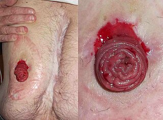
Buried bumper syndrome (BBS) is a condition that affects feeding tubes placed into the stomach through the abdominal wall. Gastrostomy tubes include an internal bumper, which secures the inner portion of the tube inside the stomach, and external bumper, which secures the outer portion of the tube and opposes the abdomen. Buried bumper syndrome occurs when the internal bumper of a gastrostomy tube erodes into the wall of the stomach. The internal bumper may become entirely buried within the fistulous tract. The main causative factor is excessive tightening of the external bumper, leading to increased pressure of the internal bumper on the wall of the stomach. Additional risk factors include: obesity, weight gain, malnutrition, corticosteroid therapy, and poor wound healing.

References

  1. "Discussion". BCM Gastroenterology Grand Rounds. Baylor College of Medicine. Archived from the original on 2012-03-03. Retrieved 2010-10-16.
  2. 1 2 3 4 Gauderer MW (2001). "Percutaneous endoscopic gastrostomy-20 years later: a historical perspective". J. Pediatr. Surg. 36 (1): 217–9. doi:10.1053/jpsu.2001.20058. PMID   11150469.
  3. Murphy LM, Lipman TO (2003). "Percutaneous endoscopic gastrostomy does not prolong survival in patients with dementia". Arch. Intern. Med. 163 (11): 1351–3. CiteSeerX   10.1.1.610.6648 . doi:10.1001/archinte.163.11.1351. PMID   12796072.
  4. AMDA – The Society for Post-Acute and Long-Term Care Medicine (February 2014), "Ten Things Physicians and Patients Should Question", Choosing Wisely: an initiative of the ABIM Foundation , AMDA – The Society for Post-Acute and Long-Term Care Medicine, retrieved 20 April 2015.
  5. Monteleoni C, Clark E (2004). "Using rapid-cycle quality improvement methodology to reduce feeding tubes in patients with advanced dementia: before and after study". BMJ. 329 (7464): 491–4. doi:10.1136/bmj.329.7464.491. PMC   515202 . PMID   15331474.
  6. Gail Waldby, "PEG-J Gastrostomy drainage jejunal feeding tubes" "Untitled Document". Archived from the original on 2011-07-16. Retrieved 2010-10-16.
  7. Deitel M, Bendago M, Spratt EH, Burul CJ, To TB (1988). "Percutaneous endoscopic gastrostomy by the "pull" and "introducer" methods". Can J Surg. 31 (2): 102–4. PMID   3349370.
  8. Abdalgaleil, Mohamed M; Shaat, Ahmed M; Elbalky, Osama S; Elnagaar, Mohamed S; Kamoun, Amr M (2018-07-01). "Early versus delayed feeding after placement of percutaneous endoscopic gastrostomy tube with safe anesthetic techniques". Menoufia Medical Journal. Medknow Publications. 31 (3): 1058–1063.
  9. Gastroenterological endoscopy. Meinhard Classen, G. N. J. Tytgat, Charles J. Lightdale. 2002. ISBN   978-1-58890-013-5
  10. Lay summary: "Feeding tubes for people with Alzheimer's disease: When you need them — and when you don't" (PDF). Consumer Reports. Archived from the original (PDF) on 12 December 2013. Retrieved 6 December 2013.
  11. Lipp A, Lusardi G, et al. (The Cochrane Collaboration) (November 2013). Lipp A (ed.). "Systemic antimicrobial prophylaxis for percutaneous endoscopic gastrostomy". The Cochrane Database of Systematic Reviews. John Wiley & Sons, Ltd (11): CD005571. doi:10.1002/14651858.cd005571.pub3. PMC   6823215 . PMID   24234575.
  12. Gurusamy KS, Koti R, Wilson P, Davidson BR, et al. (Cochrane Wounds Group) (August 2013). "Antibiotic prophylaxis for the prevention of methicillin-resistant Staphylococcus aureus (MRSA) related complications in surgical patients". The Cochrane Database of Systematic Reviews (8): CD010268. doi:10.1002/14651858.CD010268.pub2. PMID   23959704.
  13. Siamak Milanchi; Matthew T Wilson (January–March 2008). "Malposition of percutaneous endoscopic-guided gastrostomy: Guideline and management". J Minim Access Surg. 4 (1): 1–4. doi:10.4103/0972-9941.40989. PMC   2699054 . PMID   19547728.
  14. Walters G, Ramesh P, Memon MI (2005). "Buried Bumper Syndrome complicated by intra-abdominal sepsis". Age and Ageing. 34 (6): 650–1. doi: 10.1093/ageing/afi204 . PMID   16267197.
  15. 1 2 Gauderer MW, Ponsky JL, Izant RJ (1980). "Gastrostomy without laparotomy: a percutaneous endoscopic technique". J. Pediatr. Surg. 15 (6): 872–5. doi:10.1016/S0022-3468(80)80296-X. PMID   6780678.