Upper gastrointestinal series

Last updated
Upper gastrointestinal series
Normal barium swallow fluoroscopic image, showing the ingested barium sulfate being induced down the oesophagus by peristalsis.
Synonyms Upper gastrointestinal study, contrast radiography of the upper gastrointestinal tract, barium swallow, barium meal
ICD-10-PCS GroupMajor.minor

An upper gastrointestinal series, also called a barium swallow, barium study, or barium meal, is a series of radiographs used to examine the gastrointestinal tract for abnormalities. A contrast medium, usually a radiocontrast agent such as barium sulfate mixed with water, is ingested or instilled into the gastrointestinal tract, and X-rays are used to create radiographs of the regions of interest. The barium enhances the visibility of the relevant parts of the gastrointestinal tract by coating the inside wall of the tract and appearing white on the film. This in combination with other plain radiographs allows for the imaging of parts of the upper gastrointestinal tract such as the pharynx, larynx, esophagus, stomach, and small intestine such that the inside wall lining, size, shape, contour, and patency are visible to the examiner. With fluoroscopy, it is also possible to visualize the functional movement of examined organs such as swallowing, peristalsis, or sphincter closure. Depending on the organs to be examined, barium radiographs can be classified into "barium swallow", "barium meal", "barium follow-through", and "enteroclysis" ("small bowel enema"). To further enhance the quality of images, air or gas is sometimes introduced into the gastrointestinal tract in addition to barium, and this procedure is called double-contrast imaging. In this case the gas is referred to as the negative contrast medium. Traditionally the images produced with barium contrast are made with plain-film radiography, but computed tomography is also used in combination with barium contrast, in which case the procedure is called "CT enterography". [1]

Contents

Types

Barium meal examination showing the stomach and duodenum in double contrast technique with CO2 as negative contrast medium Maag.jpg
Barium meal examination showing the stomach and duodenum in double contrast technique with CO2 as negative contrast medium
Barium follow-through showing the small bowel Smallbowel.jpg
Barium follow-through showing the small bowel
Enteroclysis in double contrast technique showing stenosis of the small intestine Duenndarmstenose Morbus Crohn KM-Passage 004.jpg
Enteroclysis in double contrast technique showing stenosis of the small intestine

Various types of barium X-ray examinations are used to examine different parts of the gastrointestinal tract. These include barium swallow, barium meal, barium follow-through, and barium enema. [2] The barium swallow, barium meal, and barium follow-through are together also called an upper gastrointestinal series (or study), whereas the barium enema is called a lower gastrointestinal series (or study). [3] In upper gastrointestinal series examinations, the barium sulfate is mixed with water and swallowed orally, whereas in the lower gastrointestinal series (barium enema), the barium contrast agent is administered as an enema through a small tube inserted into the rectum. [2]

Medical uses

Barium X-ray examinations are useful tools for the study of appearance and function of the parts of the gastrointestinal tract. They are used to diagnose and monitor esophageal reflux, dysphagia, hiatus hernia, strictures, diverticula, pyloric stenosis, gastritis, enteritis, volvulus, varices, ulcers, tumors, and gastrointestinal dysmotility, as well as to detect foreign bodies. [3] [6] Although barium X-ray examinations are increasingly being replaced by more modern techniques, such as computer tomography, magnetic resonance imaging, ultrasound imaging, endoscopy and capsule endoscopy, [7] barium contrast imaging remains in common use because it offers the advantages of greater affordability, wider availability, [1] [5] and better resolution in assessing superficial mucosal lesions. [7] [8]

Mechanism

Barium sulfate is swallowed and is a radio opaque substance that does not allow the passage of X-rays. As a result, areas coated by barium sulfate will appear white on an X-ray film. The passage of barium sulfate through the gastrointestinal tract is observed by a radiologist using a fluoroscope attached to a TV monitor. The radiologist takes a series of individual X-ray images at timed intervals depending on the areas to be studied. Sometimes medication which produces gas in the gastrointestinal tract is administered together with the Barium sulfate. This gas distends the gastrointestinal lumen, providing better imaging conditions and in this case the procedure is called double-contrast imaging. [9]

Procedure

Clinical status and relevant medical history are reviewed prior to the studies. [10] Patient consent is required. [3]

Barium swallow

A barium swallow study is also known as a barium esophagram and needs little if any preparations for the study of the larynx, pharynx, and esophagus when studied alone. [11] [12]

Amongst the uses of barium swallow are: persistent dysphagia and odynophagia despite negative esophagogastroduodenoscopy (OGDS) findings, failed OGDS, esophageal motility disorder, globus pharyngis, assessment of tracheoesophageal fistula, and timed barium swallow to monitor the progress of esophageal achalasia therapy. [13] Barium sulfate suspension such as 100 ml or more of E-Z HD 200 to 250% concentration and Baritop 100% can be used. Water-soluble contrast agent such as Gastrografin (diatrizoate) and Conray (Iotalamic acid) is used instead of barium if oesophageal perforation is suspected. Low osmolar contrast medium with concentration of 300 mg/ml is used instead of gastrografin if there is risk of aspiration or there is tracheoesophageal fistula. [13]

A thick barium mixture is swallowed in supine position and fluoroscopic images of the swallowing process are made. Then several swallows of a thin barium mixture are taken and the passage is recorded by fluoroscopy and standard radiographs. The procedure is repeated several times with the examination table tilted at various angles. A total of 350–450 mL of barium is swallowed during the process. [14] [15] Normally, 90% of ingested fluid should have passed into the stomach after 15 seconds. [16]

Right anterior oblique (RAO) view is to see the oesophagus clearly, away from overlapping spine. [13] AP (anterior-posterior) view is also done to visualise the gastroesophageal junction. [13] AP and lateral views are also done to visualise the hypopharynx during swallowing at a frame rate of 3–4 per second. Left posterior oblique (LPO) position is used to identify hernias, mucosal rings, and varices. [13]

Barium meal

Intravenous injection of Buscopan (Hyoscine butylbromide) 20 mg or glucagon 0.3 mg is used to distend the stomach and slow down the emptying of the contrast into the duodenum. [13]

Right anterior oblique (RAO) view is used to demonstrate antrum and greater curve of stomach. Supine position is to demonstrate antrum and body of stomach. Left anterior oblique (LAO) view is used to see the lesser curve of stomach en face. This position is also used to check for gastroesophageal reflux when patient is asked to cough or swallow (water siphon test). Left lateral tilted with head up 45 degrees is used to demonstrate the fundus of the stomach. [13] To demonstrate the duodenal loop, the subject can lie down in prone position on a compression pad to prevent excessive barium flowing into the duodenal loop. Anterior view of duodenal loop can be seen at RAO position. [13] Duodenal cap can be visualised by taking images when subject lie down in prone position, RAO, supine, and then LAO positions or it can be seen on erect position with RAO and steep LAO views. [13] Total mucosal coating of the stomach is done by asking the subject to roll to the right side into a complete circle until RAO position. Arae gastriae in the antrum (fine reticular network of grooves) is visible if good coating is achieved. [13]

Small bowel follow-through

Indications to do this procedure are: unexplained chronic abdominal pain with weight loss, unexplained diarrhea, anemia which is caused by gastrointestinal bleeding or dependent on blood transfusion where the cause cannot be explained despite OGDS or colonoscopy investigations, partial obstruction of bowel/small bowel adhesive obstruction suspected, and unexplained malabsorption of nutrients. [13] For barium follow-through examinations, a 6-hour period of fasting is observed prior to the study. [10]

Barium is administered orally, sometimes mixed with diatrizoic acid (gastrografin) to reduce transit time in the bowel. Intravenous metoclopramide is sometimes also added to the mixture to enhance gastric emptying. [17] 600 ml of 0.5% methylcellulose can be given orally, after barium meal is given, to improve the images of small bowel follow-through by reducing the time taken for barium to pass through the small intestines, and increase the transparency of the contrast-filled small bowels. [18] Other methods to reduce transit time are to add ice cold normal saline after the administration of barium saline mixture [19] or to give a dry meal. [20]

X-ray images are then taken in a supine position at intervals of 20–30 minutes. Real-time fluoroscopy is used to assess bowel motility. The radiologist may press or palpate the abdomen during images to separate intestinal loops. The total time necessary for the test depends on the speed of bowel motility or transit time and may vary between 1 and 3 hours. [17]

Enteroclysis

Enteroclysis is also known as small bowel enema. [21] It has been largely replaced by magnetic resonance enterography/enteroclysis [13] and computed tomography enterography/enteroclysis. [22]

In addition to fasting for 8 hours prior to examination, a laxative may also be necessary for bowel preparation and cleansing. [12] The main aim of this study is to distend the proximal bowel through infusion of large amount of barium suspension. Otherwise, the distension of distal small bowel is generally similar with small bowel follow-through. Therefore, there is a need to pass a tube through the nose into the jejunum (nasojejunal tube) to administer large amount of contrast. This can be unpleasant to the subject, requires more staff, longer procedural time, and higher radiation dose when compared to small bowel follow-through. The indications for enteroclysis are generally similar to small bowel follow-through. Barium suspensions such as diluted E-Z Paque 70% and Baritop 100% can be used. After that, 600 ml of 0.5% methylcellulose is administered after 500 ml of 70% barium suspension is given. Bilbao-Dotter tube and Silk tube can be used to administer barium suspension. The subject should be fasted overnight, any antispasmodic drugs should be stopped one day before the examination, and Tetracaine lozenges can be used 30 minutes before the procedure to numb the throat for nasojejunal tube insertion. [13]

The filling of the small intestines can be viewed continuously using fluoroscopy, or viewed as standard radiographs taken at frequent intervals. The technique is a double-contrast procedure that allows detailed imaging of the entire small intestine. However, the procedure may take 6 hours or longer to complete and is quite uncomfortable to undergo. [23]

Interpretation of results

Zenker's diverticulum as seen in a barium swallow examination ZenkerSchraeg.gif
Zenker's diverticulum as seen in a barium swallow examination

Adverse effects

Barium in the lungs resulting from aspiration during a barium swallow Bariumlungfloro.PNG
Barium in the lungs resulting from aspiration during a barium swallow

Complete gastrointestinal obstruction is a contraindication for barium studies. [17]

History

Barium sulfate as a contrast medium was evolved from the prior use of bismuth preparations which were too toxic. The use of bismuth preparations had been described as early as 1898. Barium sulfate as a contrast medium in medical practice was introduced largely as a result of the works of Krause a director of the Bonn Polyclinic, now the medical faculty of the University of Bonn and his colleagues Bachem and Gunther. In a paper read in 1910 at the radiological congress they advocated for the use of barium sulfate as an opaque contrast medium in medicine. [36]

Related Research Articles

<span class="mw-page-title-main">Gastroenterology</span> Branch of medicine focused on the digestive system and its disorders

Gastroenterology is the branch of medicine focused on the digestive system and its disorders. The digestive system consists of the gastrointestinal tract, sometimes referred to as the GI tract, which includes the esophagus, stomach, small intestine and large intestine as well as the accessory organs of digestion which include the pancreas, gallbladder, and liver.

<span class="mw-page-title-main">Gastrointestinal tract</span> Organ system within humans and other animals

The gastrointestinal tract is the tract or passageway of the digestive system that leads from the mouth to the anus. The GI tract contains all the major organs of the digestive system, in humans and other animals, including the esophagus, stomach, and intestines. Food taken in through the mouth is digested to extract nutrients and absorb energy, and the waste expelled at the anus as faeces. Gastrointestinal is an adjective meaning of or pertaining to the stomach and intestines.

<span class="mw-page-title-main">Duodenum</span> First section of the small intestine

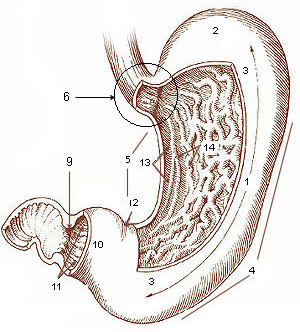
The duodenum is the first section of the small intestine in most higher vertebrates, including mammals, reptiles, and birds. In mammals it may be the principal site for iron absorption. The duodenum precedes the jejunum and ileum and is the shortest part of the small intestine.

<span class="mw-page-title-main">Lower gastrointestinal series</span> Radiographs used to examine abnormalities of the colon

A lower gastrointestinal series is a medical procedure used to examine and diagnose problems with the human colon of the large intestine. Radiographs are taken while barium sulfate, a radiocontrast agent, fills the colon via an enema through the rectum.

<span class="mw-page-title-main">Gastrointestinal stromal tumor</span> Human disease (cancer)

Gastrointestinal stromal tumors (GISTs) are the most common mesenchymal neoplasms of the gastrointestinal tract. GISTs arise in the smooth muscle pacemaker interstitial cell of Cajal, or similar cells. They are defined as tumors whose behavior is driven by mutations in the KIT gene (85%), PDGFRA gene (10%), or BRAF kinase (rare). 95% of GISTs stain positively for KIT (CD117). Most (66%) occur in the stomach and gastric GISTs have a lower malignant potential than tumors found elsewhere in the GI tract.

<span class="mw-page-title-main">Esophagogastroduodenoscopy</span> Diagnostic endoscopic procedure

Esophagogastroduodenoscopy (EGD) or oesophagogastroduodenoscopy (OGD), also called by various other names, is a diagnostic endoscopic procedure that visualizes the upper part of the gastrointestinal tract down to the duodenum. It is considered a minimally invasive procedure since it does not require an incision into one of the major body cavities and does not require any significant recovery after the procedure. However, a sore throat is common.

Radiocontrast agents are substances used to enhance the visibility of internal structures in X-ray-based imaging techniques such as computed tomography, projectional radiography, and fluoroscopy. Radiocontrast agents are typically iodine, or more rarely barium sulfate. The contrast agents absorb external X-rays, resulting in decreased exposure on the X-ray detector. This is different from radiopharmaceuticals used in nuclear medicine which emit radiation.

<span class="mw-page-title-main">Gastrointestinal disease</span> Medical condition

Gastrointestinal diseases refer to diseases involving the gastrointestinal tract, namely the esophagus, stomach, small intestine, large intestine and rectum, and the accessory organs of digestion, the liver, gallbladder, and pancreas.

<span class="mw-page-title-main">Dumping syndrome</span> Medical condition

Dumping syndrome occurs when food, especially sugar, moves too quickly from the stomach to the duodenum—the first part of the small intestine—in the upper gastrointestinal (GI) tract. This condition is also called rapid gastric emptying. It is mostly associated with conditions following gastric or esophageal surgery, though it can also arise secondary to diabetes or to the use of certain medications; it is caused by an absent or insufficiently functioning pyloric sphincter, the valve between the stomach and the duodenum.

<span class="mw-page-title-main">Virtual colonoscopy</span> Medical imaging of the colon

Virtual colonoscopy is the use of CT scanning or magnetic resonance imaging (MRI) to produce two- and three-dimensional images of the colon, from the lowest part, the rectum, to the lower end of the small intestine, and to display the images on an electronic display device. The procedure is used to screen for colon cancer and polyps, and may detect diverticulosis. A virtual colonoscopy can provide 3D reconstructed endoluminal views of the bowel. VC provides a secondary benefit of revealing diseases or abnormalities outside the colon.

<span class="mw-page-title-main">Gastrointestinal perforation</span> Medical condition

Gastrointestinal perforation, also known as gastrointestinal rupture, is a hole in the wall of the gastrointestinal tract. The gastrointestinal tract is composed of hollow digestive organs leading from the mouth to the anus. Symptoms of gastrointestinal perforation commonly include severe abdominal pain, nausea, and vomiting. Complications include a painful inflammation of the inner lining of the abdominal wall and sepsis.

<span class="mw-page-title-main">Diatrizoate</span> Chemical compound

Diatrizoate, also known as amidotrizoate, Gastrografin, is a contrast agent used during X-ray imaging. This includes visualizing veins, the urinary system, spleen, and joints, as well as computer tomography. It is given by mouth, injection into a vein, injection into the bladder, through a nasogastric tube, or rectally.

<span class="mw-page-title-main">Intestinal pseudo-obstruction</span> Medical condition

Intestinal pseudo-obstruction (IPO) is a clinical syndrome caused by severe impairment in the ability of the intestines to push food through. It is characterized by the signs and symptoms of intestinal obstruction without any lesion in the intestinal lumen. Clinical features mimic those seen with mechanical intestinal obstructions and can include abdominal pain, nausea, abdominal distension, vomiting, dysphagia and constipation depending upon the part of the gastrointestinal tract involved.

<span class="mw-page-title-main">Barium sulfate suspension</span> Contrast agent used during X-rays

Barium sulfate suspension, often simply called barium, is a contrast agent used during X-rays. Specifically it is used to improve visualization of the gastrointestinal tract on plain X-ray or computed tomography. It is taken by mouth or used rectally.

<span class="mw-page-title-main">Capsule endoscopy</span> Medical imaging procedure

Capsule endoscopy is a medical procedure used to record internal images of the gastrointestinal tract for use in disease diagnosis. Newer developments are also able to take biopsies and release medication at specific locations of the entire gastrointestinal tract. Unlike the more widely used endoscope, capsule endoscopy provides the ability to see the middle portion of the small intestine. It can be applied to the detection of various gastrointestinal cancers, digestive diseases, ulcers, unexplained bleedings, and general abdominal pains. After a patient swallows the capsule, it passes along the gastrointestinal tract, taking a number of images per second which are transmitted wirelessly to an array of receivers connected to a portable recording device carried by the patient. General advantages of capsule endoscopy over standard endoscopy include the minimally invasive procedure setup, ability to visualize more of the gastrointestinal tract, and lower cost of the procedure.

<span class="mw-page-title-main">Abdominal x-ray</span>

An abdominal x-ray is an x-ray of the abdomen. It is sometimes abbreviated to AXR, or KUB.

<span class="mw-page-title-main">Gastroparesis</span> Medical condition

Gastroparesis, also called delayed gastric emptying is a medical disorder consisting of weak muscular contractions (peristalsis) of the stomach, resulting in food and liquid remaining in the stomach for a prolonged period of time. Stomach contents thus exit more slowly into the duodenum of the digestive tract. This can result in irregular absorption of nutrients, inadequate nutrition, and poor glycemic control. The opposite of this, where stomach contents exit quickly into the duodenum is called dumping syndrome.

<span class="mw-page-title-main">Human digestive system</span> Digestive system in humans

The human digestive system consists of the gastrointestinal tract plus the accessory organs of digestion. Digestion involves the breakdown of food into smaller and smaller components, until they can be absorbed and assimilated into the body. The process of digestion has three stages: the cephalic phase, the gastric phase, and the intestinal phase.

Magnetic resonance enterography is a magnetic resonance imaging technique used to evaluate bowel wall features of both upper and lower gastro-intestinal tract, although it is usually used for small bowel evaluation. It is a less invasive technique with the advantages of no ionizing radiation exposure, multiplanarity and high contrast resolution for soft tissue.

Computed tomography enterography is a medical imaging technique which uses computed tomography scanner and contrast media to examine the small bowel. It was first introduced by Raptopoulos et al. in 1997. CT Enterography can be used to assess a variety of problems involving the small bowel, however it is mainly used to diagnose and assess severity of Crohn's disease.

References

  1. 1 2 3 4 Murphy, KP; McLaughlin, PD; O'Connor, OJ; Maher, MM (Mar 2014). "Imaging the small bowel". Current Opinion in Gastroenterology. 30 (2): 134–40. doi:10.1097/mog.0000000000000038. PMID   24419291. S2CID   41111179.
  2. 1 2 3 4 5 6 British Medical Association (2013). BMA Illustrated Medical Dictionary. Dorling Kindersley Ltd. ISBN   978-1-4093-4966-2.
  3. 1 2 3 Daniels, Rick (2010). Delmar's guide to laboratory and diagnostic tests (2nd ed.). Clifton Park, NY: Delmar/Cengage Learning. ISBN   978-1-4180-2067-5.
  4. 1 2 Kuo, P; Holloway, RH; Nguyen, NQ (May 2012). "Current and future techniques in the evaluation of dysphagia". Journal of Gastroenterology and Hepatology. 27 (5): 873–81. doi:10.1111/j.1440-1746.2012.07097.x. PMID   22369033. S2CID   5409505.
  5. 1 2 Levine, MS; Rubesin, SE; Laufer, I (Nov 2008). "Pattern approach for diseases of mesenteric small bowel on barium studies". Radiology. 249 (2): 445–60. doi:10.1148/radiol.2491071336. PMID   18812557. S2CID   15473369.
  6. Boland, Giles W (2013). Gastrointestinal imaging: the requisites (4th ed.). Philadelphia: Elsevier/Saunders. ISBN   978-0-323-10199-8.
  7. 1 2 3 Markova, I; Kluchova, K; Zboril, R; Mashlan, M; Herman, M (Jun 2010). "Small bowel imaging – still a radiologic approach?". Biomedical Papers of the Medical Faculty of the University Palacky, Olomouc, Czechoslovakia. 154 (2): 123–32. doi: 10.5507/bp.2010.019 . PMID   20668493.
  8. Sinha, Rakesh; Rawat, Sudarshan (2011-10-01). "Training the Next Generation in Luminal Gastrointestinal Radiology". American Journal of Roentgenology. 197 (4): W780. doi:10.2214/ajr.11.6870. ISSN   0361-803X. PMID   21940553.
  9. M.D, Steven R. Peikin (2014). Gastrointestinal health third edition. [S.l.]: HarperCollins e-Books. p. 29. ISBN   978-0-06-186365-3.
  10. 1 2 Hawkey, CJ (2012). Textbook of Clinical Gastroenterology and Hepatology (2nd ed.). Hoboken: John Wiley & Sons. p. 1001. ISBN   978-1-118-32140-9.
  11. Chen, Anthony; Tafti, Dawood; Tuma, Faiz (2021). "Barium Swallow". StatPearls. StatPearls Publishing. PMID   29630228 . Retrieved 23 December 2021.
  12. 1 2 3 Nightingale, Julie; Law, Robert (2012). Gastrointestinal Tract Imaging: An Evidence-Based Practice Guide. Elsevier Health Sciences. ISBN   978-0-7020-4549-3.
  13. 1 2 3 4 5 6 7 8 9 10 11 12 13 Watson N, Jones H (2018). Chapman and Nakielny's Guide to Radiological Procedures. Elsevier. pp. 49–51, 52–55, 55–60. ISBN   9780702071669.
  14. Chernecky, Cynthia; Berger, Barbara (2012). Laboratory Tests and Diagnostic Procedures. Elsevier Health Sciences. ISBN   978-1-4557-4502-9.
  15. Zare Mehrjardi, Mohammad (2013-07-16). Barium swallow and barium meal: techniques and interpretation. Monthly Student Meeting. Tehran, Iran. doi:10.13140/RG.2.2.10543.33449/1.
  16. Ziessman, Harvey A.; O'Malley, Janis P.; Thrall, James H. (2014). "Dysmotility Disorders". Nuclear Medicine. Elsevier. ISBN   978-0-323-08299-0.
  17. 1 2 3 Thomas, James; Monaghan, Tanya (2014). Oxford Handbook of Clinical Examination and Practical Skills. Oxford University Press. p. 712. ISBN   978-0-19-104454-0.
  18. Park, Kwang Bo; Ha, Hyun Kwon; Son, Se Ho; Hwang, Jae Cheul; Ji, Eun Kyung; Kim, Nam Hyeon; Kim, Pyo Nyun; Lee, Moon Kyu; Auh, Yong Ho (1996). "Use of methylcellulose in Small-Bowel Follow-Through Examination: Comparison with Enteroclysis andConventional Series in Normal Subjects". Journal of the Korean Radiological Society (in Korean). 35 (3): 351. doi: 10.3348/jkrs.1996.35.3.351 . ISSN   0301-2867. S2CID   184219822.
  19. Nolan, D J (1981-08-01). "Barium examination of the small intestine". Gut. 22 (8): 682–694. doi:10.1136/gut.22.8.682. ISSN   0017-5749. PMC   1420062 . PMID   7026379.
  20. Nijhawan, Sandeep; Kumpawat, Saket; Mallikarjun, P; Bansal, Rp; Singla, Dinesh; Ashdhir, Prachis; Mathur, Amit; Rai, Ramesh Roop (2008). "Barium meal follow-through with pneumocolon: Screening test for chronic bowel pain". World Journal of Gastroenterology. 14 (43): 6694–6698. doi: 10.3748/wjg.14.6694 . ISSN   1007-9327. PMC   2773312 . PMID   19034973.
  21. Nolan, Daniel J.; Cadman, Philip J. (May 1987). "The small bowel enema made easy". Clinical Radiology. 38 (3): 295–301. doi:10.1016/S0009-9260(87)80075-2. PMID   3581673.
  22. "American College of Radiology ACR appropriateness criteria – Chron's Disease". American College of Radiology. Archived from the original on 3 December 2020. Retrieved 7 April 2022.
  23. al.], Rene A. Day ... [et (2009). Brunner & Suddarth's textbook of Canadian medical-surgical nursing (2nd Canadian ed.). Philadelphia: Lippincott Williams & Wilkins. ISBN   978-0-7817-9989-8.
  24. 1 2 Maglinte, DD; Kohli, MD; Romano, S; Lappas, JC (Sep 2009). "Air (CO2) double-contrast barium enteroclysis". Radiology. 252 (3): 633–41. doi:10.1148/radiol.2523081972. PMID   19717748.
  25. Grant, PD; Morgan, DE; Scholz, FJ; Canon, CL (Jan–Feb 2009). "Pharyngeal dysphagia: what the radiologist needs to know". Current Problems in Diagnostic Radiology. 38 (1): 17–32. doi:10.1067/j.cpradiol.2007.08.009. PMID   19041038.
  26. 1 2 Robinson, C; Punwani, S; Taylor, S (Dec 2009). "Imaging the gastrointestinal tract in 2008". Clinical Medicine . 9 (6): 609–12. doi:10.7861/clinmedicine.9-6-609. PMC   4952308 . PMID   20095312.
  27. Brant, [edited by] William E.; Helms, Clyde A. (2007). Fundamentals of diagnostic radiology (3rd ed.). Philadelphia: Lippincott Williams & Wilkins. pp.  811. ISBN   978-0-7817-6135-2.{{cite book}}: |first1= has generic name (help)
  28. 1 2 Dambha, F; Tanner, J; Carroll, N (Jun 2014). "Diagnostic imaging in Crohn's disease: what is the new gold standard?". Best Practice & Research. Clinical Gastroenterology. 28 (3): 421–36. doi:10.1016/j.bpg.2014.04.010. PMID   24913382.
  29. Deepak, P; Bruining, DH (Aug 2014). "Radiographical evaluation of ulcerative colitis". Gastroenterology Report. 2 (3): 169–77. doi:10.1093/gastro/gou026. PMC   4124269 . PMID   24843072.
  30. Baker, ME; Einstein, DM (Mar 2014). "Barium esophagram: does it have a role in gastroesophageal reflux disease?". Gastroenterology Clinics of North America. 43 (1): 47–68. doi:10.1016/j.gtc.2013.11.008. PMID   24503359.
  31. Fidler, JL; Fletcher, JG; Bruining, DH; Trenkner, SW (Jul 2013). "Current status of CT, magnetic resonance, and barium in inflammatory bowel disease". Seminars in Roentgenology. 48 (3): 234–44. doi:10.1053/j.ro.2013.03.004. PMID   23796374.
  32. Sinha, R; Rajesh, A; Rawat, S; Rajiah, P; Ramachandran, I (May 2012). "Infections and infestations of the gastrointestinal tract. Part 1: bacterial, viral and fungal infections". Clinical Radiology. 67 (5): 484–94. doi:10.1016/j.crad.2011.10.021. PMID   22257535.
  33. 1 2 3 4 5 6 Sinha, R; Rajesh, A; Rawat, S; Rajiah, P; Ramachandran, I (May 2012). "Infections and infestations of the gastrointestinal tract. Part 2: parasitic and other infections". Clinical Radiology. 67 (5): 495–504. doi:10.1016/j.crad.2011.10.022. PMID   22169349.
  34. Engin, G; Korman, U (Sep 2011). "Gastrointestinal lymphoma: a spectrum of fluoroscopic and CT findings". Diagnostic and Interventional Radiology (Ankara, Turkey). 17 (3): 255–65. doi:10.4261/1305-3825.dir.3332-10.3. PMID   20725903. S2CID   6475386.
  35. 1 2 3 4 5 6 Baert, Henrik S. Thomsen ; Judith A.W. Webb (ed.). With contributions by P. Aspelin ... Foreword by A.L. (2009). Contrast media : safety issues and ESUR guidelines ; with 24 tables (2., rev. ed.). Berlin: Springer. ISBN   978-3-540-72783-5.{{cite book}}: CS1 maint: multiple names: authors list (link)
  36. Schott, G. D. (16 August 2012). "Some Observations on the History of the Use of Barium Salts in Medicine". Medical History. 18 (1): 9–21. doi:10.1017/S0025727300019190. PMC   1081520 . PMID   4618587.