Radioisotope renography

Last updated
Radioisotope renography
99mTc-MAG3 and 99mTc-DTPA renogram.jpg
Renal imaging using 99mTc DTPA and 99mMAG3 with renographic curves
ICD-9-CM 92.03
MeSH D011866
OPS-301 code 3-706

Radioisotope renography is a form of medical imaging of the kidneys that uses radiolabelling. A renogram, which may also be known as a MAG3 scan, allows a nuclear medicine physician or a radiologist to visualize the kidneys and learn more about how they are functioning. [1] MAG3 is an acronym for mercapto acetyl tri glycine, a compound that is chelated with a radioactive element – technetium-99m.

Contents

The two most common radiolabelled pharmaceutical agents used are 99mTc-MAG3 (MAG3 is also called "mercaptoacetyltriglycine" or "mertiatide") and 99mTc DTPA (diethylenetriaminepentacetate). Some other radiolabelled pharmaceuticals are EC (Ethylenedicysteine) and 131-iodine labelled OIH (ortho-iodohippurate). [2]

Scan procedure

After injection into the veins, the compound is excreted by the kidneys, and its progress through the renal system can be tracked with a gamma camera. A series of images are taken at regular intervals. Processing then involves drawing a region of interest (ROI) around both kidneys, and a computer program produces a graph of radioactivity inside the kidney with time, representing the quantity of tracer, from the number of counts measured inside in each image (representing a different time point). [3]

If the kidney is not getting blood for example, it will not be viewed at all, even if it looks structurally normal in medical ultrasonography or magnetic resonance imaging. If the kidney is getting blood, but there is an obstruction inferior to the kidney in the bladder or ureters, the radioisotope will not pass beyond the level of the obstruction, whereas if there is a partial obstruction then there is a delayed transit time for the MAG3 to pass. [4] More information can be gathered by calculating time activity curves; with normal kidney perfusion, peak activity should be observed after 3–5 minutes. [5] The relative quantitative information gives the differential function between each kidney's filtration activity.

Tracers

Technetium-99m MAG3 Technetium Tc 99m mertiatide.svg
Technetium-99m MAG3

MAG3 is preferred over 99mTc DTPA in neonates, patients with impaired function, and patients with suspected obstruction, due to its more efficient extraction. [2] [6] [7] The MAG3 clearance is highly correlated with the effective renal plasma flow (ERPF), and the MAG3 clearance can be used as an independent measure of kidney function. [8] After intravenous administration, about 40-50% of the MAG3 in the blood is extracted by the proximal tubules with each pass through the kidneys; the proximal tubules then secrete the MAG3 into the tubular lumen. [9]

99mTc DTPA is filtered by the glomerulus and may be used to measure the glomerular filtration rate (GFR) (in a separate test), making it theoretically the best (most accurate) choice for kidney function imaging. [10] The extraction fraction of DTPA is approximately 20%, less than half that of MAG3. [9] DTPA is the second most commonly used renal radiopharmaceutical in the United States. [11]

Clinical use

The technique is very useful in evaluating the functioning of kidneys. Radioisotopes can differentiate between passive dilatation and obstruction. It is widely used before kidney transplantation to assess the vascularity of the kidney to be transplanted and with a test dose of captopril to highlight possible renal artery stenosis in the donor's other kidney, [12] and later the performance of the transplant. [13] [14] Post-transplantation renography can be used for the diagnosis of vascular and urological complications. [15] Also, early post-transplantation renography is used for the assessment of delayed graft function. [16] [17]

The use of the test to identify reduced kidney function after test doses of captopril (an angiotensin-converting enzyme inhibitor medication) has also been used to identify the cause of hypertension in patients with kidney failure. [18] [19] Initially there was uncertainty as to the usefulness, [20] or best test parameter to identify renal artery stenosis, the eventual consensus was that the distinctive finding is of alteration in the differential function. [21]

History

In 1986, MAG3 was developed at the University of Utah by Dr. Alan R. Fritzberg, Dr. Sudhakar Kasina, and Dr. Dennis Eshima. [22] The drug underwent clinical trials in 1987 [23] and passed Phase III testing in 1988. [24]

Before the development of tracers such as 99mTc-MAG3, a range of other radiopharmaceuticals were employed. The test was first introduced in 1956, using iodine-131 diodrast. [25] [26] Later developments included iodine-131, and then iodine-123, labelled ortho-Iodohippuric acid (OIH, marketed as Hippuran). [27] [28]

99mTc-MAG3 has replaced 131I-OIH because of better quality imaging regardless of the level of kidney function, [29] and lower radiation doses. [24]

See also

Related Research Articles

<span class="mw-page-title-main">Single-photon emission computed tomography</span> Nuclear medicine tomographic imaging technique

Single-photon emission computed tomography is a nuclear medicine tomographic imaging technique using gamma rays. It is very similar to conventional nuclear medicine planar imaging using a gamma camera, but is able to provide true 3D information. This information is typically presented as cross-sectional slices through the patient, but can be freely reformatted or manipulated as required.

Technetium (<sup>99m</sup>Tc) sestamibi Pharmaceutical drug

Technetium (99mTc) sestamibi (INN) is a pharmaceutical agent used in nuclear medicine imaging. The drug is a coordination complex consisting of the radioisotope technetium-99m bound to six (sesta=6) methoxyisobutylisonitrile (MIBI) ligands. The anion is not defined. The generic drug became available late September 2008. A scan of a patient using MIBI is commonly known as a "MIBI scan".

<span class="mw-page-title-main">Scintigraphy</span> Diagnostic imaging test in nuclear medicine

Scintigraphy, also known as a gamma scan, is a diagnostic test in nuclear medicine, where radioisotopes attached to drugs that travel to a specific organ or tissue (radiopharmaceuticals) are taken internally and the emitted gamma radiation is captured by gamma cameras, which are external detectors that form two-dimensional images in a process similar to the capture of x-ray images. In contrast, SPECT and positron emission tomography (PET) form 3-dimensional images and are therefore classified as separate techniques from scintigraphy, although they also use gamma cameras to detect internal radiation. Scintigraphy is unlike a diagnostic X-ray where external radiation is passed through the body to form an image.

<span class="mw-page-title-main">Renal artery stenosis</span> Medical condition

Renal artery stenosis (RAS) is the narrowing of one or both of the renal arteries, most often caused by atherosclerosis or fibromuscular dysplasia. This narrowing of the renal artery can impede blood flow to the target kidney, resulting in renovascular hypertension – a secondary type of high blood pressure. Possible complications of renal artery stenosis are chronic kidney disease and coronary artery disease.

<span class="mw-page-title-main">Pertechnetate</span> Chemical compound or ion

The pertechnetate ion is an oxyanion with the chemical formula TcO
4
. It is often used as a convenient water-soluble source of isotopes of the radioactive element technetium (Tc). In particular it is used to carry the 99mTc isotope which is commonly used in nuclear medicine in several nuclear scanning procedures.

<span class="mw-page-title-main">Ventilation/perfusion scan</span> Medical imaging to evaluate circulation of air and blood in the lungs

A ventilation/perfusion lung scan, also called a V/Q lung scan, or ventilation/perfusion scintigraphy, is a type of medical imaging using scintigraphy and medical isotopes to evaluate the circulation of air and blood within a patient's lungs, in order to determine the ventilation/perfusion ratio. The ventilation part of the test looks at the ability of air to reach all parts of the lungs, while the perfusion part evaluates how well blood circulates within the lungs. As Q in physiology is the letter used to describe bloodflow the term V/Q scan emerged.

<span class="mw-page-title-main">Technetium-99m</span> Metastable nuclear isomer of technetium-99

Technetium-99m (99mTc) is a metastable nuclear isomer of technetium-99, symbolized as 99mTc, that is used in tens of millions of medical diagnostic procedures annually, making it the most commonly used medical radioisotope in the world.

<span class="mw-page-title-main">Myocardial perfusion imaging</span> Nuclear medicine imaging method

Myocardial perfusion imaging or scanning is a nuclear medicine procedure that illustrates the function of the heart muscle (myocardium).

Technetium 99mTc albumin aggregated (99mTc-MAA) is an injectable radiopharmaceutical used in nuclear medicine. It consists of a sterile aqueous suspension of Technetium-99m (99mTc) labeled to human albumin aggregate particles. It is commonly used for lung perfusion scanning. It is also less commonly used to visualise a peritoneovenous shunt and for isotope venography.

<span class="mw-page-title-main">Cholescintigraphy</span> Medical imaging of hepatobiliary tract using radiotracers

Cholescintigraphy or hepatobiliary scintigraphy is scintigraphy of the hepatobiliary tract, including the gallbladder and bile ducts. The image produced by this type of medical imaging, called a cholescintigram, is also known by other names depending on which radiotracer is used, such as HIDA scan, PIPIDA scan, DISIDA scan, or BrIDA scan. Cholescintigraphic scanning is a nuclear medicine procedure to evaluate the health and function of the gallbladder and biliary system. A radioactive tracer is injected through any accessible vein and then allowed to circulate to the liver, where it is excreted into the bile ducts and stored by the gallbladder until released into the duodenum.

<span class="mw-page-title-main">TCHP</span> Protein-coding gene in the species Homo sapiens

Trichoplein keratin filament-binding protein is a protein that in humans is encoded by the TCHP gene.

<span class="mw-page-title-main">Octreotide scan</span> Type of medical imaging

An octreotide scan is a type of SPECT scintigraphy used to find carcinoid, pancreatic neuroendocrine tumors, and to localize sarcoidosis. It is also called somatostatin receptor scintigraphy (SRS). Octreotide, a drug similar to somatostatin, is radiolabeled with indium-111, and is injected into a vein and travels through the bloodstream. The radioactive octreotide attaches to tumor cells that have receptors for somatostatin. A gamma camera detects the radioactive octreotide, and makes pictures showing where the tumor cells are in the body, typically by a SPECT technique. A technetium-99m based radiopharmaceutical kit is also available.

Perfusion is the passage of fluid through the lymphatic system or blood vessels to an organ or a tissue. The practice of perfusion scanning is the process by which this perfusion can be observed, recorded and quantified. The term perfusion scanning encompasses a wide range of medical imaging modalities.

Technetium (<sup>99m</sup>Tc) tetrofosmin Chemical compound

Technetium (99mTc) tetrofosmin is a drug used in nuclear medicine cardiac imaging. It is sold under the brand name Myoview. The radioisotope, technetium-99m, is chelated by two 1,2-bis[di-(2-ethoxyethyl)phosphino]ethane ligands which belong to the group of diphosphines and which are referred to as tetrofosmin.

<span class="mw-page-title-main">DMSA scan</span>

A DMSA scan is a radionuclide scan that uses dimercaptosuccinic acid (DMSA) in assessing renal morphology, structure and function. Radioactive technetium-99m is combined with DMSA and injected into a patient, followed by imaging with a gamma camera after 2-3 hours. A DMSA scan is usually static imaging, while other radiotracers like DTPA and MAG3 are usually used for dynamic imaging to assess renal excretion.

Preclinical or small-animal Single Photon Emission Computed Tomography (SPECT) is a radionuclide based molecular imaging modality for small laboratory animals. Although SPECT is a well-established imaging technique that is already for decades in use for clinical application, the limited resolution of clinical SPECT (~10 mm) stimulated the development of dedicated small animal SPECT systems with sub-mm resolution. Unlike in clinics, preclinical SPECT outperforms preclinical coincidence PET in terms of resolution and, at the same time, allows to perform fast dynamic imaging of animals.

<span class="mw-page-title-main">Gastric emptying study</span>

A gastric emptying study is a nuclear medicine study which provides an assessment of the stomach's ability to empty. It may be used if there are complications after gastric surgery, for gastric reflux, or suspected gastroparesis amongst other indications. Scintigraphy that uses gamma cameras to create two-dimensional images is generally regarded as the gold standard for gastric emptying.

<span class="mw-page-title-main">DPD scan</span> Nuclear medicine scan

A DPD scan is a type of nuclear medicine imaging test which uses radioactive technetium-99m (99mTc) and 3,3-diphosphono-1,2-propanodicarboxylic acid (DPD) to diagnose cardiac amyloidosis. The radiopharmaceutical is taken up only in patients with ATTR amyloidosis, making it a useful tool to differentiate from AL amyloidosis.

Technetium (<sup>99m</sup>Tc) mertiatide Chemical compound

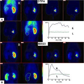
Technetium (99mTc) mertiatide is a radiopharmaceutical medication used in nuclear medicine to image the kidneys. It is a renal imaging agent that is given by intravenous injection.

Technetium (<sup>99m</sup>Tc) pentetic acid Chemical compound

Technetium (99mTc) pentetic acid, sold under the brand name Draximage DTPA among others, is a radiopharmaceutical medication used in nuclear medicine to image the brain, kidneys, or lungs. It is given by intravenous injection or via aerosol spray. It consists of technetium-99m bound to the conjugate base of pentetic acid, with sodium as an additional cation.

References

  1. "The Renogram". British Nuclear Medicine Society. Retrieved 27 April 2017.
  2. 1 2 Taylor, A. T. (18 February 2014). "Radionuclides in Nephrourology, Part 1: Radiopharmaceuticals, Quality Control, and Quantitative Indices". Journal of Nuclear Medicine. 55 (4): 608–615. doi:10.2967/jnumed.113.133447. PMC   4061739 . PMID   24549283.
  3. Elgazzar, Abdelhamid H. (2011-05-10). A Concise Guide to Nuclear Medicine. Springer. p. 15. ISBN   9783642194269.
  4. González A, Jover L, Mairal LI, Martin-Comin J, Puchal R (1994). "Evaluation of obstructed kidneys by discriminant analysis of 99mTc-MAG3 renograms". Nuklearmedizin. 33 (6): 244–7. doi:10.1055/s-0038-1629712. PMID   7854921. S2CID   24979005.
  5. Sandler, Martin P. (2003). Diagnostic Nuclear Medicine. Lippincott Williams & Wilkins. p. 868. ISBN   9780781732529.
  6. Gordon, Isky; Piepsz, Amy; Sixt, Rune (19 April 2011). "Guidelines for standard and diuretic renogram in children". European Journal of Nuclear Medicine and Molecular Imaging. 38 (6): 1175–1188. doi:10.1007/s00259-011-1811-3. PMID   21503762. S2CID   11496497.
  7. Shulkin, B. L.; Mandell, G. A.; Cooper, J. A.; Leonard, J. C.; Majd, M.; Parisi, M. T.; Sfakianakis, G. N.; Balon, H. R.; Donohoe, K. J. (14 August 2008). "Procedure Guideline for Diuretic Renography in Children 3.0" (PDF). Journal of Nuclear Medicine Technology. 36 (3): 162–168. doi: 10.2967/jnmt.108.056622 . PMID   18765635.
  8. Biersack, Hans-Jürgen; Freeman, Leonard M. (2008-01-03). Clinical Nuclear Medicine. Springer Science & Business Media. p. 173. ISBN   9783540280262.
  9. 1 2 Alazraki, Andrew Taylor, David M. Schuster, Naomi (2006). "The Genitourinary System" (PDF). A clinician's guide to nuclear medicine (2nd ed.). Reston, VA: Society of Nuclear Medicine. p. 49. ISBN   9780972647878.{{cite book}}: CS1 maint: multiple names: authors list (link)
  10. Durand, E; Prigent, A (December 2002). "The basics of renal imaging and function studies". The Quarterly Journal of Nuclear Medicine. 46 (4): 249–67. PMID   12411866.
  11. Archer, K. D.; Bolus, N. E. (27 October 2016). "Survey on the Use of Nuclear Renal Imaging in the United States". Journal of Nuclear Medicine Technology. 44 (4): 223–226. doi: 10.2967/jnmt.116.181339 . PMID   27789752.
  12. Dubovsky EV, Diethelm AG, Keller F, Russell CD (1992). "Renal transplant hypertension caused by iliac artery stenosis" (PDF). J. Nucl. Med. 33 (6): 1178–80. PMID   1534577.
  13. Kramer W, Baum RP, Scheuermann E, Hör G, Jonas D (1993). "[Follow-up after kidney transplantation. Sequential functional scintigraphy with technetium-99m-DTPA or technetium-99m-MAG3]". Urologe A (in German). 32 (2): 115–20. PMID   8475609.
  14. Li Y, Russell CD, Palmer-Lawrence J, Dubovsky EV (1994). "Quantitation of renal parenchymal retention of technetium-99m-MAG3 in renal transplants". J. Nucl. Med. 35 (5): 846–50. PMID   8176469.
  15. Benjamens S, Berger SP, Glaudemans AW, Sanders JS, Pol RA, Slart RH (2018). "Renal scintigraphy for post-transplant monitoring after kidney transplantation". Transplantation Reviews. 32 (2): 102–109. doi:10.1016/j.trre.2017.12.002. PMID   29395726.
  16. Benjamens S, Pol RA, de Geus-Oei LF, de Vries AP, Glaudemans AW, Berger SP, Slart RH (2018). "Can transplant renal scintigraphy predict the duration of delayed graft function? A dual center retrospective study". PLOS ONE. 13 (3): e0193791. Bibcode:2018PLoSO..1393791B. doi: 10.1371/journal.pone.0193791 . PMC   5862448 . PMID   29561854.
  17. Benjamens S, Pol RA, Berger SP, Glaudemans AW, Dibbets-Schneider P, Slart RH, de Geus-Oei LF (2020). "Limited clinical value of two consecutive post-transplant renal scintigraphy procedures". European Radiology. 30 (1): 452–460. doi:10.1007/s00330-019-06334-1. PMC   6890596 . PMID   31338652.
  18. Datseris IE, Bomanji JB, Brown EA, et al. (1994). "Captopril renal scintigraphy in patients with hypertension and chronic renal failure". J. Nucl. Med. 35 (2): 251–4. PMID   8294993.
  19. Kahn D, Ben-Haim S, Bushnell DL, Madsen MT, Kirchner PT (1994). "Captopril-enhanced 99Tcm-MAG3 renal scintigraphy in subjects with suspected renovascular hypertension". Nucl Med Commun. 15 (7): 515–28. doi:10.1097/00006231-199407000-00005. PMID   7970428. S2CID   36864545.
  20. Schreij G, van Es PN, van Kroonenburgh MJ, Kemerink GJ, Heidendal GA, de Leeuw PW (1996). "Baseline and postcaptopril renal blood flow measurements in hypertensives suspected of renal artery stenosis". J. Nucl. Med. 37 (10): 1652–5. PMID   8862302.
  21. Roccatello D, Picciotto G (1997). "Captopril-enhanced scintigraphy using the method of the expected renogram: improved detection of patients with renin-dependent hypertension due to functionally significant renal artery stenosis" (PDF). Nephrol. Dial. Transplant. 12 (10): 2081–6. doi: 10.1093/ndt/12.10.2081 . PMID   9351069.
  22. Fritzberg AR, Kasina S, Eshima D, Johnson DL (1986). "Synthesis and biological evaluation of technetium-99m MAG3 as a hippuran replacement". J. Nucl. Med. 27 (1): 111–6. PMID   2934521.
  23. Taylor A, Eshima D, Alazraki N (1987). "99mTc-MAG3, a new renal imaging agent: preliminary results in patients". Eur J Nucl Med. 12 (10): 510–4. doi:10.1007/BF00620476. PMID   2952506. S2CID   8632649.
  24. 1 2 Al-Nahhas AA, Jafri RA, Britton KE, et al. (1988). "Clinical experience with 99mTc-MAG3, mercaptoacetyltriglycine, and a comparison with 99mTc-DTPA". Eur J Nucl Med. 14 (9–10): 453–62. doi:10.1007/BF00252388. PMID   2975219. S2CID   23594754.
  25. TAPLIN, GV; MEREDITH, OM; KADE, H; WINTER, CC (December 1956). "The radioisotope renogram: an external test for individual kidney function and upper urinary tract patency". The Journal of Laboratory and Clinical Medicine . 48 (6): 886–901. PMID   13376984.
  26. Saterborg, N.-E. (June 1960). "The technical procedure in kidney examinations with radioactive isotopes". Acta Radiologica. 53 (6): 433–441. doi:10.3109/00016926009171695. PMID   14441893.
  27. "123I iodohippurate" (PDF). Guidelines database. Federatie Medisch Specialisten. Retrieved 26 August 2022.
  28. Biassoni, Lorenzo; Gordon, Isky (2010). "8 - Radioisotope Imaging of the Kidney and Urinary Tract". Pediatric urology (2nd ed.). Philadelphia. doi:10.1016/B978-1-4160-3204-5.00008-6. ISBN   978-1-4160-3204-5.{{cite book}}: CS1 maint: location missing publisher (link)
  29. Taylor A, Eshima D, Christian PE, Milton W (1987). "Evaluation of 99mTc mercaptoacetyltriglycine in patients with impaired renal function". Radiology. 162 (2): 365–70. doi:10.1148/radiology.162.2.2948212. PMID   2948212.