
Tears are a clear liquid secreted by the lacrimal glands found in the eyes of all land mammals. Tears are made up of water, electrolytes, proteins, lipids, and mucins that form layers on the surface of eyes. The different types of tears—basal, reflex, and emotional—vary significantly in composition.

The lacrimal bone is a small and fragile bone of the facial skeleton; it is roughly the size of the little fingernail. It is situated at the front part of the medial wall of the orbit. It has two surfaces and four borders. Several bony landmarks of the lacrimal bone function in the process of lacrimation or crying. Specifically, the lacrimal bone helps form the nasolacrimal canal necessary for tear translocation. A depression on the anterior inferior portion of the bone, the lacrimal fossa, houses the membranous lacrimal sac. Tears or lacrimal fluid, from the lacrimal glands, collect in this sac during excessive lacrimation. The fluid then flows through the nasolacrimal duct and into the nasopharynx. This drainage results in what is commonly referred to a runny nose during excessive crying or tear production. Injury or fracture of the lacrimal bone can result in posttraumatic obstruction of the lacrimal pathways.

In anatomy, the orbit is the cavity or socket of the skull in which the eye and its appendages are situated. "Orbit" can refer to the bony socket, or it can also be used to imply the contents. In the adult human, the volume of the orbit is 30 millilitres, of which the eye occupies 6.5 ml. The orbital contents comprise the eye, the orbital and retrobulbar fascia, extraocular muscles, cranial nerves II, III, IV, V, and VI, blood vessels, fat, the lacrimal gland with its sac and duct, the eyelids, medial and lateral palpebral ligaments, cheek ligaments, the suspensory ligament, septum, ciliary ganglion and short ciliary nerves.

Eye surgery, also known as ophthalmic or ocular surgery, is surgery performed on the eye or its adnexa, by an ophthalmologist. Eye surgery is synonymous with ophthalmology. The eye is a very fragile organ, and requires extreme care before, during, and after a surgical procedure to minimize or prevent further damage. An expert eye surgeon is responsible for selecting the appropriate surgical procedure for the patient, and for taking the necessary safety precautions. Mentions of eye surgery can be found in several ancient texts dating back as early as 1800 BC, with cataract treatment starting in the fifth century BC. Today it continues to be a widely practiced type of surgery, with various techniques having been developed for treating eye problems.

Dry eye syndrome (DES), also known as keratoconjunctivitis sicca (KCS), is the condition of having dry eyes. Other associated symptoms include irritation, redness, discharge, and easily fatigued eyes. Blurred vision may also occur. Symptoms range from mild and occasional to severe and continuous. Scarring of the cornea may occur in untreated cases.

The nasolacrimal duct carries tears from the lacrimal sac of the eye into the nasal cavity. The duct begins in the eye socket between the maxillary and lacrimal bones, from where it passes downwards and backwards. The opening of the nasolacrimal duct into the inferior nasal meatus of the nasal cavity is partially covered by a mucosal fold.

Scintigraphy, also known as a gamma scan, is a diagnostic test in nuclear medicine, where radioisotopes attached to drugs that travel to a specific organ or tissue (radiopharmaceuticals) are taken internally and the emitted gamma radiation is captured by external detectors to form two-dimensional images in a similar process to the capture of x-ray images. In contrast, SPECT and positron emission tomography (PET) form 3-dimensional images and are therefore classified as separate techniques from scintigraphy, although they also use gamma cameras to detect internal radiation. Scintigraphy is unlike a diagnostic X-ray where external radiation is passed through the body to form an image.

A bone scan or bone scintigraphy is a nuclear medicine imaging technique of the bone. It can help diagnose a number of bone conditions, including cancer of the bone or metastasis, location of bone inflammation and fractures, and bone infection (osteomyelitis).

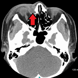
The lacrimal sac or lachrymal sac is the upper dilated end of the nasolacrimal duct, and is lodged in a deep groove formed by the lacrimal bone and frontal process of the maxilla. It connects the lacrimal canaliculi, which drain tears from the eye's surface, and the nasolacrimal duct, which conveys this fluid into the nasal cavity. Lacrimal sac occlusion leads to dacryocystitis.

The posterior lacrimal crest is a vertical bony ridge on the orbital surface of the lacrimal bone. It divides the bone into two parts. It gives origin to the lacrimal part of the orbicularis oculi muscle.
Oculoplastics, or oculoplastic surgery, includes a wide variety of surgical procedures that deal with the orbit, eyelids, tear ducts, and the face. It also deals with the reconstruction of the eye and associated structures.

Dacryocystorhinostomy (DCR) is a surgical procedure to restore the flow of tears into the nose from the lacrimal sac when the nasolacrimal duct does not function.
Dacryocystitis is an infection of the lacrimal sac, secondary to obstruction of the nasolacrimal duct at the junction of lacrimal sac. The term derives from the Greek dákryon (tear), cysta (sac), and -itis (inflammation). It causes pain, redness, and swelling over the inner aspect of the lower eyelid and epiphora. When nasolacrimal duct obstruction is secondary to a congenital barrier it is referred to as dacryocystocele. It is most commonly caused by Staphylococcus aureus and Streptococcus pneumoniae. The most common complication is corneal ulceration, frequently in association with S. pneumoniae. The mainstays of treatment are oral antibiotics, warm compresses, and relief of nasolacrimal duct obstruction by dacryocystorhinostomy.
A punctal plug, also known as tear duct plug or lacrimal plug, is a small medical device that is inserted into the tear duct (puncta) of an eye to block the duct. This prevents the drainage of liquid from the eye. They are used to treat dry eye.

The lateral palpebral raphe is a ligamentous band near the eye. Its existence is contentious, and many sources describe it as the continuation of nearby muscles. It is formed from the lateral ends of the orbicularis oculi muscle. It connects the orbicularis oculi muscle, the frontosphenoidal process of the zygomatic bone, and the tarsi of the eyelids.

Epiphora is an overflow of tears onto the face, other than caused by normal crying. It is a clinical sign or condition that constitutes insufficient tear film drainage from the eyes, in that tears will drain down the face rather than through the nasolacrimal system.
Dacryocystocele (Dacryocystitis) or timo cyst is a benign, bluish-gray mass in the inferomedial canthus that develops within a few days or weeks after birth. The uncommon condition forms as a result as a consequence of narrowing or obstruction of the nasolacrimal duct, usually during prenatal development. Nasolacrimal duct obstruction disrupts the lacrimal drainage system, eventually creating a swelling cyst in the lacrimal sac area by the nasal cavity. The location of the cyst can cause respiratory dysfunction, compromising the airway. The obstruction ultimately leads to epiphora, an abundance of tear production.

A DMSA scan is a radionuclide scan that uses dimercaptosuccinic acid (DMSA) in assessing renal morphology, structure and function. Radioactive technetium-99m is combined with DMSA and injected into a patient, followed by imaging with a gamma camera after 2-3 hours. A DMSA scan is usually static imaging, while other radiotracers like DTPA and MAG3 are usually used for dynamic imaging to assess renal excretion.

Nasolacrimal duct obstruction is the obstruction of the nasolacrimal duct and may be either congenital or acquired. Obstruction of the nasolacrimal duct leads to the excess overflow of tears called epiphora.

A gastric emptying scan is a nuclear medicine study which provides an assessment of the stomach's ability to empty. It may be used if there are complications after gastric surgery, for gastric reflux, or suspected gastroparesis amongst other indications. Scintigraphy that uses gamma cameras to create two-dimensional images is generally regarded as the gold standard for gastric emptying.
