Nasal cavity

Last updated
Nasal cavity
Illu01 head neck.jpg
Head and neck
Illu conducting passages.svg
Conducting passages
Details
Part of Nose
Identifiers
Latin cavum nasi; cavitas nasi
MeSH D009296
TA98 A06.1.02.001
TA2 3165
FMA 54378
Anatomical terminology

The nasal cavity is a large, air-filled space above and behind the nose in the middle of the face. The nasal septum divides the cavity into two cavities, [1] also known as fossae. [2] Each cavity is the continuation of one of the two nostrils. The nasal cavity is the uppermost part of the respiratory system and provides the nasal passage for inhaled air from the nostrils to the nasopharynx and rest of the respiratory tract.

Contents

The paranasal sinuses surround and drain into the nasal cavity.

Structure

Nasal cavity anatomy Anatomy of the human nasal cavity.png
Nasal cavity anatomy

The term "nasal cavity" can refer to each of the two cavities of the nose, or to the two sides combined.

CT scan in the coronal plane, showing the ostiomeatal complex (green area) CT of the ostiomeatal complex, coronal plane, with annotations.png
CT scan in the coronal plane, showing the ostiomeatal complex (green area)

The lateral wall of each nasal cavity mainly consists of the maxilla. However, there is a deficiency that is compensated for by the perpendicular plate of the palatine bone, the medial pterygoid plate, the labyrinth of ethmoid and the inferior concha. The paranasal sinuses are connected to the nasal cavity through small orifices called ostia. Most of these ostia communicate with the nose through the lateral nasal wall, via a semi-lunar depression in it known as the semilunar hiatus. The hiatus is bound laterally by a projection known as the uncinate process. This region is called the ostiomeatal complex. [3]

The roof of each nasal cavity is formed in its upper third to one half by the nasal bone and more inferiorly by the junctions of the upper lateral cartilage and nasal septum. Connective tissue and skin cover the bony and cartilaginous components of the nasal dorsum.

The floor of the nasal cavities, which also form the roof of the mouth, is made up by the bones of the hard palate: the horizontal plate of the palatine bone posteriorly and the palatine process of the maxilla anteriorly. The most anterior part of the nasal cavity is the nasal vestibule. [4] The vestibule is enclosed by the nasal cartilages and lined by the same epithelium of the skin (stratified squamous, keratinized). Within the vestibule, this changes into the typical respiratory epithelium that lines the rest of the nasal cavity and respiratory tract. Inside the nostrils of the vestibule are the nasal hair, which filter dust and other matter that are breathed in. The back of the cavity blends, via the choanae, into the nasopharynx.

The nasal cavity is divided in two by the vertical nasal septum. On the side of each nasal cavity are three horizontal outgrowths called nasal conchae (singular "concha") or turbinates. These turbinates disrupt the airflow, directing air toward the olfactory epithelium on the surface of the turbinates and the septum. The vomeronasal organ is located at the back of the septum and has a role in pheromone detection.

The nasal cavity has a nasal valve area that includes an external nasal valve and an internal nasal valve. The external nasal valve is bounded medially by the columella, laterally by the lateral nasal cartilage, and posteriorly by the nasal sill. [5] The internal nasal valve is bounded laterally by the caudal border of the lateral nasal cartilage, medially by the dorsal nasal septum, and inferiorly by the anterior border of the inferior turbinate. [6] The internal nasal valve is the narrowest region of the nasal cavity and is the primary site of nasal resistance. [7]

Segments

The nasal cavity is divided into two segments: the respiratory segment and the olfactory segment.

Blood supply

There is a rich blood supply to the nasal cavity. Blood supply comes from branches of both the internal and external carotid artery, including branches of the facial artery and maxillary artery. The named arteries of the nose are:

Nerve supply

Innervation of the nasal cavity responsible for the sense of smell is via the olfactory nerve, which sends microscopic fibers from the olfactory bulb through the cribriform plate to reach the top of the nasal cavity.

General sensory innervation is by branches of the trigeminal nerve (V1 and V2):

The nasal cavity is innervated by autonomic fibers. Sympathetic innervation to the blood vessels of the mucosa causes them to constrict, while the control of secretion by the mucous glands is carried on postganglionic parasympathetic nerve fibers originating from the facial nerve.

Function

The two nasal cavities condition the air to be received by the other areas of the respiratory tract. Owing to the large surface area provided by the nasal conchae (also known as turbinates), the air passing through the nasal cavity is warmed or cooled to within 1 degree of body temperature. In addition, the air is humidified, and dust and other particulate matter is removed by nasal hair in the nostrils. The entire mucosa of the nasal cavity is covered by a blanket of mucus, which lies superficial to the microscopic cilia and also filters inspired air. The cilia of the respiratory epithelium move the secreted mucus and particulate matter posteriorly towards the pharynx where it passes into the esophagus and is digested in the stomach. The nasal cavity also houses the sense of smell and contributes greatly to taste sensation through its posterior communication with the mouth via the choanae.

Clinical significance

Diseases of the nasal cavity include viral, bacterial and fungal infections, nasal cavity tumors, both benign and much more often malignant, as well as inflammations of the nasal mucosa. Many problems can affect the nose, including:

See also

Related Research Articles

Articles related to anatomy include:

<span class="mw-page-title-main">Rhinoplasty</span> Surgical procedure to enhance or reconstruct a human nose

Rhinoplasty, commonly called nose job, medically called nasal reconstruction is a plastic surgery procedure for altering and reconstructing the nose. There are two types of plastic surgery used – reconstructive surgery that restores the form and functions of the nose and cosmetic surgery that changes the appearance of the nose. Reconstructive surgery seeks to resolve nasal injuries caused by various traumas including blunt, and penetrating trauma and trauma caused by blast injury. Reconstructive surgery can also treat birth defects, breathing problems, and failed primary rhinoplasties. Rhinoplasty may remove a bump, narrow nostril width, change the angle between the nose and the mouth, or address injuries, birth defects, or other problems that affect breathing, such as a deviated nasal septum or a sinus condition. Surgery only on the septum is called a septoplasty.

<span class="mw-page-title-main">Ethmoid bone</span> Bone in the skull

The ethmoid bone is an unpaired bone in the skull that separates the nasal cavity from the brain. It is located at the roof of the nose, between the two orbits. The cubical bone is lightweight due to a spongy construction. The ethmoid bone is one of the bones that make up the orbit of the eye.

<span class="mw-page-title-main">Inferior nasal concha</span> Facial bone

The inferior nasal concha is one of the three paired nasal conchae in the nose. It extends horizontally along the lateral wall of the nasal cavity and consists of a lamina of spongy bone, curled upon itself like a scroll,. The inferior nasal conchae are considered a pair of facial bones. As the air passes through the turbinates, the air is churned against these mucosa-lined bones in order to receive warmth, moisture and cleansing. Superior to inferior nasal concha are the middle nasal concha and superior nasal concha which both arise from the ethmoid bone, of the cranial portion of the skull. Hence, these two are considered as a part of the cranial bones.

<span class="mw-page-title-main">Nasal concha</span> Piece of bone in the breathing passage of humans and other animals

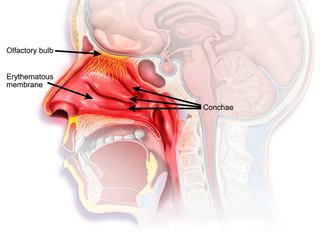
In anatomy, a nasal concha, also called a nasal turbinate or turbinal, is a long, narrow, curled shelf of bone that protrudes into the breathing passage of the nose in humans and various animals. The conchae are shaped like an elongated seashell, which gave them their name. A concha is any of the scrolled spongy bones of the nasal passages in vertebrates.

<span class="mw-page-title-main">Ethmoid sinus</span> Air-filled space near the nasal cavity

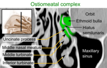
The ethmoid sinuses or ethmoid air cells of the ethmoid bone are one of the four paired paranasal sinuses. Unlike the other three pairs of paranasal sinuses which consist of one or two large cavities, the ethmoidal sinuses entail a number of small air-filled cavities. The cells are located within the lateral mass (labyrinth) of each ethmoid bone and are variable in both size and number. The cells are grouped into anterior, middle, and posterior groups; the groups differ in their drainage modalities, though all ultimately drain into either the superior or the middle nasal meatus of the lateral wall of the nasal cavity.

<span class="mw-page-title-main">Sphenoid sinus</span> One of the four paired paranasal sinuses

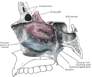
The sphenoid sinus is a paired paranasal sinus occurring within the body of the sphenoid bone. It represents one pair of the four paired paranasal sinuses. The pair of sphenoid sinuses are separated in the middle by a septum of sphenoid sinuses. Each sphenoid sinus communicates with the nasal cavity via the opening of sphenoidal sinus. The two sphenoid sinuses vary in size and shape, and are usually asymmetrical.

<span class="mw-page-title-main">Ethmoidal labyrinth</span>

The ethmoidal labyrinth or lateral mass of the ethmoid bone consists of a number of thin-walled cellular cavities, the ethmoid air cells, arranged in three groups, anterior, middle, and posterior, and interposed between two vertical plates of bone; the lateral plate forms part of the orbit, the medial plate forms part of the nasal cavity. In the disarticulated bone many of these cells are opened into, but when the bones are articulated, they are closed in at every part, except where they open into the nasal cavity.

<span class="mw-page-title-main">Anterior ethmoidal artery</span>

The anterior ethmoidal artery is a branch of the ophthalmic artery in the orbit. It exits the orbit through the anterior ethmoidal foramen alongside the anterior ethmoidal nerve. It contributes blood supply to the ethmoid sinuses, frontal sinuses, the dura mater, lateral nasal wall, and nasal septum. It issues a meningeal branch, and nasal branches.

<span class="mw-page-title-main">Anterior ethmoidal nerve</span> Nerve of the nose

The anterior ethmoidal nerve is a nerve of the head. It is a branch of the nasociliary nerve (itself a branch of the ophthalmic nerve (V1)). It arises in the orbit, and enters first the cranial cavity and then the nasal cavity. It provides sensory innervation to part of the meninges, parts of the nasal cavity, and part of the skin of the nose.

<span class="mw-page-title-main">Middle nasal concha</span>

The medial surface of the labyrinth of ethmoid consists of a thin lamella, which descends from the under surface of the cribriform plate, and ends below in a free, convoluted margin, the middle nasal concha.

<span class="mw-page-title-main">Head and neck anatomy</span>

This article describes the anatomy of the head and neck of the human body, including the brain, bones, muscles, blood vessels, nerves, glands, nose, mouth, teeth, tongue, and throat.

Chronic atrophic rhinitis, or simply atrophic rhinitis, is a chronic inflammation of the nose characterised by atrophy of nasal mucosa, including the glands, turbinate bones and the nerve elements supplying the nose. Chronic atrophic rhinitis may be primary and secondary. Special forms of chronic atrophic rhinitis are rhinitis sicca anterior and ozaena. It can also be described as the empty nose syndrome.

<span class="mw-page-title-main">Nasal cartilages</span> Supportive structures in the nose

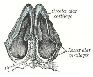
The nasal cartilages are structures within the nose that provide form and support to the nasal cavity. The nasal cartilages are made up of a flexible material called hyaline cartilage in the distal portion of the nose. There are five individual cartilages that make up the nasal cavity: septal nasal cartilage, lateral nasal cartilage, major alar cartilage, minor alar cartilage, and vomeronasal cartilage.

<span class="mw-page-title-main">Body of sphenoid bone</span>

The body of the sphenoid bone, more or less cubical in shape, is hollowed out in its interior to form two large cavities, the sphenoidal sinuses, which are separated from each other by a septum.

<span class="mw-page-title-main">Human nose</span> Feature of the human face

The human nose is the first organ of the respiratory system. It is also the principal organ in the olfactory system. The shape of the nose is determined by the nasal bones and the nasal cartilages, including the nasal septum, which separates the nostrils and divides the nasal cavity into two.

<span class="mw-page-title-main">Nose</span> Organ that smells and facilitates breathing

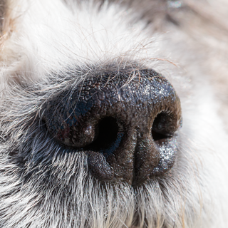
A nose is a protuberance in vertebrates that houses the nostrils, or nares, which receive and expel air for respiration alongside the mouth. Behind the nose are the olfactory mucosa and the sinuses. Behind the nasal cavity, air next passes through the pharynx, shared with the digestive system, and then into the rest of the respiratory system. In humans, the nose is located centrally on the face and serves as an alternative respiratory passage especially during suckling for infants. The protruding nose that is completely separate from the mouth part is a characteristic found only in therian mammals. It has been theorized that this unique mammalian nose evolved from the anterior part of the upper jaw of the reptilian-like ancestors (synapsids).

<span class="mw-page-title-main">Respiratory system of the horse</span> Biological system by which a horse circulates air for the purpose of gaseous exchange

The respiratory system of the horse is the biological system by which a horse circulates air for the purpose of gaseous exchange.

<span class="mw-page-title-main">Outline of human anatomy</span> Overview of and topical guide to human anatomy

The following outline is provided as an overview of and topical guide to human anatomy:

<span class="mw-page-title-main">Nasal mucosa</span> Part of the mucus membrane lining the nasal cavity

The nasal mucosa lines the nasal cavity. It is part of the respiratory mucosa, the mucous membrane lining the respiratory tract. The nasal mucosa is intimately adherent to the periosteum or perichondrium of the nasal conchae. It is continuous with the skin through the nostrils, and with the mucous membrane of the nasal part of the pharynx through the choanae. From the nasal cavity its continuity with the conjunctiva may be traced, through the nasolacrimal and lacrimal ducts; and with the frontal, ethmoidal, sphenoidal, and maxillary sinuses, through the several openings in the nasal meatuses. The mucous membrane is thickest, and most vascular, over the nasal conchae. It is also thick over the nasal septum where increased numbers of goblet cells produce a greater amount of nasal mucus. It is very thin in the meatuses on the floor of the nasal cavities, and in the various sinuses. It is one of the most commonly infected tissues in adults and children. Inflammation of this tissue may cause significant impairment of daily activities, with symptoms such as stuffy nose, headache, mouth breathing, etc.

References

  1. Standring S (2016). Gray's Anatomy: The Anatomical Basis of Clinical Practice (Forty-first ed.). Elsevier. pp. 556–565. ISBN   978-0-7020-5230-9.
  2. "Nasal fossa". TheFreeDictionary.com.
  3. Knipe H. "Ostiomeatal complex". Radiology Reference Article. Radiopaedia.org.
  4. Beitler JJ, McDonald MW, Wadsworth JT, Hudgins PA (2016). "Sinonasal Cancer". Clinical Radiation Oncology. Elsevier. pp. 673–697.e2. doi:10.1016/b978-0-323-24098-7.00036-8. ISBN   978-0-323-24098-7. The nasal vestibules are the two entry points into the nasal cavity. Each is a triangle-shaped space situated in front of the limen nasi and defined laterally by the lateral crus and alar fibrofatty tissue, medially by the medial crus of the alar cartilage and the nasal septum and the distal end of the cartilaginous septum, and columella.
  5. Hamilton, Grant S. (May 2017). "The External Nasal Valve". Facial Plastic Surgery Clinics of North America. 25 (2): 179–194. doi:10.1016/j.fsc.2016.12.010. ISSN   1558-1926. PMID   28340649.
  6. Murthy, V. Ashok; Reddy, R. Raghavendra; Pragadeeswaran, K. (August 2013). "Internal Nasal Valve and Its Significance". Indian Journal of Otolaryngology and Head & Neck Surgery. 65 (Suppl 2): 400–401. doi:10.1007/s12070-013-0618-x. ISSN   2231-3796. PMC   3738809 . PMID   24427685.
  7. Fraioli, Rebecca E.; Pearlman, Steven J. (2013-09-01). "A Patient With Nasal Valve Compromise". JAMA Otolaryngology–Head & Neck Surgery. 139 (9): 947–950. doi:10.1001/jamaoto.2013.4163. ISSN   2168-6181.
  8. Moore KL, Dalley AF (1999). Clinically Oriented Anatomy (Fourth ed.). Philadelphia: Lippincott Williams & Wilkins. ISBN   978-0-683-06141-3.
  9. Schwartz JS, Tajudeen BA, Kennedy DW (2019). "Diseases of the nasal cavity". Handbook of Clinical Neurology. 164: 285–302. doi:10.1016/B978-0-444-63855-7.00018-6. ISBN   978-0-444-63855-7. PMC   7151940 . PMID   31604553.