Paranasal sinuses are a group of four paired air-filled spaces that surround the nasal cavity. The maxillary sinuses are located under the eyes; the frontal sinuses are above the eyes; the ethmoidal sinuses are between the eyes and the sphenoidal sinuses are behind the eyes. The sinuses are named for the facial bones and sphenoid bone in which they are located. Their role is disputed and no function has been confirmed.

The nasal cavity is a large, air-filled space above and behind the nose in the middle of the face. The nasal septum divides the cavity into two cavities, also known as fossae. Each cavity is the continuation of one of the two nostrils. The nasal cavity is the uppermost part of the respiratory system and provides the nasal passage for inhaled air from the nostrils to the nasopharynx and rest of the respiratory tract.

The pyramid-shaped maxillary sinus is the largest of the paranasal sinuses, located in the maxilla. It drains into the middle meatus of the nose through the semilunar hiatus. It is located to the side of the nasal cavity, and below the orbit.

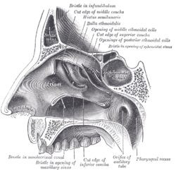
In the ethmoid bone, a sickle shaped projection, the uncinate process, projects posteroinferiorly from the ethmoid labyrinth. Between the posterior edge of this process and the anterior surface of the ethmoid bulla, there is a two-dimensional space, resembling a crescent shape. This space continues laterally as a three-dimensional slit-like space - the ethmoidal infundibulum. This is bounded by the uncinate process, medially, the orbital lamina of ethmoid bone, laterally, and the ethmoidal bulla, posterosuperiorly. This concept is easier to understand if one imagine the infundibulum as a prism so that its medial face is the hiatus semilunaris. The "lateral face" of this infundibulum contains the ostium of the maxillary sinus, which, therefore, opens into the infundibulum.
The ethmoid sinuses or ethmoid air cells of the ethmoid bone are one of the four paired paranasal sinuses. Unlike the other three pairs of paranasal sinuses which consist of one or two large cavities, the ethmoidal sinuses entail a number of small air-filled cavities. The cells are located within the lateral mass (labyrinth) of each ethmoid bone and are variable in both size and number. The cells are grouped into anterior, middle, and posterior groups; the groups differ in their drainage modalities, though all ultimately drain into either the superior or the middle nasal meatus of the lateral wall of the nasal cavity.

The ophthalmic nerve (CN V1) is a sensory nerve of the head. It is one of three divisions of the trigeminal nerve (CN V), a cranial nerve. It has three major branches which provide sensory innervation to the eye, and the skin of the upper face and anterior scalp, as well as other structures of the head.

The superior ophthalmic vein is a vein of the orbit that drains venous blood from structures of the upper orbit. It is formed by the union of the angular vein, and supraorbital vein. It passes backwards within the orbit alongside the ophthalmic artery, then exits the orbit through the superior orbital fissure to drain into the cavernous sinus.

The sphenoid sinus is a paired paranasal sinus occurring within the body of the sphenoid bone. It represents one pair of the four paired paranasal sinuses. The pair of sphenoid sinuses are separated in the middle by a septum of sphenoid sinuses. Each sphenoid sinus communicates with the nasal cavity via the opening of sphenoidal sinus. The two sphenoid sinuses vary in size and shape, and are usually asymmetrical.

The ethmoidal labyrinth or lateral mass of the ethmoid bone consists of a number of thin-walled cellular cavities, the ethmoid air cells, arranged in three groups, anterior, middle, and posterior, and interposed between two vertical plates of bone; the lateral plate forms part of the orbit, the medial plate forms part of the nasal cavity. In the disarticulated bone many of these cells are opened into, but when the bones are articulated, they are closed in at every part, except where they open into the nasal cavity.

The anterior ethmoidal artery is a branch of the ophthalmic artery in the orbit. It exits the orbit through the anterior ethmoidal foramen alongside the anterior ethmoidal nerve. It contributes blood supply to the ethmoid sinuses, frontal sinuses, the dura mater, lateral nasal wall, and nasal septum. It issues a meningeal branch, and nasal branches.

The external nasal nerve is the terminal branch of the anterior ethmoidal nerve. The external nasal nerve passes inferior-ward through the lateral nasal wall. It provides sensory innervation to the area of skin of the nose between the nasal bones superiorly and the tip of the nose inferiorly.
The anterior ethmoidal nerve is a nerve of the head. It is a branch of the nasociliary nerve (itself a branch of the ophthalmic nerve (V1)). It arises in the orbit, and enters first the cranial cavity and then the nasal cavity. It provides sensory innervation to part of the meninges, parts of the nasal cavity, and part of the skin of the nose.
The ethmoid bulla is a rounded elevation upon the lateral wall of the middle nasal meatus produced by one or more of the underlying middle ethmoidal air cells. It varies significantly based on the size of the underlying air cells.
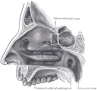
The sphenoethmoidal recess is a small triangular space of the superior nasal meatus of the nasal cavity into which the sphenoidal sinus and the posterior ethmoidal air cells open. The sphenoethmoidal recess is situated superoposterior to the superior nasal concha, between the superior nasal concha and the anterior aspect of the body of the sphenoid bone.

The maxillary hiatus is the opening of a maxillary sinus into the middle nasal meatus of the nasal cavity. It is situated superoposteriorly upon the lateral nasal wall, opening into the nasal cavity at the posterior portion of the ethmoidal infundibulum. Its opening in the maxillary sinus is present upon the superior part of the medial wall of the sinus near the roof of the sinus; because of the position, gravity cannot drain the maxillary sinus contents when the head is erect.
The posterior ethmoidal artery is an artery of the head which arises from the ophthalmic artery to supply the posterior ethmoidal air cells, and the meninges. It is smaller than the anterior ethmoidal artery.
The frontonasal duct is a duct through which either frontal sinus drains into the nasal cavity. Each frontal sinus opens into the frontonasal duct by an opening on the inferomedial part of the floor of the sinus. The frontonasal duct pases inferior-ward to open either into the middle nasal meatus at the anterior end of the ethmoidal infundibulum, or into the anterior ethmoidal air cells.
The ethmoidal infundibulum is a funnel-shaped/slit-like/curved opening/passage/space/cleft upon the anterosuperior portion of the middle nasal meatus at the hiatus semilunaris. The anterior ethmoidal air cells, and (usually) the frontonasal duct open into the ethmoidal infundibulum. The ethmoidal infundibulum extends anterosuperiorly from its opening into the nasal cavity.

The following outline is provided as an overview of and topical guide to human anatomy:

In anatomy, the term nasal meatus can refer to any of the three meatuses (passages) through the skull's nasal cavity: the superior meatus, middle meatus, and inferior meatus.