Nasal cartilages

Last updated
Nasal cartilages
Gray852.png
Cartilages of the nose. Side view. (Greater alar cartilage visible in blue at center right.)
Gray853.png
Cartilages of the nose, seen from below.
Details
Identifiers
Latin cartilagines nasi
MeSH D055171
TA98 A06.1.01.006
TA2 3136
FMA 59502
Anatomical terminology

The nasal cartilages are structures within the nose that provide form and support to the nasal cavity. [1] The nasal cartilages are made up of a flexible material called hyaline cartilage (packed collagen) in the distal portion of the nose. [2] There are five individual cartilages that make up the nasal cavity: septal nasal cartilage, lateral nasal cartilage, major alar cartilage (greater alar cartilage, or cartilage of the aperture), minor alar cartilage (lesser alar cartilage, sesamoid, or accessory cartilage), and vomeronasal cartilage (Jacobson's cartilage).

Contents

Side view of the cartilages in the nose. Gray852.png
Side view of the cartilages in the nose.

The nasal cartilages associate with other cartilage structures of the nose or with bones of the facial skeleton. These associations create vent-like structures within the nose so that air can flow from the nasal cavity to the lungs or vice versa. Therefore, the nasal cartilages are structures that aid the body in respiratory functions to intake oxygen or expire carbon dioxide.

Abnormalities or defects in the nasal cartilages affect airflow through the nasal cavity, resulting in respiratory issues. Surgical techniques have been produced to adjust the position or repair the nasal cartilages so that maximal airflow is once again accomplished. Some of these surgical techniques include: septoplasty (restructuring the septal nasal cartilage), upper lateral cartilage repositioning (restructuring the lateral nasal cartilage), and sliding alar cartilage (restructuring the major alar cartilage). Rhinoplasty, which is the surgical reconstruction of the nose, has increased in recent popularity for functional and social purposes.

Other mammals also contain nasal cartilages in order to maintain structure and function for the nasal cavity. The orientation of the nasal cartilages can produce different shapes and sizes of the nostrils and nasal cavities. For the most part, animals contain similar cartilage structures within the nose but vary in the number of different cartilage structures they have. Donkeys, buffalo, and camels have a variety of cartilage structures that are analogous to humans but they all lack septal nasal cartilages. Instead, they have multiple components merging together to form the nasal septum. Even though nasal cartilages differ between species, they all aid in the function of the respiratory system.

Structure and function

Septal nasal cartilage

Right side view of the septal nasal cartilage. Gray854.png
Right side view of the septal nasal cartilage.

The septal nasal cartilage is a flat, quadrilateral piece of hyaline cartilage that separates both nasal cavities from one another. [3] The septal nasal cartilage fits in a place between the perpendicular plate of the ethmoid and vomer bones while also being covered by an internal mucous membrane. The superior portion of the septal nasal cartilage attaches to the nasal bones, while the inferior portion attaches to the alar cartilages via fibrous tissues. The septal nasal cartilage separates both right and left nasal cavities, which allows air to pass through them. Providing two cavities generates turbulence within the tight spaces, allowing air to flow quicker bidirectionally. The septal nasal cartilage is also the main structure that provides the orientation of the nose, being the midline structure of the organ. With an offset septal nasal cartilage, the nose will appear crooked to the viewer. A crooked nose can block airflow coming from the nares to the lungs or vice versa. [4] This can lead to respiratory issues due to low oxygen but high carbon dioxide counts within the body. A surgical procedure to correct this issue is called septoplasty.

Lateral nasal cartilage

The lateral nasal cartilage is a wing-like expansion extending out from the septal nasal cartilage. [5] The lateral nasal cartilage lies inferiorly to the nasal bones while sitting superiorly to the major alar cartilage, separated by a narrow fissure. The lateral nasal cartilage and major alar cartilage curl up upon interaction with one another, forming a tight connection through fibrous tissues. Like the septal nasal cartilage, the lateral nasal cartilage is composed of hyaline cartilage. Hyaline cartilage provides form and flexibility within a specific structure.

The superior portion of the lateral nasal cartilage fuses with the septum to provide support within the nasal cavities. With a collapse of the lateral nasal cartilage, the inner nasal valve could become obstructed and prevent the movement of airflow throughout. [6] A new surgical technique to reposition the lateral nasal cartilage has been constructed to relieve the site of obstruction within the inner nasal valve and regain maximal airflow throughout the nose (upper lateral cartilage repositioning).

Major alar cartilage

The major alar cartilages are positioned with one structure on each side of the nasal tip. [7] Superiorly, the major alar cartilages are connected to the lateral nasal cartilage via fibrous tissues. Composed of hyaline cartilage, these structures are very thin and folded to form the lateral and medial crus. The medial crus is the inner portion of the major alar cartilages that are situated perpendicularly to the septal nasal cartilage. The lateral crus is the outer portion of the major alar cartilages that associate with the ala of the nose.

Major alar and minor alar cartilages, seen from below. Gray853.png
Major alar and minor alar cartilages, seen from below.

Both crus come together to form an oval tip at each nostril. Both sides of the major alar cartilages merge together to form a notch at the tip, which is referred to as the apex of the nose. With the formation of the medial and lateral walls within the nares, the major alar cartilages function to hold open each naris. This allows maximal airflow to reach the nasal valve, allowing optimal respiration. Due to weakness corresponding with the lateral crus in certain individuals, a technique called sliding alar cartilage (SAC) has been a procedure practiced to restructure and support the nasal tip. [8]

Minor alar cartilage

The minor alar cartilages are 3 to 4 small hyaline cartilage pieces on both sides of the nose that sit between the lateral nasal cartilage and the major alar cartilage. [9] Associated within the ala of the nose, these small structures are contained within the most dorsal part of the ala. Also known by its other name, "accessory cartilage", these structures aid to provide form and strength at the base of the nares in conjunction with the major alar cartilage.

Vomeronasal cartilage

The vomeronasal cartilage is a thin piece of hyaline cartilage that attaches to the vomer and extends to the septal nasal cartilage. [10] This structure is associated with the vomeronasal organ, which is part of the accessory olfactory system. This associated organ plays an important role in the sense of smell by being lined with similar epithelium to that of the olfactory region of the nose. The vomeronasal cartilage is another small component of the nose that provides strength and structure.

Reconstruction

With emerging technological advancements, reconstruction and surgical techniques have been developed to adjust the lifestyle and health of individuals. Rhinoplasty is one of the surgical practices that has become more common in the modern era. Some rhinoplastic procedures include septoplasty, sliding alar cartilage, and upper lateral cartilage repositioning. These procedures aid in cosmetic as well as functional issues involving the nose. Other reconstruction procedures are being produced and tested as the knowledge for nasal cartilages increase.

Septoplasty

External incision used during a rhinoplasty. Rhinoplasty Incisions 1.jpg
External incision used during a rhinoplasty.

Septoplasty is a surgical procedure that straightens the septal nasal cartilage within the center of the nose. [11] With a crooked septum, it is more difficult for an individual to breathe and the risk for getting a sinus infection increases. Also called a deviated septum, a crooked nose will block one or both sides of the nose, affecting the quality of life. [4] However, a deviated septum is very common and does not always create respiratory issues. Respiratory issues usually occur in more severe cases, requiring surgery to repair. [11] Surgery is also permitted to individuals that seek cosmetic changes due to moderate cases of a deviated septum. Surgery may require a surgeon to cut and remove parts of the septal nasal cartilages, replacing them later in a reconstructed format. This will allow the individual to receive more airflow through the nostrils when the surgery fully heals after 3 to 6 months. However, there are some risks correlated with this surgical procedure. These risks include a change in the shape of the nose, excessive bleeding, vacant space in the septum, trouble smelling, blood clots that need to be removed, and numbness by the facial region. Smoking can also cause further damage during the healing process of septoplastic surgery.

Upper lateral cartilage repositioning

U.S. Air Force Maj. Brent Feldt, performing a septoplasty. New Horizons 2013 130423-F-HS649-124.jpg
U.S. Air Force Maj. Brent Feldt, performing a septoplasty.

The upper lateral cartilage repositioning procedure is done to move the lateral nasal cartilage from blocking the nasal valve. [6] The nasal valve is the smallest airway within the nose and is a common site for obstruction. Other surgical procedures open up this air way by employing grafts to separate the septal nasal and lateral nasal cartilages from one another. Grafts need to be used permanently due to the complications of removing such a device. The upper lateral cartilage repositioning technique deploys a temporary stint that repositions the lateral nasal cartilage, lets it heal/be stationary due to scar tissue formation, then is removed. This open rhinoplastic procedure allows the nose to heal to an optimal position without the permanent use of man-made hardware. This procedure is just one way to resolve issues involving lateral nasal cartilage deformities. Other procedures are being produced and improved upon in order to generate the simplest and most safe surgical procedure.

Sliding alar cartilage (SAC)

Patient with metal split after a rhinoplastic procedure. RhinoplastySplint3.jpg
Patient with metal split after a rhinoplastic procedure.

The sliding alar cartilage is a procedure to strengthen and support the nasal tip. [8] This medical practice is completed on the greater alar cartilage in order to reshape this structure. The greater alar cartilages can become very weak or have deformities, creating respiratory issues. Other medical procedures that reshape the greater alar cartilages use grafts or cartilage re-sectioning. The SAC procedure is completed within two to three minutes. In that timeframe, the tip of the nose is cut open, the greater alar cartilage is manipulated to preserve the scroll area, providing strength and structure, then the incision is sutured back up. [12] This simple technique creates tip definition while maintaining airway function. While there are other procedures to strengthen the greater alar cartilage, the SAC procedure is gaining momentum into common rhinoplastic operations.

Related Research Articles

<span class="mw-page-title-main">Otorhinolaryngology</span> Medical specialty

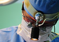
Otorhinolaryngology is a surgical subspeciality within medicine that deals with the surgical and medical management of conditions of the head and neck. Doctors who specialize in this area are called otorhinolaryngologists, otolaryngologists, head and neck surgeons, or ENT surgeons or physicians. Patients seek treatment from an otorhinolaryngologist for diseases of the ear, nose, throat, base of the skull, head, and neck. These commonly include functional diseases that affect the senses and activities of eating, drinking, speaking, breathing, swallowing, and hearing. In addition, ENT surgery encompasses the surgical management of cancers and benign tumors and reconstruction of the head and neck as well as plastic surgery of the face, scalp, and neck.

<span class="mw-page-title-main">Rhinoplasty</span> Surgical procedure to enhance or reconstruct a human nose

Rhinoplasty, commonly called nose job, medically called nasal reconstruction is a plastic surgery procedure for altering and reconstructing the nose. There are two types of plastic surgery used – reconstructive surgery that restores the form and functions of the nose and cosmetic surgery that changes the appearance of the nose. Reconstructive surgery seeks to resolve nasal injuries caused by various traumas including blunt, and penetrating trauma and trauma caused by blast injury. Reconstructive surgery can also treat birth defects, breathing problems, and failed primary rhinoplasties. Rhinoplasty may remove a bump, narrow nostril width, change the angle between the nose and the mouth, or address injuries, birth defects, or other problems that affect breathing, such as a deviated nasal septum or a sinus condition. Surgery only on the septum is called a septoplasty.

<span class="mw-page-title-main">Vomer</span> Unpaired facial bone of the skull

The vomer is one of the unpaired facial bones of the skull. It is located in the midsagittal line, and articulates with the sphenoid, the ethmoid, the left and right palatine bones, and the left and right maxillary bones. The vomer forms the inferior part of the nasal septum in humans, with the superior part formed by the perpendicular plate of the ethmoid bone. The name is derived from the Latin word for a ploughshare and the shape of the bone.

<span class="mw-page-title-main">Nasal cavity</span> Large, air-filled space above and behind the nose in the middle of the face

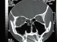
The nasal cavity is a large, air-filled space above and behind the nose in the middle of the face. The nasal septum divides the cavity into two cavities, also known as fossae. Each cavity is the continuation of one of the two nostrils. The nasal cavity is the uppermost part of the respiratory system and provides the nasal passage for inhaled air from the nostrils to the nasopharynx and rest of the respiratory tract.

<span class="mw-page-title-main">Nasal concha</span> Piece of bone in the breathing passage of humans and other animals

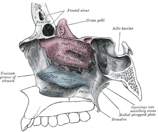
In anatomy, a nasal concha, also called a nasal turbinate or turbinal, is a long, narrow, curled shelf of bone that protrudes into the breathing passage of the nose in humans and various animals. The conchae are shaped like an elongated seashell, which gave them their name. A concha is any of the scrolled spongy bones of the nasal passages in vertebrates.

<span class="mw-page-title-main">Septoplasty</span> Corrective surgical procedure

Septoplasty [ˈsɛp.toˌplæ.sti] (Etymology: L, saeptum, septum; Gk, πλάσσειν plassein – to shape), or alternatively submucous septal resection and septal reconstruction, is a corrective surgical procedure done to straighten a deviated nasal septum – the nasal septum being the partition between the two nasal cavities. Ideally, the septum should run down the center of the nose. When it deviates into one of the cavities, it narrows that cavity and impedes airflow. Deviated nasal septum or “crooked” internal nose can occur at childbirth or as the result of an injury or other trauma. If the wall that functions as a separator of both sides of the nose is tilted towards one side at a degree greater than 50%, it might cause difficulty breathing. Often the inferior turbinate on the opposite side enlarges, which is termed compensatory hypertrophy. Deviations of the septum can lead to nasal obstruction. Most surgeries are completed in 60 minutes or less, while the recovery time could be up to several weeks. Put simply, septoplasty is a surgery that helps repair the passageways in the nose making it easier to breathe. This surgery is usually performed on patients with a deviated septum, recurrent rhinitis, or sinus issues.

<span class="mw-page-title-main">Nasal septum</span> Separator of the left and right airways in the nose

The nasal septum separates the left and right airways of the nasal cavity, dividing the two nostrils.

<span class="mw-page-title-main">Nasal septum deviation</span> Disorder of the nose

Nasal septum deviation is a physical disorder of the nose, involving a displacement of the nasal septum. Some displacement is common, affecting 80% of people, mostly without their knowledge.

<span class="mw-page-title-main">Nasal septum perforation</span> Medical condition

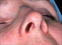
A nasal septum perforation is a medical condition in which the nasal septum, the bony/cartilaginous wall dividing the nasal cavities, develops a hole or fissure.

<span class="mw-page-title-main">Nasalis muscle</span>

The nasalis muscle is a sphincter-like muscle of the nose. It has a transverse part and an alar part. It compresses the nasal cartilages, and can "flare" the nostrils. It can be used to test the facial nerve (VII), which supplies it.

<span class="mw-page-title-main">Dilator naris muscle</span> Facial muscle that controls the nostril opening

The dilator naris muscle is a part of the nasalis muscle. It has an anterior and a posterior part. It has origins from the nasal notch of the maxilla and the major alar cartilage, and a single insertion near the margin of the nostril. It controls nostril width, including changes during breathing. Its function can be tested as an analogue for the function of the facial nerve (VII), which supplies it.

<span class="mw-page-title-main">Empty nose syndrome</span> Medical condition

Empty nose syndrome (ENS) is a clinical syndrome, the hallmark symptom of which is a sensation of suffocation despite a clear airway. This syndrome is often referred to as a form of secondary atrophic rhinitis. ENS is a potential complication of nasal turbinate surgery or injury. Patients have usually undergone a turbinectomy or other surgical procedures that injure the nasal turbinates.

<span class="mw-page-title-main">Major alar cartilage</span> Cartilaginous plate in the nose

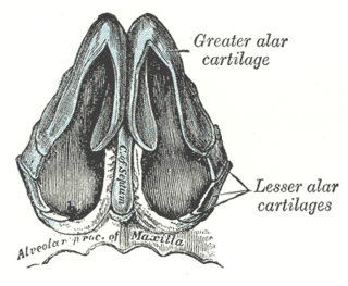
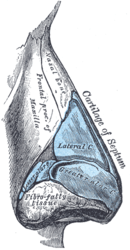
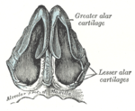
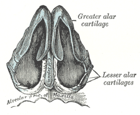
The major alar cartilage is a thin, flexible plate, situated immediately below the lateral nasal cartilage, and bent upon itself in such a manner as to form the medial wall and lateral wall of the nostril of its own side.

<span class="mw-page-title-main">Septal nasal cartilage</span> Hyaline cartilage in the nose

The septal nasal cartilage is composed of hyaline cartilage. It is somewhat quadrilateral in form, thicker at its margins than at its center, and completes the separation between the nasal cavities in front.

<span class="mw-page-title-main">Human nose</span> Feature of the human face

The human nose is the most protruding part of the human face. It bears the nostrils and is the first organ of the respiratory system. It is also the principal organ in the olfactory system. The shape of the nose is determined by the nasal bones and the nasal cartilages, including the nasal septum which separates the nostrils and divides the nasal cavity into two.

<span class="mw-page-title-main">Nasal septal hematoma</span> Medical condition

Nasal septal hematoma is a condition affecting the nasal septum. It can be associated with trauma.

<span class="mw-page-title-main">Respiratory system of the horse</span> Biological system by which a horse circulates air for the purpose of gaseous exchange

The respiratory system of the horse is the biological system by which a horse circulates air for the purpose of gaseous exchange.

<span class="mw-page-title-main">Turbinectomy</span> Surgical removal of the turbinate bones in the nasal passage

A turbinectomy or turbinoplasty is a surgical procedure, that removes tissue, and sometimes bone, of the turbinates in the nasal passage, particularly the inferior nasal concha. The procedure is usually performed to relieve nasal obstructions. In most cases, turbinate hypertrophy is accompanied by some septum deviation, so the surgery is done along with septoplasty.

Nasal chondrocytes (NC) are present in the hyaline cartilage of the nasal septum and in fact are the only cell type within the tissue. Similar to chondrocytes present in articular cartilage, NC express extracellular matrix proteins such as glycosaminoglycans and collagen.

Nasal surgery is a medical procedure designed to treat various conditions that cause nasal blockages in the upper respiratory tract, for example nasal polyps, inferior turbinate hypertrophy, and chronic rhinosinusitis. It encompasses several types of techniques, including rhinoplasty, septoplasty, sinus surgery, and turbinoplasty, each with its respective postoperative treatments. Furthermore, nasal surgery is also conducted for cosmetic purposes. While there are potential risks and complications associated, the advancement of medical instruments and enhanced surgical skills have helped mitigate them.

References

  1. Lang, Johannes (1989). Clinical Anatomy of the Nose, Nasal Cavity and Paranasal Sinuses. Thieme. pp. 10–15. ISBN   978-3-13-738401-4 . Retrieved March 11, 2021.
  2. Krishnan, Yamini; Grodzinsky, Alan J. (2018). "Cartilage Diseases". Matrix Biology. 71–72: 51–69. doi:10.1016/j.matbio.2018.05.005. ISSN   0945-053X. PMC   6146013 . PMID   29803938.
  3. Gleason, Edward Baldwin (1918). A Manual of Diseases of the Nose, Throat, and Ear. W.B. Saunders Company. pp. 56–57. Retrieved March 25, 2021.
  4. 1 2 BOCCIERI, A. (2013). "The crooked nose". Acta Otorhinolaryngologica Italica. 33 (3): 163–168. ISSN   0392-100X. PMC   3709523 . PMID   23853411.
  5. Schünke, Michael; Ross, Lawrence M.; Schulte, Erik; Lamperti, Edward D.; Schumacher, Udo (2007). Thieme Atlas of Anatomy: Head and Neuroanatomy. Thieme. p. 18. ISBN   978-1-58890-441-6 . Retrieved March 25, 2021.
  6. 1 2 Stupak, Howard D. (2011-02-01). "Endonasal Repositioning of the Upper Lateral Cartilage and the Internal Nasal Valve". Annals of Otology, Rhinology & Laryngology. 120 (2): 88–94. doi:10.1177/000348941112000203. ISSN   0003-4894. PMID   21391419 . Retrieved March 30, 2021.
  7. Morris, Sir Henry; McMurrich, James Playfair (1907). Morris's Human Anatomy; a Complete Systematic Treatise. Vol. 3. P. Blakiston's son & Company. pp. 1068–1069. Retrieved April 6, 2021.
  8. 1 2 Ozmen, Selahattin; Eryilmaz, Tolga; Sencan, Ayse; Cukurluoglu, Onur; Uygur, Safak; Ayhan, Suhan; Atabay, Kenan (2009). "Sliding Alar Cartilage (SAC) Flap: A New Technique for Nasal Tip Surgery". Annals of Plastic Surgery. 63 (5): 480–485. doi:10.1097/SAP.0b013e31819538a8. ISSN   0148-7043. PMID   19801923 . Retrieved March 30, 2021.
  9. Toldt, Carl; Rosa, Alois Dalla (1919). An Atlas of Human Anatomy for Students and Physicians. Rebman Company. p. 470. Retrieved April 7, 2021.
  10. "Vomeronasal cartilage". IMAIOS. Retrieved 2021-04-09.
  11. 1 2 "Septoplasty – Mayo Clinic". www.mayoclinic.org. Retrieved 2021-03-23.
  12. Racy, Emmanuel; Fanous, Amanda; Pressat-Laffouilhere, Thibaut; Benmoussa, Nadia (2019). "The Modified Sliding Alar Cartilage Flap: A Novel Way to Preserve the Internal Nasal Valve as Illustrated by Three-Dimensional Modeling". Plastic and Reconstructive Surgery. 144 (3): 593–599. doi:10.1097/PRS.0000000000005991. ISSN   0032-1052. PMID   31461010 . Retrieved April 8, 2021.