Articles related to anatomy include:

The lacrimal bone is a small and fragile bone of the facial skeleton; it is roughly the size of the little fingernail. It is situated at the front part of the medial wall of the orbit. It has two surfaces and four borders. Several bony landmarks of the lacrimal bone function in the process of lacrimation or crying. Specifically, the lacrimal bone helps form the nasolacrimal canal necessary for tear translocation. A depression on the anterior inferior portion of the bone, the lacrimal fossa, houses the membranous lacrimal sac. Tears or lacrimal fluid, from the lacrimal glands, collect in this sac during excessive lacrimation. The fluid then flows through the nasolacrimal duct and into the nasopharynx. This drainage results in what is commonly referred to a runny nose during excessive crying or tear production. Injury or fracture of the lacrimal bone can result in posttraumatic obstruction of the lacrimal pathways.

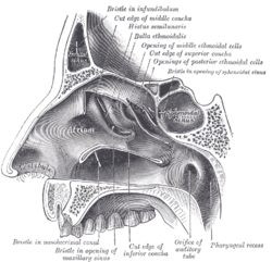
The inferior nasal concha is one of the three paired nasal conchae in the nose. It extends horizontally along the lateral wall of the nasal cavity and consists of a lamina of spongy bone, curled upon itself like a scroll,. The inferior nasal conchae are considered a pair of facial bones. As the air passes through the turbinates, the air is churned against these mucosa-lined bones in order to receive warmth, moisture and cleansing. Superior to inferior nasal concha are the middle nasal concha and superior nasal concha which both arise from the ethmoid bone, of the cranial portion of the skull. Hence, these two are considered as a part of the cranial bones.

The nasal cavity is a large, air-filled space above and behind the nose in the middle of the face. The nasal septum divides the cavity into two cavities, also known as fossae. Each cavity is the continuation of one of the two nostrils. The nasal cavity is the uppermost part of the respiratory system and provides the nasal passage for inhaled air from the nostrils to the nasopharynx and rest of the respiratory tract.

The ophthalmic artery (OA) is an artery of the head. It is the first branch of the internal carotid artery distal to the cavernous sinus. Branches of the ophthalmic artery supply all the structures in the orbit around the eye, as well as some structures in the nose, face, and meninges. Occlusion of the ophthalmic artery or its branches can produce sight-threatening conditions.

The pyramid-shaped maxillary sinus is the largest of the paranasal sinuses, located in the maxilla. It drains into the middle meatus of the nose through the semilunar hiatus. It is located to the side of the nasal cavity, and below the orbit.

The orbital or horizontal part of the frontal bone consists of two thin triangular plates, the orbital plates, which form the vaults of the orbits, and are separated from one another by a median gap, the ethmoidal notch.

In the ethmoid bone, a sickle shaped projection, the uncinate process, projects posteroinferiorly from the ethmoid labyrinth. Between the posterior edge of this process and the anterior surface of the ethmoid bulla, there is a two-dimensional space, resembling a crescent shape. This space continues laterally as a three-dimensional slit-like space - the ethmoidal infundibulum. This is bounded by the uncinate process, medially, the orbital lamina of ethmoid bone, laterally, and the ethmoidal bulla, posterosuperiorly. This concept is easier to understand if one imagine the infundibulum as a prism so that its medial face is the hiatus semilunaris. The "lateral face" of this infundibulum contains the ostium of the maxillary sinus, which, therefore, opens into the infundibulum.

The ethmoid sinuses or ethmoid air cells of the ethmoid bone are one of the four paired paranasal sinuses. Unlike the other three pairs of paranasal sinuses which consist of one or two large cavities, the ethmoidal sinuses entail a number of small air-filled cavities. The cells are located within the lateral mass (labyrinth) of each ethmoid bone and are variable in both size and number. The cells are grouped into anterior, middle, and posterior groups; the groups differ in their drainage modalities, though all ultimately drain into either the superior or the middle nasal meatus of the lateral wall of the nasal cavity.

The semilunar hiatus is a crescent-shaped/semicircular/ curved slit/groove upon the lateral wall of the nasal cavity at the middle nasal meatus just inferior to the ethmoidal bulla. It is the location of the openings for the frontal sinus, maxillary sinus, and anterior ethmoidal sinus. It is bounded inferiorly and anteriorly by the sharp concave margin of the uncinate process of the ethmoid bone, superiorly by the ethmoidal bulla, and posteriorly by the ethmoidal process of the inferior nasal concha. It leads into the ethmoidal infundibulum; it marks the medial limit of the ethmoidal infundibulum.

The ethmoidal labyrinth or lateral mass of the ethmoid bone consists of a number of thin-walled cellular cavities, the ethmoid air cells, arranged in three groups, anterior, middle, and posterior, and interposed between two vertical plates of bone; the lateral plate forms part of the orbit, the medial plate forms part of the nasal cavity. In the disarticulated bone many of these cells are opened into, but when the bones are articulated, they are closed in at every part, except where they open into the nasal cavity.
The anterior ethmoidal artery is a branch of the ophthalmic artery in the orbit. It exits the orbit through the anterior ethmoidal foramen alongside the anterior ethmoidal nerve. It contributes blood supply to the ethmoid sinuses, frontal sinuses, the dura mater, lateral nasal wall, and nasal septum. It issues a meningeal branch, and nasal branches.
The ethmoid bulla is a rounded elevation upon the lateral wall of the middle nasal meatus produced by one or more of the underlying middle ethmoidal air cells. It varies significantly based on the size of the underlying air cells.
The frontonasal duct is a duct through which either frontal sinus drains into the nasal cavity. Each frontal sinus opens into the frontonasal duct by an opening on the inferomedial part of the floor of the sinus. The frontonasal duct pases inferior-ward to open either into the middle nasal meatus at the anterior end of the ethmoidal infundibulum, or into the anterior ethmoidal air cells.

The perpendicular plate of palatine bone is the vertical part of the palatine bone, and is thin, of an oblong form, and presents two surfaces and four borders.

The frontal process of the maxilla is a strong plate, which projects upward, medialward, and backward from the maxilla, forming part of the lateral boundary of the nose.

The body of the sphenoid bone, more or less cubical in shape, is hollowed out in its interior to form two large cavities, the sphenoidal sinuses, which are separated from each other by a septum.
The ethmoidal infundibulum is a funnel-shaped/slit-like/curved opening/passage/space/cleft upon the anterosuperior portion of the middle nasal meatus at the hiatus semilunaris. The anterior ethmoidal air cells, and (usually) the frontonasal duct open into the ethmoidal infundibulum. The ethmoidal infundibulum extends anterosuperiorly from its opening into the nasal cavity.

The human nose is the first organ of the respiratory system. It is also the principal organ in the olfactory system. The shape of the nose is determined by the nasal bones and the nasal cartilages, including the nasal septum which separates the nostrils and divides the nasal cavity into two.

In anatomy, the term nasal meatus can refer to any of the three meatuses (passages) through the skull's nasal cavity: the superior meatus, middle meatus, and inferior meatus.