Computed tomography of the abdomen and pelvis

Last updated
Computed tomography of the abdomen and pelvis
CT of a normal abdomen and pelvis, thumbnail.png
CT scan of a normal abdomen and pelvis
ICD-9-CM 88.01
OPS-301 code 3-225-3-226
A CT scan image showing a ruptured abdominal aortic aneurysm. RupturedAAA.png
A CT scan image showing a ruptured abdominal aortic aneurysm.
CT Scan of 11 cm Wilms' tumor of right kidney in 13-month-old patient.

Computed tomography of the abdomen and pelvis is an application of computed tomography (CT) and is a sensitive method for diagnosis of abdominal diseases. It is used frequently to determine stage of cancer and to follow progress. It is also a useful test to investigate acute abdominal pain (especially of the lower quadrants, whereas ultrasound is the preferred first line investigation for right upper quadrant pain). Renal stones, appendicitis, pancreatitis, diverticulitis, abdominal aortic aneurysm, and bowel obstruction are conditions that are readily diagnosed and assessed with CT. CT is also the first line for detecting solid organ injury after trauma.

Contents

Advantages

Multidetector CT (MDCT) can clearly delineate anatomic structures in the abdomen, which is critical in the diagnosis of internal diaphragmatic and other nonpalpable or unsuspected hernias. MDCT also offers clear detail of the abdominal wall allowing wall hernias to be identified accurately. [1]

Contrast administration

Abdominal imaging is associated with many potential uses for the different phases of contrast CT. The majority of abdominal and pelvic CT’s can be performed using a single-phase, but the evaluation of some tumor types (hepatic/pancreatic/renal), the urinary collecting system, and trauma patients among others, may be best performed with multiple phases.[ citation needed ]

In discussing the numerous phases and indications for CT, best patient care requires individualized CT protocols based upon each patient’s specific symptoms, pathology, and underlying co-morbidities. Although labor intensive, this provides the highest likelihood of an accurate diagnosis with the lowest necessary radiation dose. The following discussion will provide a basic outline of current best practice, but not all clinical scenarios can be accounted for. [2]

Contrast enhanced CT examinations can be acquired at a variety of specific time points after intravenous contrast injection (timing is dependent on the phase of contrast enhancement needed and organ system being evaluated). The timing should be chosen specifically to optimize contrast distribution within the solid organ parenchyma in question.[ citation needed ]

In cases of suspected bowel leak or perforation, gastrointestinal fistula, interloop abscess or other fluid collection, oncologic staging and surveillance, and CT colonography, oral positive contrast is useful in delineating the lesions. [3] 1% dilute barium solution can be administered orally for bowel preparation for CT scan of the abdomen. [4]

Unenhanced CT

FIGURE 1. Non-contrast CT demonstrating multiple bilateral renal calculi (arrows), which can be obscured on contrast-enhanced images, particularly delayed images when there is excreted contrast in the renal collecting system; axial left, coronal reformat on right. Non-contrast CT of multiple bilateral renal calculi.jpg
FIGURE 1. Non-contrast CT demonstrating multiple bilateral renal calculi (arrows), which can be obscured on contrast-enhanced images, particularly delayed images when there is excreted contrast in the renal collecting system; axial left, coronal reformat on right.

Non-contrast CT scans Figure 1a (left) and 1b (right) are of limited use for the differentiation of soft tissue structures. However, materials like blood, calcium (renal stones, vascular atherosclerosis), bone, and pulmonary parenchyma are highly visible and can usually be adequately assessed with non-contrast CT. For example, in the abdomen and pelvis, there are several indications for non-contrast imaging. These include: evaluation of renal calculi; assessment for gross intra-abdominal hemorrhage; and post-endostent volume measurements. In addition, non-contrast images are often obtained in conjunction with contrast enhanced images in evaluating potential renal transplant donors and in the evaluation of the pancreas (in combination with contrast phases). Of note, dual-energy CT and the development of virtual “non-contrast” images (VNC imaging) may ultimately obviate the combination scans. Additionally, CT angiography examinations performed for pathologies like aneurysms and dissection are frequently performed in conjunction with non-contrast imaging. The non-contrast images facilitate the differentiation of active extravasation or acute bleeding from vascular calcifications.[ citation needed ]

Portal venous phase

The most common technique is to perform portal venous phase imaging in the abdomen and pelvis (approximately 60–90 seconds after contrast administration, figure 2). This results in near optimal contrast opacification of the majority of the solid abdominal organs and it is used for a wide variety of indications: nonspecific abdominal pain; hernia; infection; masses (with a few exceptions such as hypervascular, renal, and some hepatic tumors); and in most follow-up examinations. As a general rule, this single phase is adequate unless there is a specific clinical indication that has been shown to benefit from other phases.[ citation needed ]

Early arterial phase (CT angiography)

FIGURE 3. Axial (left) and coronal (right) CT angiography images of the abdominal aorta evaluating for aortic aneurysm. Abdominal CT angiography.jpg
FIGURE 3. Axial (left) and coronal (right) CT angiography images of the abdominal aorta evaluating for aortic aneurysm.

CT angiography (CTA) is highly effective for evaluation of the arterial system, and has largely replaced conventional angiography due to the lower risk profile and ability to survey the entire abdomen. Images are acquired after a rapid bolus of intravenous contrast material (3-7 cc/s) during the arterial phase (15–35 seconds after injection) when the concentration of contrast material in the arterial system is high (figures 3). Images are usually acquired using narrow collimation (<1 mm) and can be retrospectively reconstructed using dedicated 3-dimensional workstations and software. CTA is commonly used in the head and chest in the evaluation of pulmonary emboli, aneurysms, vascular malformations, dissection, bleeding and ischemia. Indications for early arterial phase imaging include: evaluation of aneurysms or dissections (cerebral, aortic, etc.), hepatic, splanchnic or renal arterial anatomy, and arterial imaging in liver or kidney transplantation. Single phase arterial imaging is often used in the evaluation of trauma patients either a complete chest/abdomen/pelvis examination with arterial phase imaging of the chest and portal venous phase imaging of the abdomen/pelvis or just a portal venous phase of abdomen and pelvis depending on the mechanism and severity of the trauma. CTA is also commonly performed in the abdomen and pelvis for evaluating vascular malformations and in the evaluation of bleeding. Mesenteric ischemia can also be evaluated using CT angiography. CTA of the abdomen and pelvis is often performed in combination with a CTA for evaluating the extremity vasculature.[ citation needed ]

Late arterial phase

FIGURE 4: Arterial and portal venous phase CT of cholangiocarcinoma. Selected images from a biphasic CT demonstrating early arterial enhancement of a posterior right hepatic lobe mass with mild wash out on delayed phase images in the setting of cirrhosis characteristic of hepatocellular carcinoma. Arterial and portal venous phase CT of cholangiocarcinoma.jpg
FIGURE 4: Arterial and portal venous phase CT of cholangiocarcinoma. Selected images from a biphasic CT demonstrating early arterial enhancement of a posterior right hepatic lobe mass with mild wash out on delayed phase images in the setting of cirrhosis characteristic of hepatocellular carcinoma.

The late arterial phase is timed to correspond to the peak concentration of contrast material in highly vascular tumors and is performed approximately 20–35 seconds after the injection of intravenous contrast. Early arterial phase imaging is predominantly utilized for angiography and will be discussed separately. Late arterial phase imaging is almost always performed in conjunction with other phases (e.g. portal venous phase) to allow more complete characterization of any identified abnormalities (figure 4). The primary indication for a late arterial phase is for the evaluation of hypervascular tumors of the liver such as hepatocellular carcinoma or hypervascular metastases (figure 4). Typical hypervascular tumors for which this would be used include: hepatocellular carcinoma; renal cell carcinoma; melanoma; carcinoid/neuroendocrine tumors; some sarcomas; choriocarcinoma; and thyroid carcinoma. Although a “hypervascular”, biphasic evaluation would generally be used for these patients, note that a single phase is often adequate for follow up imaging.[ citation needed ]

Systemic venous phase

CT imaging specific for the venous structures is performed uncommonly. Most venous structures are partially opacified on the routine contrast enhancing images and suffice for most examinations. However, occasionally evaluation of the inferior vena cava is desired, such as prior to IVC filter placement/removal or evaluation of IVC thrombosis.[ citation needed ]

Delayed phase

FIGURE 5. Selected images form CT performed using a Cholangiocarcinoma specific protocol. 5a is a portal venous phase image demonstrating a single low attenuation mass which does not appear to enhance. 5b is a 15 minute delayed image which demonstrates delayed enhancement of the liver mass (arrow) characteristic of Cholangiocarcinoma. Several other enhancing masses (arrowheads) are also seen which were not evident on the portal venous phase images. Arterial and portal venous phase CT of cholangiocarcinoma.jpg
FIGURE 5. Selected images form CT performed using a Cholangiocarcinoma specific protocol. 5a is a portal venous phase image demonstrating a single low attenuation mass which does not appear to enhance. 5b is a 15 minute delayed image which demonstrates delayed enhancement of the liver mass (arrow) characteristic of Cholangiocarcinoma. Several other enhancing masses (arrowheads) are also seen which were not evident on the portal venous phase images.

Delayed phase imaging (figure 5) encompasses scanning at a variety of different times following contrast administration, and depends on the pathology in question. Typical delayed imaging times range from a few minutes to up to 15 minutes or longer. The most common indications for delayed phase imaging are evaluation of the kidneys, collecting system (ureters and bladder) and specific kidney, liver, and adrenal tumors. Evaluation of the kidneys, ureters and bladder are discussed separately in the renal imaging section. Cholangiocarcinoma occurring within the extrahepatic biliary tree or intrahepatic cholangiocarcinomas are a common reason for delayed imaging. Cholangiocarcinomas are fibrotic tumors which enhance slowly, and are usually imaged following a 10-15 minute delay. Similarly, adrenal masses can be evaluated with multiphase imaging including an unenhanced CT, portal venous phase and a 10 minute delay CT which allows for evaluation and calculation of the enhancement and washout characteristics aiding in distinguishing benign adrenal adenomas from other adrenal masses.[ citation needed ]

Outside of the evaluation of masses, delayed phase images can be used in the evaluation of active vascular extravasation in trauma patients, vascular malformations, and aneurysm disruption.

Organ-specific considerations

Liver masses

FIGURE 6. Selected images from a biphasic CT of Focal Nodular Hyperplasia in the left hepatic lobe (arrow). These masses have characteristic early arterial enhancement (6a) with contrast wash out on the portal venous phase images (6b) from the mass making these lesions difficult to identify on portal venous phase images alone. Late arterial and portal venous phase CT of focal nodular hyperplasia.jpg
FIGURE 6. Selected images from a biphasic CT of Focal Nodular Hyperplasia in the left hepatic lobe (arrow). These masses have characteristic early arterial enhancement (6a) with contrast wash out on the portal venous phase images (6b) from the mass making these lesions difficult to identify on portal venous phase images alone.

When evaluating hepatic masses, it can be advantageous to have both late arterial and portal venous phase images (biphasic imaging, figure 4) since some tumors enhance briskly during the arterial phase (hepatocellular carcinoma, hepatic adenoma, follicular nodular hyperplasia (FNH), and hypervascular metastasis), but may be occult or difficult to characterize on portal venous phase imaging alone (figure 6). However, it should be stressed that the addition of late arterial phase images is only indicated if one of these tumors is suspected, or if there is a need for further characterization of a hepatic mass, since the large majority of patients will not benefit from the addition of this phase. In addition, if there is a need to definitively characterize a hepatic mass, MRI is generally more sensitive and specific, with no associated radiation dose.[ citation needed ]

Transient hepatic attenuation differences in the arterial phase may mimic diseases of the liver.

Kidney masses

Detection and characterization of renal parenchymal masses is a frequent indication for CT. An initial noncontrast CT is important for detecting calcium or fat in a lesion, and to provide baseline attenuation of any renal masses. Following noncontrast scanning, intravenous contrast is injected and a corticomedullary phase is obtained at approximately 70 seconds (figure 7a, 7b). The corticomedullary phase is characterized by enhancement of the renal cortex as well as the renal vasculature. This phase is valuable in the evaluation of benign renal variants, lymphadenopathy and vasculature, however certain medullary renal masses may not be visible during this phase due to minimal enhancement of the medulla and collecting system. The parenchymal phase is obtained approximately 100–200 seconds after the injection of contrast material (figure 7c). Parenchymal phase imaging demonstrates continued enhancement of the cortex, enhancement of the medulla, and various levels of contrast material in the collecting system. The parenchymal phase is highly important for the detection and characterization of renal masses, parenchymal abnormalities, and the renal collecting system. This method of imaging does not evaluate for abnormalities of the collecting system.[ citation needed ]

Common renal masses can occasionally be differentiated from each other using this imaging technique. Renal cell carcinomas and oncocytomas typically demonstrate intense heterogeneous enhancement on the parenchymal phase images and cannot be reliably differentiated from each other but can be distinguished from other renal masses. Angiomyolipomas (AML’s) also demonstrate intense contrast enhancement but characteristically contain macroscopic fat which can be detected on the noncontrast images, and can help to differentiate AML’s from renal cell carcinomas and oncocytomas. Renal lymphoma on the other hand, will often have decreased enhancement when compared to the renal parenchyma on the parenchymal phase images.[ citation needed ]

CT urography

FIGURE 8. Selected images from a CT Urography protocol CT. 8a is an axial CT image from the renal parenchymal phase. There is a mildly enhancing soft tissue mass in the left renal pelvis (arrow) consistent with a transitional cell carcinoma. Figure 8b (coronal reformats) and 8c (left oblique coronal reformats) demonstrate the double bolus technique of CT Urography. These images confirm soft tissue mass (arrows) in the renal pelvis with contrast excretion into the collecting system (arrowheads). Renal parenchymal phase CT of transitional cell carcinoma.jpg
FIGURE 8. Selected images from a CT Urography protocol CT. 8a is an axial CT image from the renal parenchymal phase. There is a mildly enhancing soft tissue mass in the left renal pelvis (arrow) consistent with a transitional cell carcinoma. Figure 8b (coronal reformats) and 8c (left oblique coronal reformats) demonstrate the double bolus technique of CT Urography. These images confirm soft tissue mass (arrows) in the renal pelvis with contrast excretion into the collecting system (arrowheads).

CT urography (CTU) is commonly used in the evaluation of hematuria, and specifically tailored to image the renal collecting system, ureters and bladder in addition to the renal parenchyma. Initial imaging includes a noncontrast phase to detect renal calculi as a source of hematuria. Note that dual energy CT may eventually allow the noncontrast phase to be eliminated. Contrast enhancement techniques for CTU vary from institution to institution. A common technique is a double bolus, single phase imaging algorithm. Excretory phase imaging allows for not only evaluation of the ureteral lumen, but also periureteral abnormalities including external masses and lymphadenopathy.[ citation needed ]

Pancreatic masses

FIGURE 9. Selected images from a pancreatic protocol. 9a is a noncontrast CT image demonstrating subtle fullness in the region of the pancreatic neck (arrow). 9b is a CT image performed during the early arterial phase during which there is opacification of the arterial structure with subtle fullness in the pancreatic neck (arrow). The pancreas is not enhancing during this phase. 9c was performed in a late arterial/pancreatic phase demonstrating normal enhancement of the pancreas (arrowhead) with a hypoenhancing mass (arrow) in the pancreatic neck. The pancreatic mass is more visible during this phase. Non-contrast, early arterial, and late arterial phase CT of pancreas with hypoenhancing mass.jpg
FIGURE 9. Selected images from a pancreatic protocol. 9a is a noncontrast CT image demonstrating subtle fullness in the region of the pancreatic neck (arrow). 9b is a CT image performed during the early arterial phase during which there is opacification of the arterial structure with subtle fullness in the pancreatic neck (arrow). The pancreas is not enhancing during this phase. 9c was performed in a late arterial/pancreatic phase demonstrating normal enhancement of the pancreas (arrowhead) with a hypoenhancing mass (arrow) in the pancreatic neck. The pancreatic mass is more visible during this phase.

Pancreatic masses are often evaluated using both an early arterial (to evaluate for vascular involvement and thus resectability, figure 9a) and a later “pancreatic” phase (which optimizes pancreatic parenchymal enhancement and thus is best at differentiating pancreatic tumors from pancreatic parenchyma, figure 9b). Pancreatic adenocarcinoma typically is hypoenhancing when compared to the surrounding parenchyma. Most other common pancreatic tumors are hypervascular with avid enhancement (such as pancreatic neuroendocrine tumors) and appear brighter than the surrounding pancreatic parenchyma after the injection of intravenous contrast material.[ citation needed ]

Incidental findings

CT imaging should be performed to evaluate the specific clinical question, however incidental findings are noted in approximately 5-16 % of patients scanned for an unrelated reasons. It is not acceptable practice to anticipate the possibility of incidental lesions given their low incidence and prospectively add additional phases to routine protocols. Unfortunately, several recent surveys demonstrated that this practice is more common than might be anticipated, and contributes to unnecessary medical radiation exposure to a large population of patients. Even more egregious is the fact that many of these findings could potentially be more accurately evaluated with other non-radiation imaging modalities such as MRI or ultrasound.[ citation needed ]

Although the management of incidental findings is not the focus of this chapter, some of these findings will require complete characterization with further CT phases such as arterial phase (certain liver tumors) or delayed images (adrenal lesions). Management of incidental findings has been controversial since they are relatively common, especially in the elderly, and more CT scanning may be required for further characterization of what is frequently a benign finding. In an effort to provide guidance on which incidental findings should be appropriately further evaluated and what the appropriate imaging modality should be, the ACR published a white paper on management of incidental findings detected at CT of the abdomen in 2010.[ citation needed ]

Conclusion

Volume rendering of an abdominal CT. Abdominal CT with scan range and field of view, without annotations.jpg
Volume rendering of an abdominal CT.

Multiphase CT examinations are very important for the detection and characterization of certain clinical conditions, but should not be generalized for every patient undergoing CT of the abdomen and pelvis. A recent survey demonstrated that many physicians are routinely performing multiphase CT for the majority of patients in an attempt to prospectively characterize potential lesions detected during the scan. However, unindicated multiphase CT examinations are an important source of medical radiation that does not contribute to the care of patients. Adherence to published standards such as the ACR appropriateness criteria can both decrease medical radiation and optimize imaging for the specific clinical indication.[ citation needed ]

Related Research Articles

Liver tumors are abnormal growth of liver cells on or in the liver. Several distinct types of tumors can develop in the liver because the liver is made up of various cell types. Liver tumors can be classified as benign (non-cancerous) or malignant (cancerous) growths. They may be discovered on medical imaging, and the diagnosis is often confirmed with liver biopsy. Signs and symptoms of liver masses vary from being asymptomatic to patients presenting with an abdominal mass, hepatomegaly, abdominal pain, jaundice, or some other liver dysfunction. Treatment varies and is highly specific to the type of liver tumor.

<span class="mw-page-title-main">Renal cell carcinoma</span> Medical condition

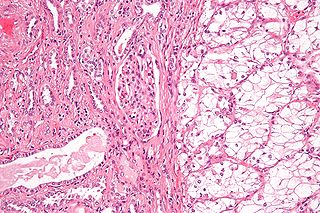
Renal cell carcinoma (RCC) is a kidney cancer that originates in the lining of the proximal convoluted tubule, a part of the very small tubes in the kidney that transport primary urine. RCC is the most common type of kidney cancer in adults, responsible for approximately 90–95% of cases. It is more common in men. It is most commonly diagnosed in the elderly.

<span class="mw-page-title-main">Kidney cancer</span> Medical condition

Kidney cancer, also known as renal cancer, is a group of cancers that starts in the kidney. Symptoms may include blood in the urine, a lump in the abdomen, or back pain. Fever, weight loss, and tiredness may also occur. Complications can include spread to the lungs or brain.

<span class="mw-page-title-main">Interventional radiology</span> Medical subspecialty

Interventional radiology (IR) is a medical specialty that performs various minimally-invasive procedures using medical imaging guidance, such as x-ray fluoroscopy, computed tomography, magnetic resonance imaging, or ultrasound. IR performs both diagnostic and therapeutic procedures through very small incisions or body orifices. Diagnostic IR procedures are those intended to help make a diagnosis or guide further medical treatment, and include image-guided biopsy of a tumor or injection of an imaging contrast agent into a hollow structure, such as a blood vessel or a duct. By contrast, therapeutic IR procedures provide direct treatment—they include catheter-based medicine delivery, medical device placement, and angioplasty of narrowed structures.

In medical or research imaging, an incidental imaging finding is an unanticipated finding which is not related to the original diagnostic inquiry. As with other types of incidental medical findings, they may represent a diagnostic, ethical, and philosophical dilemma because their significance is unclear. While some coincidental findings may lead to beneficial diagnoses, others may lead to overdiagnosis that results in unnecessary testing and treatment, sometimes called the "cascade effect".

<span class="mw-page-title-main">Adrenocortical carcinoma</span> Medical condition

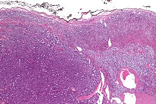
Adrenocortical carcinoma (ACC) is an aggressive cancer originating in the cortex of the adrenal gland.

Transcatheter arterial chemoembolization (TACE) is a minimally invasive procedure performed in interventional radiology to restrict a tumor's blood supply. Small embolic particles coated with chemotherapeutic drugs are injected selectively through a catheter into an artery directly supplying the tumor. These particles both block the blood supply and induce cytotoxicity, attacking the tumor in several ways.

<span class="mw-page-title-main">Portal vein thrombosis</span> Disease of the liver

Portal vein thrombosis (PVT) is a vascular disease of the liver that occurs when a blood clot occurs in the hepatic portal vein, which can lead to increased pressure in the portal vein system and reduced blood supply to the liver. The mortality rate is approximately 1 in 10.

<span class="mw-page-title-main">Focal nodular hyperplasia</span> Medical condition

Focal nodular hyperplasia is a benign tumor of the liver, which is the second most prevalent tumor of the liver after hepatic hemangioma. It is usually asymptomatic, rarely grows or bleeds, and has no malignant potential. This tumour was once often resected because it was difficult to distinguish from hepatic adenoma, but with modern multiphase imaging it is usually now diagnosed by strict imaging criteria and not resected.

<span class="mw-page-title-main">Neuroendocrine tumor</span> Medical condition

Neuroendocrine tumors (NETs) are neoplasms that arise from cells of the endocrine (hormonal) and nervous systems. They most commonly occur in the intestine, where they are often called carcinoid tumors, but they are also found in the pancreas, lung, and the rest of the body.

<span class="mw-page-title-main">Abdominal ultrasonography</span> Type of medical scan

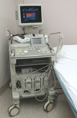
Abdominal ultrasonography is a form of medical ultrasonography to visualise abdominal anatomical structures. It uses transmission and reflection of ultrasound waves to visualise internal organs through the abdominal wall. For this reason, the procedure is also called a transabdominal ultrasound, in contrast to endoscopic ultrasound, the latter combining ultrasound with endoscopy through visualize internal structures from within hollow organs.

<span class="mw-page-title-main">Axitinib</span> Chemical compound

Axitinib, sold under the brand name Inlyta, is a small molecule tyrosine kinase inhibitor developed by Pfizer. It has been shown to significantly inhibit growth of breast cancer in animal (xenograft) models and has shown partial responses in clinical trials with renal cell carcinoma (RCC) and several other tumour types.

Urologic diseases or conditions include urinary tract infections, kidney stones, bladder control problems, and prostate problems, among others. Some urologic conditions do not affect a person for that long and some are lifetime conditions. Kidney diseases are normally investigated and treated by nephrologists, while the specialty of urology deals with problems in the other organs. Gynecologists may deal with problems of incontinence in women.

<span class="mw-page-title-main">Kidney tumour</span> Medical condition

Kidney tumours are tumours, or growths, on or in the kidney. These growths can be benign or malignant.

<span class="mw-page-title-main">Renal cyst</span> Medical condition

A renal cyst is a fluid collection in or on the kidney. There are several types based on the Bosniak classification. The majority are benign, simple cysts that can be monitored and not intervened upon. However, some are cancerous or are suspicious for cancer and are commonly removed in a surgical procedure called nephrectomy.

<span class="mw-page-title-main">Contrast CT</span> Medical imaging technique

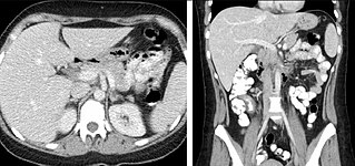
Contrast CT, or contrast enhanced computed tomography (CECT), is X-ray computed tomography (CT) using radiocontrast. Radiocontrasts for X-ray CT are generally iodine-based types. This is useful to highlight structures such as blood vessels that otherwise would be difficult to delineate from their surroundings. Using contrast material can also help to obtain functional information about tissues. Often, images are taken both with and without radiocontrast. CT images are called precontrast or native-phase images before any radiocontrast has been administered, and postcontrast after radiocontrast administration.

<span class="mw-page-title-main">Renal ultrasonography</span> Examination of one or both kidneys using medical ultrasound

Renal ultrasonography is the examination of one or both kidneys using medical ultrasound.

Ultrasonography of liver tumors involves two stages: detection and characterization.

Transarterial bland embolization is a catheter-based tumor treatment of the liver. In this procedure, embolizing agents can be delivered through the tumor's feeding artery in order to completely occlude the tumor's blood supply. The anti-tumor effects are solely based on tumor ischemia and infarction of tumor tissue, as no chemotherapeutic agents are administered. The rationale for the use of bland embolization for hepatocellular carcinoma (HCC) and/or other hyper-vascular tumors is based on the fact that a normal liver receives a dual blood supply from the hepatic artery (25%) and the portal vein (75%). As the tumor grows, it becomes increasingly dependent on the hepatic artery for blood supply. Once a tumor nodule reaches a diameter of 2 cm or more, most of the blood supply is derived from the hepatic artery. Therefore, bland embolization and transarterial chemoembolization (TACE) consist of the selective angiographic occlusion of the tumor arterial blood supply with a variety of embolizing agents, with or without the precedence of local chemotherapy infusion. The occlusion by embolic particles results in tumor hypoxia and necrosis, without affecting the normal hepatic parenchyma.

A computed tomography urography is a computed tomography scan that examines the urinary tract after contrast dye is injected into a vein.

References

  1. Lee HK, Park SJ, Yi BH. Multidetector CT reveals diverse variety of abdominal hernias. Archived 2010-06-18 at the Wayback Machine Diagnostic Imaging. 2010;32(5):27-31.
  2. Note that the ACR appropriateness criteria can be found on the ACR website (http://www.acr.org/ac)
  3. Pickhardt, Perry J. (July 2020). "Positive Oral Contrast Material for Abdominal CT: Current Clinical Indications and Areas of Controversy". American Journal of Roentgenology. 215 (1): 69–78. doi:10.2214/AJR.19.21989. ISSN   0361-803X. PMID   31913069. S2CID   210087445.
  4. Chambers, S.E.; Best, J.J.K. (January 1984). "A comparison of dilute barium and dilute water-soluble contrast in opacification of the bowel for abdominal computed tomography". Clinical Radiology. 35 (6): 463–464. doi:10.1016/S0009-9260(84)80054-9. PMID   6499385.