High-resolution computed tomography

Last updated
High-resolution computed tomography
Pulmon fibrosis.PNG
HRCT of lung showing extensive fibrosis possibly from usual interstitial pneumonitis. There is also a large emphysematous bulla.
ICD-9-CM 87.41

High-resolution computed tomography (HRCT) is a type of computed tomography (CT) with specific techniques to enhance image resolution. It is used in the diagnosis of various health problems, though most commonly for lung disease, by assessing the lung parenchyma. [1] On the other hand, HRCT of the temporal bone is used to diagnose various middle ear diseases such as otitis media, cholesteatoma, and evaluations after ear operations. [2]

Contents

Technique

Low-dose high-resolution (1.25 mm) chest CT

HRCT is performed using a conventional CT scanner. However, imaging parameters are chosen so as to maximize spatial resolution: [1] a narrow slice width is used (usually 1–2 mm), a high spatial resolution image reconstruction algorithm is used, field of view is minimized, so as to minimize the size of each pixel, and other scan factors (e.g. focal spot) may be optimized for resolution at the expense of scan speed.

Depending on the suspected diagnosis, the scan may be performed in both inspiration and expiration. In inspiration images are taken in the prone position. [3] In expiratory HRCT the scan is taken in the supine position (face up). [4]

As HRCT's aim is to assess a generalized lung disease, the test is conventionally performed by taking thin sections which are 10–40 mm apart from each other. The result is a few images that should be representative of the lungs in general, but that cover only approximately one tenth of the lungs. [5]

Intravenous contrast agents are not used for HRCT [6] as the lung inherently has very high contrast (soft tissue against air), and the technique itself is unsuitable for assessment of the soft tissues and blood vessels, which are the major targets of contrast agents.

Impact of modern CT technology

HRCT of a normal thorax, taken in the axial, coronal and sagittal planes, respectively.
Click here to scroll through the image stacks. High-resolution computed tomographs of a normal thorax (thumbnail).jpg
HRCT of a normal thorax, taken in the axial, coronal and sagittal planes, respectively.

The technique of HRCT was developed with relatively slow CT scanners, which did not make use of multi-detector (MDCT) technology. The parameters of scan duration, z-axis resolution and coverage were interdependent. To cover the chest in a reasonable time period with a conventional chest CT scan required thick sections (e.g., 10mm thick) to ensure contiguous coverage. As performing contiguous thin sections required unacceptably prolonged scan time, HRCT examination was performed with widely spaced sections. Because of the different scan parameters for conventional and HRCT examinations, if a patient required both, they had to be performed sequentially.

Modern MDCT scanners are able to overcome this interdependence, and are capable of imaging at full resolution yet retain very fast coverage - images can then be reconstructed retrospectively from the volumetric raw data. Because of this, it may be possible to reconstruct inspiratory HRCT-like images from the data taken from a 'normal' chest CT scan. [7]

Alternatively, the scanner could be configured to perform contiguous 1mm sections for a HRCT examination - this provides greater diagnostic information as it examines the entire lung, and permits the use of multi-planar reconstruction techniques. However, it brings the expense of irradiating the entire chest (instead of approximately 10%) when performed using widely spaced sections. [8]

Lung disease

HRCT is used for diagnosis and assessment of interstitial lung disease, such as pulmonary fibrosis, and other generalized lung diseases such as emphysema and bronchiectasis.

Applications

Airways diseases, such as emphysema or bronchiolitis obliterans, cause air trapping on expiration, even though they may cause only minor changes to lung structure in their early stages. To enhance sensitivity for these conditions, the scan may be performed in both inspiration and expiration.

HRCT may be diagnostic for conditions such as emphysema or bronchiectasis. While HRCT may be able to identify pulmonary fibrosis, it may not always be able to further categorize the fibrosis to a specific pathological type (e.g., non-specific interstitial pneumonitis or desquamative interstitial pneumonitis). The major exception is UIP, which has very characteristic features, and may be confidently diagnosed on HRCT alone. [9]

Where HRCT is unable to reach a definitive diagnosis, it helps locate an abnormality, and so helps planning a biopsy, which may provide the final diagnosis.

Other miscellaneous conditions where HRCT is useful include lymphangitis carcinomatosa, fungal, or other atypical, infections, chronic pulmonary vascular disease, lymphangioleiomyomatosis, and sarcoidosis.

Organ transplant patients, particularly lung, or heart-lung transplant recipients, are at relatively high risk of developing pulmonary complications of the long-term drug and immunosuppressive treatment. The major pulmonary complication is bronchiolitis obliterans, which may be a sign of lung graft rejection.

HRCT has better sensitivity for bronchiolitis obliterans than conventional radiography. [10] [11] [12] Some transplant centers may arrange annual HRCT to screen for this.

Diagnostic imaging, including HRCT, is one of the main diagnostic tools for COVID-19. [13] There is some debate about the usefulness of CT compared to other methods and imaging modalities for diagnosis. [14] Under HRCT scan, infected individuals generally showed a multifocal or unifocal involvement of ground-glass opacity (GGO). [15]

Nodularity

The presence of lung nodules on high resolution CT is a keystone in understanding the appropriate differential. Typically, the distribution of nodules is divided into perilymphatic, centrilobular and random categories. Furthermore, nodules can be ill-defined, implying they are in the alveoli, or well defined, suggesting an interstitial position. Distribution and appearance allow understanding of the disease process relative to the secondary lobule of the lung, the smallest anatomic unit with surrounding connective tissue, usually 1–2 cm across. [16]

Perilymphatic nodularity deposits at the periphery of the secondary lobule and tends to respect pleural surfaces and fissures. Sarcoidosis, lymphangitic spread of carcinoma, silicosis, coal worker's pneumoconiosis, and more rare diagnoses such as lymphoid interstitial pneumonitis and amyloidosis are included in the differential. Centrilobular nodularity deposits at the center of the secondary lobule, but spares pleural surfaces. Differential includes endobronchial tuberculosis, bronchopneumonia, endobronchial spread of tumor, and again silicosis or coal workers’ pneumoconiosis. For randomly distributed nodules, the differential includes miliary tuberculosis, fungal pneumonia, hematogenous metastasis and diffuse sarcoidosis. [16]

Prone versus supine position

Because the bases of the lungs lie posteriorly in the chest, a mild degree of collapse under the lungs' own weight can occur when the patient lies on their back. As the very base of the lungs may be the first region affected in several lung diseases, most notably asbestosis or usual interstitial pneumonia (UIP), the patient may be asked to lie prone to improve sensitivity to early changes of these conditions.

The lung bases are often inconsistent in appearance in patients due to the potential for atelectasis causing positional ground glass or consolidative opacities. When the patient is positioned prone, or on their belly, the lung bases can expand further and help distinguish atelectasis from early fibrosis. In patients with normal chest radiographs, prone scans have been found useful in 17% of cases, particularly in excluding posterior lung abnormalities. In patients with abnormal findings on chest radiographs, prone scans are only useful in 4% of cases. The scans may be more useful in patients with basilar predominant disease processes, such as asbestosis and idiopathic pulmonary fibrosis. [16]

Temporal bone diseases

HRCT temporal bone is used to determine the anatomy of chronic otitis media (middle ear is located inside the temporal bone), congenital abnormalities, and loss of surgical landmarks due to previous operation. Thus, HRCT is useful in surgical planning and management of temporal bone diseases. [2]

See also

Related Research Articles

<span class="mw-page-title-main">Interstitial lung disease</span> Group of diseases

Interstitial lung disease (ILD), or diffuse parenchymal lung disease (DPLD), is a group of respiratory diseases affecting the interstitium (the tissue and space around the alveoli of the lungs. It concerns alveolar epithelium, pulmonary capillary endothelium, basement membrane, and perivascular and perilymphatic tissues. It may occur when an injury to the lungs triggers an abnormal healing response. Ordinarily, the body generates just the right amount of tissue to repair damage, but in interstitial lung disease, the repair process is disrupted, and the tissue around the air sacs becomes scarred and thickened. This makes it more difficult for oxygen to pass into the bloodstream. The disease presents itself with the following symptoms: shortness of breath, nonproductive coughing, fatigue, and weight loss, which tend to develop slowly, over several months. The average rate of survival for someone with this disease is between three and five years. The term ILD is used to distinguish these diseases from obstructive airways diseases.

<span class="mw-page-title-main">Chest radiograph</span> Projection X-ray of the chest

A chest radiograph, called a chest X-ray (CXR), or chest film, is a projection radiograph of the chest used to diagnose conditions affecting the chest, its contents, and nearby structures. Chest radiographs are the most common film taken in medicine.

<span class="mw-page-title-main">Hypersensitivity pneumonitis</span> Medical condition

Hypersensitivity pneumonitis (HP) or extrinsic allergic alveolitis (EAA) is a syndrome caused by the repetitive inhalation of antigens from the environment in susceptible or sensitized people. Common antigens include molds, bacteria, bird droppings, bird feathers, agricultural dusts, bioaerosols and chemicals from paints or plastics. People affected by this type of lung inflammation (pneumonitis) are commonly exposed to the antigens by their occupations, hobbies, the environment and animals. The inhaled antigens produce a hypersensitivity immune reaction causing inflammation of the airspaces (alveoli) and small airways (bronchioles) within the lung. Hypersensitivity pneumonitis may eventually lead to interstitial lung disease.

<span class="mw-page-title-main">Pneumonitis</span> General inflammation of lung tissue

Pneumonitis describes general inflammation of lung tissue. Possible causative agents include radiation therapy of the chest, exposure to medications used during chemo-therapy, the inhalation of debris, aspiration, herbicides or fluorocarbons and some systemic diseases. If unresolved, continued inflammation can result in irreparable damage such as pulmonary fibrosis.

<span class="mw-page-title-main">Cryptogenic organizing pneumonia</span> Medical condition

Cryptogenic organizing pneumonia (COP), formerly known as bronchiolitis obliterans organizing pneumonia (BOOP), is an inflammation of the bronchioles (bronchiolitis) and surrounding tissue in the lungs. It is a form of idiopathic interstitial pneumonia.

<span class="mw-page-title-main">Bronchiolitis obliterans</span> Medical condition

Bronchiolitis obliterans (BO), also known as obliterative bronchiolitis, constrictive bronchiolitis and popcorn lung, is a disease that results in obstruction of the smallest airways of the lungs (bronchioles) due to inflammation. Symptoms include a dry cough, shortness of breath, wheezing and feeling tired. These symptoms generally get worse over weeks to months. It is not related to cryptogenic organizing pneumonia, previously known as bronchiolitis obliterans organizing pneumonia.

Air trapping, also called gas trapping, is an abnormal retention of air in the lungs where it is difficult to exhale completely. It is observed in obstructive lung diseases such as asthma, bronchiolitis obliterans syndrome and chronic obstructive pulmonary diseases such as emphysema and chronic bronchitis.

<span class="mw-page-title-main">Idiopathic pulmonary fibrosis</span> Medical condition

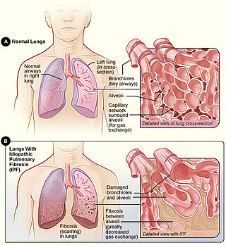
Idiopathic pulmonary fibrosis (IPF), or (formerly) fibrosing alveolitis, is a rare, progressive illness of the respiratory system, characterized by the thickening and stiffening of lung tissue, associated with the formation of scar tissue. It is a type of chronic scarring lung disease characterized by a progressive and irreversible decline in lung function. The tissue in the lungs becomes thick and stiff, which affects the tissue that surrounds the air sacs in the lungs. Symptoms typically include gradual onset of shortness of breath and a dry cough. Other changes may include feeling tired, and abnormally large and dome shaped finger and toenails. Complications may include pulmonary hypertension, heart failure, pneumonia or pulmonary embolism.

<span class="mw-page-title-main">CT pulmonary angiogram</span>

A CT pulmonary angiogram (CTPA) is a medical diagnostic test that employs computed tomography (CT) angiography to obtain an image of the pulmonary arteries. Its main use is to diagnose pulmonary embolism (PE). It is a preferred choice of imaging in the diagnosis of PE due to its minimally invasive nature for the patient, whose only requirement for the scan is an intravenous line.

<span class="mw-page-title-main">Usual interstitial pneumonia</span> Medical condition

Usual interstitial pneumonia (UIP) is a form of lung disease characterized by progressive scarring of both lungs. The scarring (fibrosis) involves the pulmonary interstitium. UIP is thus classified as a form of interstitial lung disease.

<span class="mw-page-title-main">Idiopathic interstitial pneumonia</span> Medical condition

Idiopathic interstitial pneumonia (IIP), or noninfectious pneumonia are a class of diffuse lung diseases. These diseases typically affect the pulmonary interstitium, although some also have a component affecting the airways. There are seven recognized distinct subtypes of IIP.

Pulmonary toxicity is the medical name for side effects on the lungs.

<span class="mw-page-title-main">Lung nodule</span> Medical condition

A lung nodule or pulmonary nodule is a relatively small focal density in the lung. A solitary pulmonary nodule (SPN) or coin lesion, is a mass in the lung smaller than three centimeters in diameter. A pulmonary micronodule has a diameter of less than three millimetres. There may also be multiple nodules.

<span class="mw-page-title-main">Respiratory bronchiolitis</span> Medical condition

Respiratory bronchiolitis is a lung disease associated with tobacco smoking. In pathology, it is defined by the presence of "smoker's macrophages". When manifesting significant clinical symptoms it is referred to as respiratory bronchiolitis interstitial lung disease (RB-ILD).

Rheumatoid lung disease is a disease of the lung associated with RA, rheumatoid arthritis. Rheumatoid lung disease is characterized by pleural effusion, pulmonary fibrosis, lung nodules and pulmonary hypertension. Common symptoms associated with the disease include shortness of breath, cough, chest pain and fever. It is estimated that about one quarter of people with rheumatoid arthritis develop this disease, which are more likely to develop among elderly men with a history of smoking.

<span class="mw-page-title-main">Ground-glass opacity</span> Radiologic sign on radiographs and computed tomography scans

Ground-glass opacity (GGO) is a finding seen on chest x-ray (radiograph) or computed tomography (CT) imaging of the lungs. It is typically defined as an area of hazy opacification (x-ray) or increased attenuation (CT) due to air displacement by fluid, airway collapse, fibrosis, or a neoplastic process. When a substance other than air fills an area of the lung it increases that area's density. On both x-ray and CT, this appears more grey or hazy as opposed to the normally dark-appearing lungs. Although it can sometimes be seen in normal lungs, common pathologic causes include infections, interstitial lung disease, and pulmonary edema.

<span class="mw-page-title-main">Flock worker's lung</span> Occupational disease

Flock worker's lung is an occupational lung disease caused by exposure to flock, small fibers that are glued to a backing in order to create a specific texture. People who work in flocking are at risk of inhaling small pieces of the flock fibers, which causes interstitial lung disease. The disease was initially described in 1998, when a group of workers at a flocking plant developed interstitial lung disease of unknown cause.

Diffuse idiopathic pulmonary neuroendocrine cell hyperplasia (DIPNECH) is a diffuse parenchymal lung disease which often presents with symptoms of cough and shortness of breath. The pathological definition published by the World Health Organization is “a generalized proliferation of scattered single cells, small nodules, or linear proliferations of pulmonary neuroendocrine (PNE) cells that may be confined to the bronchial and bronchiolar epithelium.” The true prevalence of this disease is not known. To date, just under 200 cases have been reported in the literature. However, with an increase in recognition of this disease by radiologists and pulmonologists, the number of cases has been increasing. DIPNECH predominantly affects middle-aged women with slowly progressive lung obstruction. DIPNECH is usually discovered in one of two ways: 1) as an unexpected finding following a lung surgery; or 2) by evaluation of a patient in a pulmonary clinic with longstanding, unexplained symptoms.

<span class="mw-page-title-main">Emphysema</span> Medical condition

Emphysema, in general is any air-filled enlargement in the body's tissues. Specifically emphysema refers to the enlargement of air spaces (alveoli) in the lungs, and is also known as pulmonary emphysema.

<span class="mw-page-title-main">Smoking-related interstitial fibrosis (SRIF)</span> Abnormal amount of collagen in the lung (fibrosis) caused by cigarette smoking

Smoking-related interstitial fibrosis (SRIF) is an abnormality in the lungs characterized by excessive collagen deposition within the walls of the air sacs. This abnormality can be seen with a microscope and diagnosed by pathologists. It is caused by cigarette smoking.

References

  1. 1 2 "ACR–STR Practice Parameter for the Performance of High-Resolution Computed Tomography (HRCT) of the Lungs in Adults" (PDF). American College of Radiology. 2015. Retrieved 27 June 2017.
  2. 1 2 Thukral CL, Singh A, Singh S, Sood AS, Singh K (September 2015). "Role of High Resolution Computed Tomography in Evaluation of Pathologies of Temporal Bone". Journal of Clinical and Diagnostic Research. 9 (9): TC07–TC10. doi:10.7860/JCDR/2015/12268.6508. PMC   4606307 . PMID   26500978.
  3. Di Muzzy B. "HRCT chest: prone images". Radiology Reference Article. Radiopaedia.org. Retrieved 7 November 2021.
  4. Mudgal P. "HRCT chest: expiration (technique)". Radiology Reference Article. Radiopaedia.org. Retrieved 7 November 2021.
  5. Worthy S (March 1995). "High resolution computed tomography of the lungs". BMJ. 310 (6980): 615–6. doi:10.1136/bmj.310.6980.616. PMC   2549004 . PMID   7703739.
  6. Peroés-Golubiéciâc T, Sharma OP (2007). Clinical Atlas of Interstitial Lung Disease. Springer. p. 19. ISBN   978-1-84628-326-0. OCLC   780404335.
  7. Schoepf US (2005). Multidetector-Row CT of the Thorax. Springer. p. 84. ISBN   978-3-540-30006-9.
  8. Schwarz MI, King TE (2011). Interstitial Lung Disease. Shelton, CT: PMPH. p. 106. ISBN   978-1-60795-024-0.
  9. Gotway MB, Freemer MM, King TE (June 2007). "Challenges in pulmonary fibrosis. 1: Use of high resolution CT scanning of the lung for the evaluation of patients with idiopathic interstitial pneumonias". Thorax. 62 (6): 546–53. doi:10.1136/thx.2004.040022. PMC   2117220 . PMID   17536033.
  10. Zare Mehrjardi M, Kahkouee S, Pourabdollah M (March 2017). "Radio-pathological correlation of organizing pneumonia (OP): a pictorial review". The British Journal of Radiology. 90 (1071): 20160723. doi:10.1259/bjr.20160723. PMC   5601538 . PMID   28106480.
  11. Chan A, Allen R (March 2004). "Bronchiolitis obliterans: an update". Current Opinion in Pulmonary Medicine. 10 (2): 133–41. doi:10.1097/00063198-200403000-00008. PMID   15021183. S2CID   6767627.
  12. Khan AN, Hanley S, Macdonald S, Chandramohan M, Ghanem SA, Irion KL (6 January 2017). "Imaging in Bronchiolitis Obliterans Organizing Pneumonia: Overview, Radiography, Computed Tomography". Medscape. WebMD. Retrieved 29 June 2017.
  13. "Clinical management of severe acute respiratory infection when novel coronavirus (2019-nCoV) infection is suspected" (PDF). World Health Organization. 28 January 2020.
  14. "RCR position on the role of CT in patients suspected with COVID-19 infection". The Royal College of Radiologists. 12 March 2020.
  15. Chung M, Bernheim A, Mei X, Zhang N, Huang M, Zeng X, et al. (April 2020). "CT Imaging Features of 2019 Novel Coronavirus (2019-nCoV)". Radiology. 295 (1): 202–207. doi: 10.1093/pcmedi/pbaa004 . PMC   7107051 .
  16. 1 2 3 Webb WR, Müller NL, Naidich DP (2014). High-resolution CT of the lung (5th ed.). ISBN   978-1-4511-7601-8.