Interstitial lung disease

Last updated
Interstitial lung disease
Other namesDiffuse parenchymal lung disease (DPLD)
End-stage interstitial lung disease (honeycomb lung).jpg
End-stage pulmonary fibrosis of unknown origin, taken from an autopsy
Specialty Pulmonology
Complications Respiratory failure
Usual onsetMonths – years, depending on cause/type
DurationMonths – years, depending on cause/type
Frequency1.9 million (2015) [1]
Deaths122,000 (2015) [2]

Interstitial lung disease (ILD), or diffuse parenchymal lung disease (DPLD), [3] is a group of respiratory diseases affecting the interstitium (the tissue) and space around the alveoli (air sacs) of the lungs. [4] It concerns alveolar epithelium, pulmonary capillary endothelium, basement membrane, and perivascular and perilymphatic tissues. It may occur when an injury to the lungs triggers an abnormal healing response. Ordinarily, the body generates just the right amount of tissue to repair damage, but in interstitial lung disease, the repair process is disrupted, and the tissue around the air sacs (alveoli) becomes scarred and thickened. This makes it more difficult for oxygen to pass into the bloodstream. The disease presents itself with the following symptoms: shortness of breath, nonproductive coughing, fatigue, and weight loss, which tend to develop slowly, over several months. The average rate of survival for someone with this disease is between three and five years. [5] The term ILD is used to distinguish these diseases from obstructive airways diseases.

Contents

There are specific types in children, known as children's interstitial lung diseases. The acronym ChILD is sometimes used for this group of diseases. [6]

Thirty to 40% of those with interstitial lung disease eventually develop pulmonary fibrosis which has a median survival of 2.5-3.5 years. [7] Idiopathic pulmonary fibrosis is interstitial lung disease for which no obvious cause can be identified (idiopathic) and is associated with typical findings both radiographic (basal and pleural-based fibrosis with honeycombing) and pathologic (temporally and spatially heterogeneous fibrosis, histopathologic honeycombing, and fibroblastic foci).

In 2015, interstitial lung disease, together with pulmonary sarcoidosis, affected 1.9 million people. [1] They resulted in 122,000 deaths. [2]

Causes

Interstitial lung disease affects gas flow in the alveoli 2 SVH Lung Health Interstitial Lung Disease final 1080.jpg
Interstitial lung disease affects gas flow in the alveoli
The alveoli Alveolus diagram.svg
The alveoli
Micrograph of usual interstitial pneumonia (UIP). UIP is the most common pattern of idiopathic interstitial pneumonia (a type of interstitial lung disease) and usually represents idiopathic pulmonary fibrosis. H&E stain. Autopsy specimen. Usual interstitial pneumonia (1).JPG
Micrograph of usual interstitial pneumonia (UIP). UIP is the most common pattern of idiopathic interstitial pneumonia (a type of interstitial lung disease) and usually represents idiopathic pulmonary fibrosis. H&E stain. Autopsy specimen.

ILD may be classified as to whether its cause is not known (idiopathic) or known (secondary). [8]

Idiopathic

Idiopathic interstitial pneumonia is the term given to ILDs with an unknown cause. They represent the majority of cases of interstitial lung diseases (up to two-thirds of cases). [9] They were subclassified by the American Thoracic Society in 2002 into 7 subgroups: [10]

Secondary

Secondary ILDs are those diseases with a known etiology, including:[ citation needed ]

Connective tissue and autoimmune diseases

Connective tissue related disease represents approximately 25% of all cases of ILD. [7]

Inhaled substances (pneumoconiosis)

Drug-induced

Infection

Malignancy

Childhood interstitial lung disease and ILD predominately in children

Diagnosis

Pneumocystis pneumonia can present with interstitial lung disease, as seen in the reticular markings on this AP chest x-ray. PCP CAP CXR.JPG
Pneumocystis pneumonia can present with interstitial lung disease, as seen in the reticular markings on this AP chest x-ray.
A chest X-ray demonstrating pulmonary fibrosis due to amiodarone IPF amiodarone.JPG
A chest X-ray demonstrating pulmonary fibrosis due to amiodarone

Diagnosis of ILD involves assessing the signs and symptoms as well as a detailed history investigating occupational exposures. ILD usually presents with dyspnea, worsening exercise intolerance and 30-50% of those with ILD have a chronic cough. On examination, velcro crackles, in which the crackles compare to the sound of velcro being unfastened, are common in ILD. [7] Pulmonary function tests usually show a restrictive defect with decreased diffusion capacity of carbon monoxide (DLCO) indicating reduced alveolar to blood capillary transport. [7] Pulmonary function testing is indicated for all people with ILD and the FVC loss and DLCO is prognostic, with an FVC loss of greater than 5% per year associated with a poor prognosis in fibrosis subtypes of ILD. [7]

A chest x-ray is 63% sensitive and 93% specific for ILD. [7] With advances in computed tomography, CT scans of the chest have supplanted lung biopsy as the preferred diagnostic test for ILD. A thoracic CT scan is 91% sensitive and 71% specific for ILD. [7] In higher income countries, less than 10% of people with ILD undergo a lung biopsy as part of the diagnostic evaluation. [13]

A lung biopsy may be required if the clinical history and imaging are not clearly suggestive of a specific diagnosis or malignancy cannot otherwise be ruled out. Surgical lung biopsy or via a video-assisted thoracoscopic surgery (VATS) biopsy is associated with a mortality rate up to 1-2%. A bronchoscopic transbronchial cryobiopsy, in which a camera is introduced into the airways followed by rapid freezing of an area of lung tissue prior to biopsy is associated a lower complication rate and a much lower mortality rate compared to VATS or surgical biopsy with near comparable diagnostic accuracy. [14] There are four types of histopathologic patterns seen in ILD: usual interstitial pneumonia, non-specific interstitial pneumonia, organizing pneumonia, and diffuse alveolar damage. [7] There is significant overlap of the histopathological and radiologic features of each ILD type making diagnosis challenging; even with lung biopsy, 15% of cases of ILD cannot be classified. [7]

Pulmonary function testing

Most patients with suspected ILD are likely to undergo complete pulmonary function testing. These tests are useful in diagnosis and determining severity of the disease. [15]

Although there is large diversity in interstitial lung disease, most follow a restrictive pattern. Restrictive defects are defined by decreased TLC (total lung capacity), RV (residual volume), FVC (forced vital capacity) and FEV1 (forced expiratory volume in one second). As both FVC and FEV1 are reduced, the FVC to FEV1 ratio remains normal or is increased. [15]

As disease progression increases and the lungs become stiffer lung volumes will continue to decrease; lower TLC, RV, FVC and FEV1 scores are associated with a more severe disease progression and poorer prognosis. [15]

X-ray and CT (computed tomography)

Chest radiography is usually the first test to detect interstitial lung diseases, but the chest radiograph can be normal in up to 10% of patients, especially early in the disease process. [16] [17]

High-resolution CT of the chest is the preferred modality and differs from routine CT of the chest. Conventional (regular) CT chest examines 7–10 mm slices obtained at 10 mm intervals; high resolution CT examines 1–1.5 mm slices at 10 mm intervals using a high-spatial-frequency reconstruction algorithm. The HRCT therefore provides approximately 10 times more resolution than the conventional CT chest, allowing the HRCT to elicit details that cannot otherwise be visualized. [16] [18]

Radiologic appearance alone, however, is not adequate and should be interpreted in the clinical context, keeping in mind the temporal profile of the disease process. [16]

Interstitial lung diseases can be classified according to radiologic patterns. [16]

Pattern of opacities

Consolidation
  • Acute:
    • Alveolar hemorrhage syndromes
    • Acute eosinophilic pneumonia
    • Acute interstitial pneumonia
    • Cryptogenic organizing pneumonia
  • Chronic:
Linear or reticular opacities
  • Acute:
    • Pulmonary edema
  • Chronic:
    • Idiopathic pulmonary fibrosis
    • Connective tissue-associated interstitial lung diseases
    • Asbestosis
    • Sarcoidosis
    • Hypersensitivity pneumonitis
    • Drug-induced lung disease
Small nodules
  • Acute:
    • Hypersensitivity pneumonitis
  • Chronic:
    • Hypersensitivity pneumonitis
    • Sarcoidosis
    • Silicosis
    • Coal workers pneumoconiosis
    • Respiratory bronchiolitis
    • Alveolar microlithiasis
Cystic airspaces
  • Chronic:
    • Pulmonary Langerhans cell histiocytosis
    • Pulmonary lymphangioleiomyomatosis
    • Honeycomb lung caused by idiopathic pulmonary fibrosis (IPF) or other diseases
Ground glass opacities
  • Acute:
    • Alveolar hemorrhage syndromes
    • Pulmonary edema
    • Hypersensitivity pneumonitis
    • Acute inhalational exposures
    • Drug-induced lung diseases
    • Acute interstitial pneumonia
  • Chronic:
    • Nonspecific interstitial pneumonia
    • Respiratory bronchiolitis-associated interstitial lung disease
    • Desquamative interstitial pneumonia
    • Drug-induced lung diseases
    • Pulmonary alveolar proteinosis
Thickened alveolar septa
  • Acute:
    • Pulmonary edema
  • Chronic:
    • Lymphangitic carcinomatosis
    • Pulmonary alveolar proteinosis
    • Sarcoidosis
    • Pulmonary veno-occlusive disease [16]

Distribution

Upper lung predominance
  • Pulmonary Langerhans cell histiocytosis
  • Silicosis
  • Coal workers pneumoconiosis
  • Carmustine-related pulmonary fibrosis
  • Respiratory broncholitis associated with interstitial lung disease
Lower lung predominance
  • Idiopathic pulmonary fibrosis
  • Pulmonary fibrosis associated with connective tissue diseases (ILD-CTD)
  • Asbestosis
  • Chronic aspiration
Central predominance (perihilar)
  • Sarcoidosis
  • Berylliosis
Peripheral predominance
  • Idiopathic pulmonary fibrosis
  • Chronic eosinophilic pneumonia
  • Cryptogenic organizing pneumonia [16]

Associated findings

Pleural effusion or thickening
  • Pulmonary edema
  • Connective tissue diseases
  • Asbestosis
  • Lymphangitic carcinomatosis
  • Lymphoma
  • Lymphangioleiomyomatosis
  • Drug-induced lung diseases
Lymphadenopathy
  • Sarcoidosis
  • Silicosis
  • Berylliosis
  • Lymphangitic carcinomatosis
  • Lymphoma
  • Lymphocytic interstitial pneumonia [16]

Genetic testing

For some types of paediatric ILDs and few forms adult ILDs, genetic causes have been identified. These may be identified by blood tests. For a limited number of cases, this is a definite advantage, as a precise molecular diagnosis can be done; frequently then there is no need for a lung biopsy. Testing is available for

  • Surfactant protein B deficiency (mutations in SFTPB )
  • Surfactant protein C deficiency (mutations in SFTPC )
  • ABCA3 deficiency (mutations in ABCA3 )
  • Brain–lung–thyroid syndrome (Mutations in TTF1 )
  • Congenital pulmonary alveolar proteinosis (mutations in CSFR2A and/or CSFR2B)

Diffuse developmental disorder

  • Alveolar capillary dysplasia (mutations in FoxF1 )

Idiopathic pulmonary fibrosis

  • Mutations in telomerase reverse transcriptase ( TERT )
  • Mutations in telomerase RNA component (TERC)
  • Mutations in the regulator of telomere elongation helicase 1 (RTEL1)
  • Mutations in poly(A)-specific ribonuclease (PARN)

Treatment

ILD is not a single disease but encompasses many different pathological processes, hence treatment is different for each disease. If a specific occupational exposure cause is found, the person should avoid that environment. If a drug cause is suspected, that drug should be discontinued.[ citation needed ]

Oxygen therapy

Oxygen therapy at home is recommended in those with significantly low oxygen levels. [19] Oxygen therapy in ILD is associated with improvements in quality of life but reductions in mortality are uncertain. [7]

Pulmonary rehabilitation

Pulmonary rehabilitation appears to be useful with the benefits being sustainable longer term with improvements in exercise capacity (as measured by a six minute walking test), dyspnea, and quality of life. [20] [7]

Lung transplantation

Lung transplantation is an option if the ILD progresses despite therapy in appropriately selected patients with no other contraindications. [21] [22] Life expectancy after lung transplant is 5.2 years in those with idiopathic interstitial pneumonias (including idiopathic pulmonary fibrosis) and 6.7 years in those with other types of ILD. [7]

Medications

The antifibrotics pirfenidone and nintedanib have been shown to slow the decline in lung function (as measured by forced vital capacity [FVC]) in those with ILD compared to placebo. [7] Pirfenidone was associated with a 45% less decline in FVC at 52 weeks compared to placebo in a trial involving people with idiopathic pulmonary fibrosis, and was associated with a slower FVC decline in those with progressive pulmonary fibrosis. [23] Nintedanib was also associated with a slower FVC decline and increased mean survival in people with ILD. [7]

The immunomodulator tocilizumab has a benefit in scleroderma associated ILD by helping to preserve lung function (as measured by FVC) at 48 weeks. [7] The immunomodulators cyclophosphamide, mycophenolate mofetil and rituximab all showed improved lung function (as measured by % predicted FVC) compared to placebo in systemic sclerosis or scleroderma associated ILD. [7]

The inhaled vasodilator treprostinil (a synthetic prostacyclin which acts as a prostaglandin I2 analogue) is indicated in the treatment of pulmonary hypertension secondary to interstitial lung disease and is associated with improved exercise capacity as measured by a 6-minute walk test. [24]

Supportive care

Those with ILD should stop smoking cigarettes if they smoke. Vaccinations against pneumococcus, Covid-19, RSV and influenza are indicated in all those with ILD. [7] Short acting opiates are known to improve breathlessness symptoms in those with end stage lung disease. The opiate agonist-antagonist nalbuphine and morphine are also known to improve coughing in those with ILD and other end stage lung diseases. [7]

Prognosis

The median survival in idiopathic pulmonary fibrosis is 3-3.5 years. In those who receive a lung transplant, the medial survival in idiopathic pulmonary fibrosis is 5.2 years, as compared to 6.7 years in those with other types of ILD. [7] ILD is associated with a 3-fold increased risk of lung cancer. [7]

Related Research Articles

<span class="mw-page-title-main">Berylliosis</span> Medical condition

Berylliosis, or chronic beryllium disease (CBD), is a chronic allergic-type lung response and chronic lung disease caused by exposure to beryllium and its compounds, a form of beryllium poisoning. It is distinct from acute beryllium poisoning, which became rare following occupational exposure limits established around 1950. Berylliosis is an occupational lung disease.

<span class="mw-page-title-main">Asbestosis</span> Pneumoconiosis caused by inhalation and retention of asbestos fibers

Asbestosis is long-term inflammation and scarring of the lungs due to asbestos fibers. Symptoms may include shortness of breath, cough, wheezing, and chest tightness. Complications may include lung cancer, mesothelioma, and pulmonary heart disease.

<span class="mw-page-title-main">Chest radiograph</span> Projection X-ray of the chest

A chest radiograph, chest X-ray (CXR), or chest film is a projection radiograph of the chest used to diagnose conditions affecting the chest, its contents, and nearby structures. Chest radiographs are the most common film taken in medicine.

<span class="mw-page-title-main">Pulmonary fibrosis</span> Disease that causes scarring of the lungs

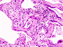
Pulmonary fibrosis is a condition in which the lungs become scarred over time. Symptoms include shortness of breath, a dry cough, feeling tired, weight loss, and nail clubbing. Complications may include pulmonary hypertension, respiratory failure, pneumothorax, and lung cancer.

<span class="mw-page-title-main">Hypersensitivity pneumonitis</span> Medical condition

Hypersensitivity pneumonitis (HP) or extrinsic allergic alveolitis (EAA) is a syndrome caused by the repetitive inhalation of antigens from the environment in susceptible or sensitized people. Common antigens include molds, bacteria, bird droppings, bird feathers, agricultural dusts, bioaerosols and chemicals from paints or plastics. People affected by this type of lung inflammation (pneumonitis) are commonly exposed to the antigens by their occupations, hobbies, the environment and animals. The inhaled antigens produce a hypersensitivity immune reaction causing inflammation of the airspaces (alveoli) and small airways (bronchioles) within the lung. Hypersensitivity pneumonitis may eventually lead to interstitial lung disease.

Neuroendocrine hyperplasia is rare and poorly understood lung condition which is characterized by an abnormal growth of pulmonary neuroendocrine cells in the lungs. It is a non-progressive disease of the interstitial tissues of the lungs. Prior to the findings of the hyperplasia of neuroendocrine cells it was known as tachypnea of infancy, as most children outgrow the need for oxygen supplementation within two to seven years. It is characterized by tachypnea, hypoxemia, and retractions. It is typically diagnosed in infants and children younger than one year of age. There is no currently recognized treatment, infants and children are given oxygen supplementation until they outgrow the need; since neuroendocrine cells do not multiply or get larger in size while the lungs continue to grow. This allows the lung disease to have less effect on lung function with age, although they will always have the same amount of neuroendocrine cells as they were born with.

<span class="mw-page-title-main">Pneumonitis</span> General inflammation of lung tissue

Pneumonitis describes general inflammation of lung tissue. Possible causative agents include radiation therapy of the chest, exposure to medications used during chemo-therapy, the inhalation of debris, aspiration, herbicides or fluorocarbons and some systemic diseases. If unresolved, continued inflammation can result in irreparable damage such as pulmonary fibrosis.

<span class="mw-page-title-main">Cryptogenic organizing pneumonia</span> Medical condition

Cryptogenic organizing pneumonia (COP), formerly known as bronchiolitis obliterans organizing pneumonia (BOOP), is an inflammation of the bronchioles (bronchiolitis) and surrounding tissue in the lungs. It is a form of idiopathic interstitial pneumonia.

<span class="mw-page-title-main">Acute interstitial pneumonitis</span> Medical condition

Acute interstitial pneumonitis is a rare, severe lung disease that usually affects otherwise healthy individuals. There is no known cause or cure.

<span class="mw-page-title-main">Alveolar lung disease</span> Medical condition

Alveolar lung diseases, are a group of diseases that mainly affect the alveoli of the lungs.

Occupational lung diseases comprise a broad group of diseases, including occupational asthma, industrial bronchitis, chronic obstructive pulmonary disease (COPD), bronchiolitis obliterans, inhalation injury, interstitial lung diseases, infections, lung cancer and mesothelioma. These can be caused directly or due to immunological response to an exposure to a variety of dusts, chemicals, proteins or organisms. Occupational cases of interstitial lung disease may be misdiagnosed as COPD, idiopathic pulmonary fibrosis, or a myriad of other diseases; leading to a delay in identification of the causative agent.

<span class="mw-page-title-main">Idiopathic pulmonary fibrosis</span> Medical condition

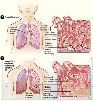
Idiopathic pulmonary fibrosis (IPF), or (formerly) fibrosing alveolitis, is a rare, progressive illness of the respiratory system, characterized by the thickening and stiffening of lung tissue, associated with the formation of scar tissue. It is a type of chronic scarring lung disease characterized by a progressive and irreversible decline in lung function. The tissue in the lungs becomes thick and stiff, which affects the tissue that surrounds the air sacs in the lungs. Symptoms typically include gradual onset of shortness of breath and a dry cough. Other changes may include feeling tired, and abnormally large and dome shaped finger and toenails. Complications may include pulmonary hypertension, heart failure, pneumonia or pulmonary embolism.

<span class="mw-page-title-main">Bird fancier's lung</span> Type of hypersensitivity pneumonitis

Bird fancier's lung (BFL), also known as bird breeder's lung, is a type of hypersensitivity pneumonitis. It can cause shortness of breath, fever, dry cough, chest pain, anorexia and weight loss, fatigue, and progressive pulmonary fibrosis. It is triggered by exposure to avian proteins present in the dry dust of droppings or feathers of a variety of birds. The lungs become inflamed, with granuloma formation. It mostly affects people who work with birds or own many birds.

<span class="mw-page-title-main">Usual interstitial pneumonia</span> Medical condition

Usual interstitial pneumonia (UIP) is a form of lung disease characterized by progressive scarring of both lungs. The scarring (fibrosis) involves the pulmonary interstitium. UIP is thus classified as a form of interstitial lung disease.

<span class="mw-page-title-main">High-resolution computed tomography</span> Diagnostic imaging test

High-resolution computed tomography (HRCT) is a type of computed tomography (CT) with specific techniques to enhance image resolution. It is used in the diagnosis of various health problems, though most commonly for lung disease, by assessing the lung parenchyma. On the other hand, HRCT of the temporal bone is used to diagnose various middle ear diseases such as otitis media, cholesteatoma, and evaluations after ear operations.

Restrictive lung diseases are a category of extrapulmonary, pleural, or parenchymal respiratory diseases that restrict lung expansion, resulting in a decreased lung volume, an increased work of breathing, and inadequate ventilation and/or oxygenation. Pulmonary function test demonstrates a decrease in the forced vital capacity.

<span class="mw-page-title-main">Idiopathic interstitial pneumonia</span> Medical condition

Idiopathic interstitial pneumonia (IIP), or noninfectious pneumonia are a class of diffuse lung diseases. These diseases typically affect the pulmonary interstitium, although some also have a component affecting the airways. There are seven recognized distinct subtypes of IIP.

<span class="mw-page-title-main">Respiratory bronchiolitis</span> Medical condition

Respiratory bronchiolitis is a lung disease associated with tobacco smoking. In pathology, it is defined by the presence of "smoker's macrophages". When manifesting significant clinical symptoms it is referred to as respiratory bronchiolitis interstitial lung disease (RB-ILD).

<span class="mw-page-title-main">Ground-glass opacity</span> Radiologic sign on radiographs and computed tomography scans

Ground-glass opacity (GGO) is a finding seen on chest x-ray (radiograph) or computed tomography (CT) imaging of the lungs. It is typically defined as an area of hazy opacification (x-ray) or increased attenuation (CT) due to air displacement by fluid, airway collapse, fibrosis, or a neoplastic process. When a substance other than air fills an area of the lung it increases that area's density. On both x-ray and CT, this appears more grey or hazy as opposed to the normally dark-appearing lungs. Although it can sometimes be seen in normal lungs, common pathologic causes include infections, interstitial lung disease, and pulmonary edema.

References

  1. 1 2 GBD 2015 Disease and Injury Incidence and Prevalence Collaborators (8 October 2016). "Global, regional, and national incidence, prevalence, and years lived with disability for 310 diseases and injuries, 1990–2015: a systematic analysis for the Global Burden of Disease Study 2015". Lancet. 388 (10053): 1545–1602. doi:10.1016/S0140-6736(16)31678-6. PMC   5055577 . PMID   27733282.
  2. 1 2 GBD 2015 Mortality and Causes of Death Collaborators (8 October 2016). "Global, regional, and national life expectancy, all-cause mortality, and cause-specific mortality for 249 causes of death, 1980–2015: a systematic analysis for the Global Burden of Disease Study 2015". Lancet. 388 (10053): 1459–1544. doi:10.1016/S0140-6736(16)31012-1. PMC   5388903 . PMID   27733281.
  3. King TE (August 2005). "Clinical advances in the diagnosis and therapy of the interstitial lung diseases". American Journal of Respiratory and Critical Care Medicine. 172 (3): 268–79. doi:10.1164/rccm.200503-483OE. PMID   15879420. S2CID   29591585.
  4. "Frequently Asked Questions About Interstitial Lung Disease". University of Chicago Medical Center.
  5. Meyer, Keith C; Decker, Catherine A (2017-04-03). "Role of pirfenidone in the management of pulmonary fibrosis". Therapeutics and Clinical Risk Management. 13: 427–437. doi: 10.2147/TCRM.S81141 . ISSN   1176-6336. PMC   5388201 . PMID   28435277.
  6. Bush A, Cunningham S, de Blic J, Barbato A, Clement A, Epaud R, Hengst M, Kiper N, Nicholson AG, Wetzke M, Snijders D, Schwerk N, Griese M (November 2015). "European protocols for the diagnosis and initial treatment of interstitial lung disease in children". review. Thorax. 70 (11): 1078–84. doi: 10.1136/thoraxjnl-2015-207349 . hdl: 10044/1/24599 . PMID   26135832.
  7. 1 2 3 4 5 6 7 8 9 10 11 12 13 14 15 16 17 18 19 20 21 22 Maher, Toby M. (22 April 2024). "Interstitial Lung Disease: A Review". JAMA. doi:10.1001/jama.2024.3669.
  8. Bourke SJ (August 2006). "Interstitial lung disease: progress and problems". Postgraduate Medical Journal. 82 (970): 494–9. doi:10.1136/pgmj.2006.046417. PMC   2585700 . PMID   16891438.
  9. Kreuter, Michael; Herth, Felix J. F.; Wacker, Margarethe; Leidl, Reiner; Hellmann, Andreas; Pfeifer, Michael; Behr, Jürgen; Witt, Sabine; Kauschka, Dagmar (2015). "Exploring Clinical and Epidemiological Characteristics of Interstitial Lung Diseases: Rationale, Aims, and Design of a Nationwide Prospective Registry—The EXCITING-ILD Registry". BioMed Research International. 2015: 123876. doi: 10.1155/2015/123876 . PMC   4657073 . PMID   26640781.
  10. "Interstitial Lung Diseases". The Lecturio Medical Concept Library. 23 September 2020. Retrieved 27 July 2021.
  11. Vehar SJ, Yadav R, Mukhopadhyay S, Nathani A, Tolle LB (Dec 2022). "Smoking-related interstitial fibrosis (SRIF) in patients presenting with diffuse parenchymal lung disease". American Journal of Clinical Pathology. 159 (2): 146–157. doi:10.1093/ajcp/aqac144. PMC   9891418 . PMID   36495281.
  12. "Dr. Daniele Macchini, Treating COVID-19 Patients in Bergamo, Italy, Describes Horrible Situation". 11 March 2020.
  13. Cottin, Vincent (November 2016). "Lung biopsy in interstitial lung disease: balancing the risk of surgery and diagnostic uncertainty". European Respiratory Journal. 48 (5): 1274–1277. doi:10.1183/13993003.01633-2016.
  14. Troy, Lauren K; Grainge, Christopher; Corte, Tamera J (February 2020). "Diagnostic accuracy of transbronchial lung cryobiopsy for interstitial lung disease diagnosis (COLDICE): a prospective, comparative study". The Lancet Respiratory Medicine. 8 (2): 171–181. doi:10.1016/S2213-2600(19)30342-X.
  15. 1 2 3 King, Talmadge E Jr (February 27, 2023). "Approach to the adult with interstitial lung disease: Diagnostic testing" . www.uptodate.com.
  16. 1 2 3 4 5 6 7 Ryu JH, Olson EJ, Midthun DE, Swensen SJ (November 2002). "Diagnostic approach to the patient with diffuse lung disease". Mayo Clinic Proceedings. 77 (11): 1221–7, quiz 1227. doi: 10.4065/77.11.1221 . PMID   12440558.
  17. "What Is Pulmonary Fibrosis?". Northwestern Medicine. Archived from the original on 2014-02-26. Retrieved 2014-02-22.
  18. Zare Mehrjardi M, Kahkouee S, Pourabdollah M (March 2017). "Radio-pathological correlation of organizing pneumonia (OP): a pictorial review". The British Journal of Radiology. 90 (1071): 20160723. doi:10.1259/bjr.20160723. PMC   5601538 . PMID   28106480.
  19. Hayes D, Jr; Wilson, KC; Krivchenia, K; Hawkins, SMM; Balfour-Lynn, IM; Gozal, D; Panitch, HB; Splaingard, ML; Rhein, LM; Kurland, G; Abman, SH; Hoffman, TM; Carroll, CL; Cataletto, ME; Tumin, D; Oren, E; Martin, RJ; Baker, J; Porta, GR; Kaley, D; Gettys, A; Deterding, RR (1 February 2019). "Home Oxygen Therapy for Children. An Official American Thoracic Society Clinical Practice Guideline". American Journal of Respiratory and Critical Care Medicine. 199 (3): e5–e23. doi:10.1164/rccm.201812-2276ST. PMC   6802853 . PMID   30707039.
  20. Dowman, Leona; Hill, Catherine J.; May, Anthony; Holland, Anne E. (2021-02-01). "Pulmonary rehabilitation for interstitial lung disease". The Cochrane Database of Systematic Reviews. 2021 (2): CD006322. doi:10.1002/14651858.CD006322.pub4. ISSN   1469-493X. PMC   8094410 . PMID   34559419.
  21. Kotloff RM, Thabut G (July 2011). "Lung transplantation". American Journal of Respiratory and Critical Care Medicine. 184 (2): 159–71. doi:10.1164/rccm.201101-0134CI. PMID   21471083.
  22. Whelan TP (March 2012). "Lung transplantation for interstitial lung disease". Clinics in Chest Medicine. 33 (1): 179–89. doi:10.1016/j.ccm.2011.12.003. PMID   22365254.
  23. King, Talmadge E.; Bradford, Williamson Z.; Castro-Bernardini, Socorro; Fagan, Elizabeth A.; Glaspole, Ian; Glassberg, Marilyn K.; Gorina, Eduard; Hopkins, Peter M.; Kardatzke, David; Lancaster, Lisa; Lederer, David J.; Nathan, Steven D.; Pereira, Carlos A.; Sahn, Steven A.; Sussman, Robert; Swigris, Jeffrey J.; Noble, Paul W. (29 May 2014). "A Phase 3 Trial of Pirfenidone in Patients with Idiopathic Pulmonary Fibrosis". New England Journal of Medicine. 370 (22): 2083–2092. doi:10.1056/NEJMoa1402582.
  24. Hassoun, Paul M. (16 December 2021). "Pulmonary Arterial Hypertension". New England Journal of Medicine. 385 (25): 2361–2376. doi:10.1056/NEJMra2000348.