Related Research Articles

Pulmonology, pneumology or pneumonology is a medical specialty that deals with diseases involving the respiratory tract. It is also known as respirology, respiratory medicine, or chest medicine in some countries and areas.

Pulmonary hypertension is a condition of increased blood pressure in the arteries of the lungs. Symptoms include shortness of breath, fainting, tiredness, chest pain, swelling of the legs, and a fast heartbeat. The condition may make it difficult to exercise. Onset is typically gradual. According to the definition at the 6th World Symposium of Pulmonary Hypertension in 2018, a patient is deemed to have pulmonary hypertension if the pulmonary mean arterial pressure is greater than 20mmHg at rest, revised down from a purely arbitrary 25mmHg, and pulmonary vascular resistance (PVR) greater than 3 Wood units.

Pulmonary alveolar proteinosis (PAP) is a rare lung disorder characterized by an abnormal accumulation of surfactant-derived lipoprotein compounds within the alveoli of the lung. The accumulated substances interfere with the normal gas exchange and expansion of the lungs, ultimately leading to difficulty breathing and a predisposition to developing lung infections. The causes of PAP may be grouped into primary, secondary, and congenital causes, although the most common cause is a primary autoimmune condition in an individual.

Interstitial lung disease (ILD), or diffuse parenchymal lung disease (DPLD), is a group of respiratory diseases affecting the interstitium and space around the alveoli of the lungs. It concerns alveolar epithelium, pulmonary capillary endothelium, basement membrane, and perivascular and perilymphatic tissues. It may occur when an injury to the lungs triggers an abnormal healing response. Ordinarily, the body generates just the right amount of tissue to repair damage, but in interstitial lung disease, the repair process is disrupted, and the tissue around the air sacs (alveoli) becomes scarred and thickened. This makes it more difficult for oxygen to pass into the bloodstream. The disease presents itself with the following symptoms: shortness of breath, nonproductive coughing, fatigue, and weight loss, which tend to develop slowly, over several months. The average rate of survival for someone with this disease is between three and five years. The term ILD is used to distinguish these diseases from obstructive airways diseases.

Pulmonary fibrosis is a condition in which the lungs become scarred over time. Symptoms include shortness of breath, a dry cough, feeling tired, weight loss, and nail clubbing. Complications may include pulmonary hypertension, respiratory failure, pneumothorax, and lung cancer.
Neuroendocrine hyperplasia is rare and poorly understood lung condition which is characterized by an abnormal growth pulmonary neuroendocrine cells in the lungs. It is a non-progressive disease of the interstitial tissues of the lungs. Prior to the findings of the hyperplasia of neuroendocrine cells it was known as tachypnea of infancy, as most children outgrow the need for oxygen supplementation within two to seven years. It is characterized by tachypnea, hypoxemia, and retractions. It is typically diagnosed in infants and children younger than one year of age. There is no currently recognized treatment, infants and children are given oxygen supplementation until they outgrow the need; since neuroendocrine cells do not multiply or get larger in size while the lungs continue to grow. This allows the lung disease to have less effect on lung function with age, although they will always have the same amount of neuroendocrine cells as they were born with.

Cryptogenic organizing pneumonia (COP), formerly known as bronchiolitis obliterans organizing pneumonia (BOOP), is an inflammation of the bronchioles (bronchiolitis) and surrounding tissue in the lungs. It is a form of idiopathic interstitial pneumonia.

Respiratory diseases, or lung diseases, are pathological conditions affecting the organs and tissues that make gas exchange difficult in air-breathing animals. They include conditions of the respiratory tract including the trachea, bronchi, bronchioles, alveoli, pleurae, pleural cavity, the nerves and muscles of respiration. Respiratory diseases range from mild and self-limiting, such as the common cold, influenza, and pharyngitis to life-threatening diseases such as bacterial pneumonia, pulmonary embolism, tuberculosis, acute asthma, lung cancer, and severe acute respiratory syndromes, such as COVID-19. Respiratory diseases can be classified in many different ways, including by the organ or tissue involved, by the type and pattern of associated signs and symptoms, or by the cause of the disease.

Bronchopulmonary dysplasia is a chronic lung disease in which premature infants, usually those who were treated with supplemental oxygen, require long-term oxygen. The alveoli that are present tend to not be mature enough to function normally. It is more common in infants with low birth weight (LBW) and those who receive prolonged mechanical ventilation to treat respiratory distress syndrome (RDS). It results in significant morbidity and mortality. The definition of BPD has continued to evolve primarily due to changes in the population, such as more survivors at earlier gestational ages, and improved neonatal management including surfactant, antenatal glucocorticoid therapy, and less aggressive mechanical ventilation.
Idiopathic pulmonary haemosiderosis (IPH) is a lung disease of unknown cause that is characterized by alveolar capillary bleeding and accumulation of haemosiderin in the lungs. It is rare, with an incidence between 0.24 and 1.23 cases per million people.
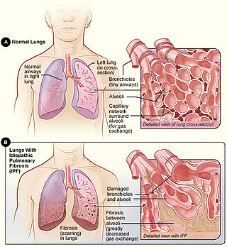
Idiopathic pulmonary fibrosis (IPF), or (formerly) fibrosing alveolitis, is a rare, progressive illness of the respiratory system, characterized by the thickening and stiffening of lung tissue, associated with the formation of scar tissue. It is a type of chronic scarring lung disease characterized by a progressive and irreversible decline in lung function. The tissue in the lungs becomes thick and stiff, which affects the tissue that surrounds the air sacs in the lungs. Symptoms typically include gradual onset of shortness of breath and a dry cough. Other changes may include feeling tired, and abnormally large and dome shaped finger and toenails. Complications may include pulmonary hypertension, heart failure, pneumonia or pulmonary embolism.
Williams–Campbell syndrome (WCS) is a disease of the airways where cartilage in the bronchi is defective. It is a form of congenital cystic bronchiectasis. This leads to collapse of the airways and bronchiectasis. It acts as one of the differential to allergic bronchopulmonary aspergillosis. WCS is a deficiency of the bronchial cartilage distally.
Pulmonary hygiene, formerly referred to as pulmonary toilet, is a set of methods used to clear mucus and secretions from the airways. The word pulmonary refers to the lungs. The word toilet, related to the French toilette, refers to body care and hygiene; this root is used in words such as toiletry that also relate to cleansing.
Alveolar capillary dysplasia (ACD) is a rare, congenital diffuse lung disease characterized by abnormal blood vessels in the lungs that cause highly elevated pulmonary blood pressure and an inability to effectively oxygenate and remove carbon dioxide from the blood. ACD typically presents in newborn babies within hours of birth as rapid and labored breathing, blue-colored lips or skin, quickly leading to respiratory failure and death. Atypical forms of ACD have been reported with initially milder symptoms and survival of many months before the onset of respiratory failure or lung transplantation.
Rheumatoid lung disease is a disease of the lung associated with RA, rheumatoid arthritis. Rheumatoid lung disease is characterized by pleural effusion, pulmonary fibrosis, lung nodules and pulmonary hypertension. Common symptoms associated with the disease include shortness of breath, cough, chest pain and fever. It is estimated that about one quarter of people with rheumatoid arthritis develop this disease, which are more likely to develop among elderly men with a history of smoking.
Surfactant metabolism dysfunction is a condition where pulmonary surfactant is insufficient for adequate respiration. Surface tension at the liquid-air interphase in the alveoli makes the air sacs prone to collapsing post expiration. This is due to the fact that water molecules in the liquid-air surface of alveoli are more attracted to one another than they are to molecules in the air. For sphere-like structures like alveoli, water molecules line the inner walls of the air sacs and stick tightly together through hydrogen bonds. These intermolecular forces put great restraint on the inner walls of the air sac, tighten the surface all together, and unyielding to stretch for inhalation. Thus, without something to alleviate this surface tension, alveoli can collapse and cannot be filled up again. Surfactant is essential mixture that is released into the air-facing surface of inner walls of air sacs to lessen the strength of surface tension. This mixture inserts itself among water molecules and breaks up hydrogen bonds that hold the tension. Multiple lung diseases, like ISD or RDS, in newborns and late-onsets cases have been linked to dysfunction of surfactant metabolism.

Ground-glass opacity (GGO) is a finding seen on chest x-ray (radiograph) or computed tomography (CT) imaging of the lungs. It is typically defined as an area of hazy opacification (x-ray) or increased attenuation (CT) due to air displacement by fluid, airway collapse, fibrosis, or a neoplastic process. When a substance other than air fills an area of the lung it increases that area's density. On both x-ray and CT, this appears more grey or hazy as opposed to the normally dark-appearing lungs. Although it can sometimes be seen in normal lungs, common pathologic causes include infections, interstitial lung disease, and pulmonary edema.

Emphysema is any air-filled enlargement in the body's tissues. Most commonly emphysema refers to the enlargement of air spaces (alveoli) in the lungs, and is also known as pulmonary emphysema.
Multisystemic smooth muscle dysfunction syndrome (MSMDS) is a genetic disorder caused by R179 missense mutations in the ACTA2 gene. Initially described as a case report in 1999, it was characterized in 2010 as a syndrome of congenital mydriasis, patent ductus arteriosus, and aneurysmal arterial disease—in particular aortic and thoracic aneurysms. The disorder has variable penetrance, ranging from severely symptomatic and fatal in early neonatal period to a more benign and manageable course with surgical intervention.

Underrepresented populations, especially black and hispanic populations with cystic fibrosis are often not successfully diagnosed. This is in part due to the minimal dissemination of existing data on patients from these underrepresented groups. While white populations do appear to experience a higher frequency of cystic fibrosis, other ethnicities are also affected and not always by the same biological mechanisms. Thus, many healthcare and treatment options are less reliable or unavailable to underrepresented populations. This issue affects the level at which public health needs are being met across the world.
References
- 1 2 Hime, Neil J.; Zurynski, Yvonne; Fitzgerald, Dominic; Selvadurai, Hiran; Phu, Amy; Deverell, Marie; Elliott, Elizabeth J.; Jaffe, Adam (December 24, 2015). "Childhood interstitial lung disease: A systematic review". Pediatric Pulmonology. 50 (12): 1383–1392. doi:10.1002/ppul.23183. PMID 25931270. S2CID 23161366.
- ↑ Moore K (2018). Clinically oriented anatomy. Wolters Kluwer. p. 336. ISBN 9781496347213.
- ↑ Clement, Annick; Nathan, Nadia; Epaud, Ralph; Fauroux, Brigitte; Corvol, Harriet (August 20, 2010). "Interstitial lung diseases in children". Orphanet Journal of Rare Diseases. 5 (22): 22. doi: 10.1186/1750-1172-5-22 . PMC 2939531 . PMID 20727133.
- 1 2 3 Guillerman, R. Paul (March 24, 2010). "Imaging of Childhood Interstitial Lung Disease". Pediatric Allergy, Immunology, and Pulmonology. 23 (1): 43–68. doi:10.1089/ped.2010.0010. PMC 3269227 . PMID 22332031.
- 1 2 Kurland, G.; Deterding, R. R.; Hagood, J. S.; Young, L. R.; Brody, A. S.; Castile, R. G.; Dell, S.; Fan, L. L.; Hamvas, A.; Hilman, B. C.; Langston, C.; Nogee, L. M.; Redding, G. J.; American Thoracic Society Committee on Childhood Interstitial Lung Disease (chILD) the chILD Research Network (2013). "An official American Thoracic Society clinical practice guideline: classification, evaluation, and management of childhood interstitial lung diseas... - PubMed - NCBI". American Journal of Respiratory and Critical Care Medicine. 188 (3): 376–94. doi:10.1164/rccm.201305-0923ST. PMC 3778735 . PMID 23905526.
- 1 2 Bromley, Susan; Vizcaya, David (May 24, 2017). "Pulmonary hypertension in childhood interstitial lung disease: A systematic review of the literature". Pediatric Pulmonology. 52 (5): 689–698. doi:10.1002/ppul.23632. PMID 27774750. S2CID 22728341.
- ↑ Cleveland, Robert H.; Lee, Edward Y. (2019-09-24). Imaging in Pediatric Pulmonology. Springer Nature. pp. 145–148. ISBN 978-3-030-23979-4.
- ↑ "Childhood Interstitial Lung Disease". National Heart, Lung, and Blood Institute. Retrieved 30 November 2020.