Chest tube

Last updated
Chest tube
Cross-section Blake Drain.jpg
The free end of the Chest Drainage Device is usually attached to an underwater seal, below the level of the chest. This allows the air or fluid to escape from the pleural space, and prevents anything returning to the chest.
Other namesIntercostal drain
Specialty pulmonology
ICD-9-CM 34.04
MeSH D013907

A chest tube (also chest drain, thoracic catheter, tube thoracostomy or intercostal drain) is a surgical drain that is inserted through the chest wall and into the pleural space or the mediastinum. The insertion of the tube is sometimes a lifesaving procedure. The tube can be used to remove clinically undesired substances such as air (pneumothorax), [1] excess fluid (pleural effusion or hydrothorax), blood (hemothorax), chyle (chylothorax) or pus (empyema) from the intrathoracic space. An intrapleural chest tube is also known as a Bülau drain or an intercostal catheter (ICC), and can either be a thin, flexible silicone tube (known as a "pigtail" drain), or a larger, semi-rigid, fenestrated plastic tube, which often involves a flutter valve or underwater seal.

Contents

The concept of chest drainage was first advocated by Hippocrates when he described the treatment of empyema by means of incision, cautery and insertion of metal tubes. [2] However, the technique was not widely used until the influenza epidemic of 1918 to evacuate post-pneumonic empyema, which was first documented by Dr. C. Pope, on a 22-month-old infant. [3] The use of chest tubes in postoperative thoracic care was reported in 1922, [4] and they were regularly used post-thoracotomy in World War II, though they were not routinely used for emergency tube thoracostomy following acute trauma until the Korean War. [5]

Uses

Left-sided pneumothorax (right side of image) on CT scan of the chest with chest tube in place. Pneumothorax CT.jpg
Left-sided pneumothorax (right side of image) on CT scan of the chest with chest tube in place.

Medical uses of chest tube are as follows: [6]

Contraindications

Contraindications to chest tube placement include refractory coagulopathy and presence of a diaphragmatic hernia, as well as hepatic hydrothorax. [7] Additional contraindications include scarring in the pleural space (adhesions).

Complications

Complications that are sometimes associated with chest tubes include the potential for clogging, air leaks, infection, hemorrhage, re-expansion pulmonary edema. Injury to the liver, spleen or diaphragm is also possible if the tube is placed behind (inferior) to the pleural cavity or is mispositioned. Injuries to the thoracic aorta and heart can also occur. [5] [8] The rate of complications of chest tubes inserted for trauma-related treatment needs has been estimated at approximately 19%. [9] The rate of complications is variable and other estimations have been made that share a rate of closer to 40%. [10]

Insertional complications

Complications that arise while the chest tube is being inserted or within the first day of the insertional procedure include a risk of injury to organs near the insertional site. [10]

Positional complications

Complications that arise after the tube has been inserted for one day or longer include the potential for tube blockages (obstruction), air leaks, kinking, or entrapment in the lung fissure once the lung has been expanded. Chest tube clogging can lead to retained blood around the heart and lungs that can contribute to complications and increase mortality. [11] A common complication after thoracic surgery that arises within 30–50% of patients are air leaks. If a chest tube clogs when there is an air leak the patient will develop a pneumothorax. This can be life-threatening. [12] Here, digital chest drainage systems can provide real time information as they monitor intra-pleural pressure and air leak flow, constantly. [13] Keeping vigilant about chest tube clogging is imperative for the team taking care of the patient in the early postoperative period.

Minor complications include a subcutaneous hematoma or seroma, anxiety, shortness of breath, and cough (after removing large volume of fluid). In most cases, the chest tube related pain goes away after the chest tube is removed, however, chronic pain related to chest tube induced scarring of the intercostal space is not uncommon.[ citation needed ]

Subcutaneous emphysema indicates backpressure created by undrained air, often caused by a clogged chest tube or insufficient negative pressure.[ citation needed ] If a person has subcutaneous emphysema, it is likely their chest tube is not draining and consideration should be given if it should be unclogged or another tube should be placed so that the air leaking from the lung can be adequately drained.

Infections

Problems keeping the site clean or with sterilizing instruments can lead to infections. [10] When chest tubes are placed due to either blunt or penetrating trauma, antibiotics may decrease the risks of infectious complications. [14]

Removal complications

There is also a risk of complications after the chest tube has been removed. [10] Potential complications include problems with re-sealing the chest that can lead to trapped air or if a foreign object is retained in the chest after the procedure. [10]

Device

Characteristics

Size of chest tube:
Adult male = 28-32 Fr
Pp Adult female = 28 Fr
Child = 18 Fr
Newborn = 12-14 Fr Dreno toracico tubular multiperfurado.JPG
Size of chest tube:
Adult male = 28–32 Fr
Pp Adult female = 28 Fr
Child = 18 Fr
Newborn = 12–14 Fr
Chest tube drainage holes Chest Tube Drainage Holes.jpg
Chest tube drainage holes

Chest tubes are commonly made from clear plastics like PVC and soft silicone. Chest tubes are made in a range of sizes measured by their external diameter from 6 Fr to 40 Fr. Chest tubes, like most catheters, are measured in French catheter scale. For adults, 20 Fr to 40 Fr (6.7 to 13.3mm external diameter) are commonly used, and 6 Fr to 26 Fr for children. Conventional chest tubes feature multiple drainage fenestrations in the section of the tube which resides inside the patient, as well as distance markers along the length of the tube, and a radiopaque stripe which outlines the first drainage hole. [5] Chest tubes are also provided in right angle, trocar, flared, and tapered configurations for different drainage needs. As well, some chest tubes are coated with heparin to help prevent thrombus formation, though the effect of this is disputed. [16]

Chest tube have an end hole (proximal, toward the patient) and a series of side holes. The number of side holes is generally 6 on most chest tubes. The length of tube that has side holes is the effective drainage length (EDL). In chest tubes designed for pediatric heart surgery, the EDL is shorter, generally by only having 4 side holes. [17]

Channel style chest drains, also called Blake drains, are so-called silastic drains made of silicone and feature open flutes that reside inside the patient. Drainage is thought to be achieved by capillary action, allowing the fluids to travel through the open grooves into a closed cross section, which contains the fluid and allows it to be suctioned through the tube. [18] Though these chest tubes are more expensive than conventional ones, they are theoretically less painful. [19]

Chest drainage system

Portable electronic system P4053296 logo.jpg
Portable electronic system
Chest tube drainage system diagram, with parts labeled in Labelled chest tube drainage system.png
Chest tube drainage system diagram, with parts labeled in

A chest drainage system is typically used to collect chest drainage (air, blood, effusions). Most commonly, drainage systems use three chambers which are based on the three-bottle system. The first chamber allows fluid that is drained from the chest to be collected. The second chamber functions as a "water seal", which acts as a one way valve allowing gas to escape, but not reenter the chest. Air bubbling through the water seal chamber is usual when the patient coughs or exhales but may indicate, if continual, a pleural or system leak that should be evaluated critically. It can also indicate a leak of air from the lung. The third chamber is the suction control chamber. The height of the water in this chamber regulates the negative pressure applied to the system. A gentle bubbling through the water column minimizes evaporation of the fluid and indicates that the suction is being regulated to the height of the water column. In this way, increased wall suction does not increase the negative pressure of the system. Newer drainage systems eliminate the water seal using a mechanical check-valve, and some also use a mechanical regulator to regulate the suction pressure. Systems which employ both these are dubbed "dry" systems, whereas systems that retain the water seal but use a mechanical regulator are called "wet-dry" systems. Systems which use a water seal and water column regulator are called "wet" systems. Dry systems are advantageous as tip-overs of wet systems can spill and mix with blood, mandating the replacement of the system. Even newer systems are smaller and more ambulatory so the patient can be sent home for drainage if indicated. [5]

The free end of the tube is usually attached to an underwater seal, below the level of the chest. This allows the air or fluid to escape from the pleural space, and prevents anything returning to the chest. Alternatively, the tube can be attached to a flutter valve. This allows patients with pneumothorax to remain more mobile. [20]

More recently digital or electronic chest drainage systems have been introduced. An onboard motor is used as vacuum source along with an integrated suction control canister and water seal. These systems monitor the patient and will alert if the measured data are out of range. Due to the digital control of the negative pressure, the system is able to objectively quantify the presence of a pleural or system leak. Digital drainage systems allow clinicians to mobilize patients early, even for those on continuous suction, which is difficult to accomplish with the traditional water-seal system under suction. [12] [21] Application of such systems can also lead to a reduction in complications. [22] [23]

Technique

Thoracostomy

It can be inserted in an area described as the "safe zone", which is a region bordered by the lateral border of pectoralis major, a horizontal line inferior to the axilla, the anterior border of latissimus dorsi and a horizontal line superior to the nipple. [24] This should translate to the tube being inserted into the fifth intercostal space slightly anterior to the mid axillary line. [25]

Chest tubes are usually inserted under local anesthesia. The skin over the area of insertion is first cleansed with antiseptic solution, such as iodine, before sterile drapes are placed around the area. The local anesthetic is injected into the skin and down to the muscle, and after the area is numb a small incision is made in the skin and a passage made through the skin and muscle into the chest. The tube is placed through this passage. If necessary, patients may be given additional analgesics for the procedure. Once the tube is in place it is sutured to the skin to prevent it falling out and a dressing applied to the area. Once the drain is in place, a chest radiograph will be taken to check the location of the drain. The tube stays in for as long as there is air or fluid to be removed, or risk of air gathering.

Chest tubes can also be placed using a trocar, which is a pointed metallic bar used to guide the tube through the chest wall. This method is less popular due to an increased risk of iatrogenic lung injury. Placement using the Seldinger technique, in which a blunt guidewire is passed through a needle (over which the chest tube is then inserted) has been described.

Protocols to maintain chest tube patency by preventing chest tube clogging are necessary.

Postoperative drainage

The placement technique for postoperative drainage (e.g. cardiac surgery) differs from the technique used for emergency situations. At the completion of open cardiac procedures, chest tubes are placed through separate stab incisions, typically near the inferior aspect of the sternotomy incision. In some instances multiple drains may be used to evacuate the mediastinal, pericardial, and pleural spaces. The drainage holes are placed inside the patient and the chest tube is passed out through the incision. Once the tube is in place, it is sutured to the skin to prevent movement. The chest tube is then connected to the drainage canister using additional tubing and connectors and connected to a suction source, typically regulated to -20 cm of water. [18]

Dressings

After suturing, dressings are applied for hygienical reasons covering the wound. First, a y-slit compress is used around the tube. Second, a compress (10 x 10 cm) is placed on top and finally an adhesive plaster is added in a way that tension is avoided. A bridle rein is recommended to fix the tube to the skin. This tape bridge will prevent the tube from moving backwards and the possibility to cause clogging. It also prevents pain as it reduces tension on the fixation stitch. Alternatively, a large adhesive plaster that functions like a tape bridge may be used. [26]

Management

Chest tubes should be kept free of dependent loops, kinks, and obstructions which may prevent drainage. [27] In general, chest tubes are not clamped except during insertion, removal, or when diagnosing air leaks.[ citation needed ]

Chest tube clogging with blood clots of fibrinous material is common. When this occurs, it can result in retained blood around the heart or lungs that can lead to complications such as hematoma that needs to be drained, effusions, empyema, or, in the long term, fibrothorax. Thus its critical to maintain chest tube patency. Manual manipulation, often called milking, stripping, fan folding, or tapping, of chest tubes is commonly performed to clear chest tube obstructions. However these approaches are controversial. No conclusive evidence has demonstrated that any of these techniques are more effective than the others, and no method has shown to improve chest tube drainage. [28] Furthermore, chest tube manipulation has proved to increase negative pressure, which may be detrimental, and painful to the patient. [28] For these reasons, many hospitals do not allow these types of manual tube manipulations. [29]

One option is active chest tube clearance without breaking the sterile field. According to a consensus of multiple experts in cardiac surgery, anesthesia and critical care in 2019 the ERAS Guidelines for Perioperative Care recommends active clearance of chest tubes to prevent retained blood and other complications. [30] Makeshift efforts such as open chest tube clearing that involves breaking the sterile environment separating the chest tube from the drainage canister tubing to suction it out should not be performed. [31]

The chest tube can only be removed when the subject clinical condition is stable, the lungs are fully aerated as seen on chest X-ray, chest tube drainage is less than 200 cc per day, and there is no air leak into the lungs pleura. [32]

Site of placement

In December 2018 the European Respiratory Journal published correspondences that raise the possibility of improving mobility as well as patient outcomes by placing a chest tube more optimally. [33] [34]

Related Research Articles

<span class="mw-page-title-main">Pneumothorax</span> Abnormal collection of air in the pleural space

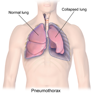
A pneumothorax is an abnormal collection of air in the pleural space between the lung and the chest wall. Symptoms typically include sudden onset of sharp, one-sided chest pain and shortness of breath. In a minority of cases, a one-way valve is formed by an area of damaged tissue, and the amount of air in the space between chest wall and lungs increases; this is called a tension pneumothorax. This can cause a steadily worsening oxygen shortage and low blood pressure. This leads to a type of shock called obstructive shock, which can be fatal unless reversed. Very rarely, both lungs may be affected by a pneumothorax. It is often called a "collapsed lung", although that term may also refer to atelectasis.

<span class="mw-page-title-main">Pleurisy</span> Disease of the lungs

Pleurisy, also known as pleuritis, is inflammation of the membranes that surround the lungs and line the chest cavity (pleurae). This can result in a sharp chest pain while breathing. Occasionally the pain may be a constant dull ache. Other symptoms may include shortness of breath, cough, fever, or weight loss, depending on the underlying cause. Pleurisy can be caused by a variety of conditions, including viral or bacterial infections, autoimmune disorders, and pulmonary embolism.

<span class="mw-page-title-main">Nasogastric intubation</span> Feeding tube going into the stomach through the nose and throat

Nasogastric intubation is a medical process involving the insertion of a plastic tube through the nose, down the esophagus, and down into the stomach. Orogastric intubation is a similar process involving the insertion of a plastic tube through the mouth. Abraham Louis Levin invented the NG tube. Nasogastric tube is also known as Ryle's tube in Commonwealth countries, after John Alfred Ryle.

<span class="mw-page-title-main">Pleural effusion</span> Accumulation of excess fluid in the pleural cavity

A pleural effusion is accumulation of excessive fluid in the pleural space, the potential space that surrounds each lung. Under normal conditions, pleural fluid is secreted by the parietal pleural capillaries at a rate of 0.6 millilitre per kilogram weight per hour, and is cleared by lymphatic absorption leaving behind only 5–15 millilitres of fluid, which helps to maintain a functional vacuum between the parietal and visceral pleurae. Excess fluid within the pleural space can impair inspiration by upsetting the functional vacuum and hydrostatically increasing the resistance against lung expansion, resulting in a fully or partially collapsed lung.

<span class="mw-page-title-main">Pleural empyema</span> Medical condition

Pleural empyema is a collection of pus in the pleural cavity caused by microorganisms, usually bacteria. Often it happens in the context of a pneumonia, injury, or chest surgery. It is one of the various kinds of pleural effusion. There are three stages: exudative, when there is an increase in pleural fluid with or without the presence of pus; fibrinopurulent, when fibrous septa form localized pus pockets; and the final organizing stage, when there is scarring of the pleura membranes with possible inability of the lung to expand. Simple pleural effusions occur in up to 40% of bacterial pneumonias. They are usually small and resolve with appropriate antibiotic therapy. If however an empyema develops additional intervention is required.

<span class="mw-page-title-main">Pleurodesis</span> Medical procedure on pleural cavity

Pleurodesis is a medical procedure in which part of the pleural space is artificially obliterated. It involves the adhesion of the visceral and the costal pleura. The mediastinal pleura is spared.

<span class="mw-page-title-main">Thoracotomy</span> Surgical procedure

A thoracotomy is a surgical procedure to gain access into the pleural space of the chest. It is performed by surgeons to gain access to the thoracic organs, most commonly the heart, the lungs, or the esophagus, or for access to the thoracic aorta or the anterior spine. A thoracotomy is the first step in thoracic surgeries including lobectomy or pneumonectomy for lung cancer or to gain thoracic access in major trauma.

<span class="mw-page-title-main">Hemothorax</span> Blood accumulation in the pleural cavity

A hemothorax is an accumulation of blood within the pleural cavity. The symptoms of a hemothorax may include chest pain and difficulty breathing, while the clinical signs may include reduced breath sounds on the affected side and a rapid heart rate. Hemothoraces are usually caused by an injury, but they may occur spontaneously due to cancer invading the pleural cavity, as a result of a blood clotting disorder, as an unusual manifestation of endometriosis, in response to pneumothorax, or rarely in association with other conditions.

<span class="mw-page-title-main">Chylothorax</span> Medical condition

A chylothorax is an abnormal accumulation of chyle, a type of lipid-rich lymph, in the space surrounding the lung. The lymphatics of the digestive system normally returns lipids absorbed from the small bowel via the thoracic duct, which ascends behind the esophagus to drain into the left brachiocephalic vein. If normal thoracic duct drainage is disrupted, either due to obstruction or rupture, chyle can leak and accumulate within the negative-pressured pleural space. In people on a normal diet, this fluid collection can sometimes be identified by its turbid, milky white appearance, since chyle contains emulsified triglycerides.

<span class="mw-page-title-main">Thoracentesis</span> Removal of fluids/air from the pleural cavity of the lungs

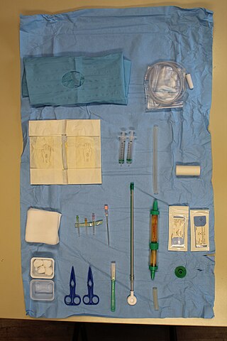
Thoracentesis, also known as thoracocentesis, pleural tap, needle thoracostomy, or needle decompression, is an invasive medical procedure to remove fluid or air from the pleural space for diagnostic or therapeutic purposes. A cannula, or hollow needle, is carefully introduced into the thorax, generally after administration of local anesthesia. The procedure was first performed by Morrill Wyman in 1850 and then described by Henry Ingersoll Bowditch in 1852.

Hemopneumothorax, or haemopneumothorax, is the condition of having both air (pneumothorax) and blood (hemothorax) in the chest cavity. A hemothorax, pneumothorax, or the combination of both can occur due to an injury to the lung or chest.

<span class="mw-page-title-main">Drain (surgery)</span> Tube used to remove pus, blood or other fluids from a wound

A surgical drain is a tube used to remove pus, blood or other fluids from a wound, body cavity, or organ. They are commonly placed by surgeons or interventional radiologists after procedures or some types of injuries, but they can also be used as an intervention for decompression. There are several types of drains, and selection of which to use often depends on the placement site and how long the drain is needed.

<span class="mw-page-title-main">Flutter valve</span> Valve used in respiratory medicine

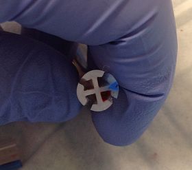
In respiratory medicine, a flutter valve is a one-way check valve used to prevent airflow back into a chest tube, and usually is applied to drain air from a pneumothorax. The design of the flutter valve features a rubber sleeve in a plastic case, where the rubber sleeve is arranged so that when air flows through the valve the sleeve opens and allows the outwards airflow from the body of the patient; however, when the airflow is reversed, the rubber sleeve closes and halts backwards airflow into the body of the patient.

A thoracostomy is a small incision of the chest wall, with maintenance of the opening for drainage. It is most commonly used for the treatment of a pneumothorax. This is performed by physicians, paramedics, and nurses usually via needle thoracostomy or an incision into the chest wall with the insertion of a thoracostomy tube or with a hemostat and the provider's finger.

<span class="mw-page-title-main">Pulmonary laceration</span> Medical condition

A pulmonary laceration is a chest injury in which lung tissue is torn or cut. An injury that is potentially more serious than pulmonary contusion, pulmonary laceration involves disruption of the architecture of the lung, while pulmonary contusion does not. Pulmonary laceration is commonly caused by penetrating trauma but may also result from forces involved in blunt trauma such as shear stress. A cavity filled with blood, air, or both can form. The injury is diagnosed when collections of air or fluid are found on a CT scan of the chest. Surgery may be required to stitch the laceration, to drain blood, or even to remove injured parts of the lung. The injury commonly heals quickly with few problems if it is given proper treatment; however it may be associated with scarring of the lung or other complications.

<span class="mw-page-title-main">Subcutaneous emphysema</span> Medical condition

Subcutaneous emphysema occurs when gas or air accumulates and seeps under the skin, where normally no gas should be present. Subcutaneous refers to the subcutaneous tissue, and emphysema refers to trapped air pockets. Since the air generally comes from the chest cavity, subcutaneous emphysema usually occurs around the upper torso, such as on the chest, neck, face, axillae and arms, where it is able to travel with little resistance along the loose connective tissue within the superficial fascia. Subcutaneous emphysema has a characteristic crackling-feel to the touch, a sensation that has been described as similar to touching warm Rice Krispies. This sensation of air under the skin is known as subcutaneous crepitation, a form of crepitus.

<span class="mw-page-title-main">Eloesser flap</span> Surgical procedure

The Eloesser flap is a surgical procedure developed by Dr. Leo Eloesser in 1935 at the San Francisco General Hospital. It was originally intended to aid with drainage of tuberculous empyemas, since at the time there were no effective medications to treat tuberculosis. The procedure was used extensively until the development of effective chemotherapy for tuberculosis in the late 1940s and early 1950s. It is still used occasionally for chronic empyemas.

<span class="mw-page-title-main">Chest drainage</span>

Chest drains are surgical drains placed within the pleural space to facilitate removal of unwanted substances in order to preserve respiratory functions and hemodynamic stability. Some chest drains may utilize a flutter valve to prevent retrograde flow, but those that do not have physical valves employ a water trap seal design, often aided by continuous suction from a wall suction or a portable vacuum pump.

<span class="mw-page-title-main">Mediastinal shift</span> Medical condition

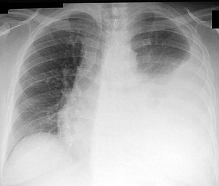
Mediastinal shift is an abnormal movement of the mediastinal structures toward one side of the chest cavity. A shift indicates a severe imbalance of pressures inside the chest. Mediastinal shifts are generally caused by increased lung volume, decreased lung volume, or abnormalities in the pleural space. Additionally, masses inside the mediastinum or musculoskeletal abnormalities can also lead to abnormal mediastinal arrangement. Typically, these shifts are observed on x-ray but also on computed tomography (CT) or magnetic resonance imaging (MRI). On chest x-ray, tracheal deviation, or movement of the trachea away from its midline position can be used as a sign of a shift. Other structures like the heart can also be used as reference points. Below are examples of pathologies that can cause a mediastinal shift and their appearance.

<span class="mw-page-title-main">Lung surgery</span>

Lung surgery is a type of thoracic surgery involving the repair or removal of lung tissue, and can be used to treat a variety of conditions ranging from lung cancer to pulmonary hypertension. Common operations include anatomic and nonanatomic resections, pleurodesis and lung transplants. Though records of lung surgery date back to the Classical Age, new techniques such as VATS continue to be developed.

References

  1. Noppen M, Alexander P, Driesen P, Slabbynck H, Verstraeten A (May 2002). "Manual aspiration versus chest tube drainage in first episodes of primary spontaneous pneumothorax: a multicenter, prospective, randomized pilot study". American Journal of Respiratory and Critical Care Medicine. 165 (9): 1240–1244. doi:10.1164/rccm.200111-078OC. PMID   11991872.
  2. Hippocrates (1847). Genuine Works of Hippocrates. Sydenham Society.
  3. Graham ME, Bell CR (1918). "Open Pneumothorax: Its relation to the treatment of empyema". J Med Sci. 156 (6): 839–871. doi:10.1097/00000441-191812000-00007.
  4. Lilienthal H (March 1922). "Resection of the lung for supportive infections with a report based on 31 consecutive operative cases in which resection was done or intended". Annals of Surgery. 75 (3): 257–320. doi:10.1097/00000658-192203000-00001. PMC   1399898 . PMID   17864604.
  5. 1 2 3 4 Miller KS, Sahn SA (February 1987). "Chest tubes. Indications, technique, management and complications". Chest. 91 (2): 258–264. doi:10.1378/chest.91.2.258. PMID   3542404.
  6. Kwiatt M, Tarbox A, Seamon MJ, Swaroop M, Cipolla J, Allen C, et al. (April 2014). "Thoracostomy tubes: A comprehensive review of complications and related topics". International Journal of Critical Illness and Injury Science. 4 (2): 143–155. doi: 10.4103/2229-5151.134182 . PMC   4093965 . PMID   25024942.
  7. Runyon BA, Greenblatt M, Ming RH (July 1986). "Hepatic hydrothorax is a relative contraindication to chest tube insertion". The American Journal of Gastroenterology. 81 (7): 566–567. PMID   3717119.
  8. Mohrsen S, McMahon N, Corfield A, McKee S (December 2021). "Complications associated with pre-hospital open thoracostomies: a rapid review". Scandinavian Journal of Trauma, Resuscitation and Emergency Medicine. 29 (1): 166. doi: 10.1186/s13049-021-00976-1 . PMC   8643006 . PMID   34863280.
  9. Hernandez, Matthew C; El Khatib, Moustafah; Prokop, Larry; Zielinski, Martin D.; Aho, Johnathon M. (2018). "Complications in Tube Thoracostomy: Systematic review and Meta-analysis". The Journal of Trauma and Acute Care Surgery. 85 (2): 410–416. doi:10.1097/TA.0000000000001840. ISSN   2163-0755. PMC   6081248 . PMID   29443856.
  10. 1 2 3 4 5 Anderson, Devon; Chen, Sarah A.; Godoy, Luis A.; Brown, Lisa M.; Cooke, David T. (2022-03-01). "Comprehensive Review of Chest Tube Management: A Review". JAMA Surgery. 157 (3): 269–274. doi:10.1001/jamasurg.2021.7050. ISSN   2168-6254. PMID   35080596.
  11. Balzer F, von Heymann C, Boyle EM, Wernecke KD, Grubitzsch H, Sander M (August 2016). "Impact of retained blood requiring reintervention on outcomes after cardiac surgery". The Journal of Thoracic and Cardiovascular Surgery. 152 (2): 595–601.e4. doi: 10.1016/j.jtcvs.2016.03.086 . PMID   27210474.
  12. 1 2 Brunelli A, Cassivi SD, Salati M, Fibla J, Pompili C, Halgren LA, et al. (April 2011). "Digital measurements of air leak flow and intrapleural pressures in the immediate postoperative period predict risk of prolonged air leak after pulmonary lobectomy". European Journal of Cardio-Thoracic Surgery. 39 (4): 584–588. doi: 10.1016/j.ejcts.2010.07.025 . PMID   20801054.
  13. "Compact Digital Thoracic Drain Systems for the Management of Thoracic Surgical Patients: A Review of the Clinical Effectiveness, Safety, and Cost-Effectiveness". Canadian Agency for Drugs and Technologies in Health. October 1, 2014. Archived from the original on May 13, 2019. Retrieved May 13, 2019.
  14. Ayoub F, Quirke M, Frith D (2019). "Use of prophylactic antibiotic in preventing complications for blunt and penetrating chest trauma requiring chest drain insertion: a systematic review and meta-analysis". Trauma Surgery & Acute Care Open. 4 (1): e000246. doi: 10.1136/tsaco-2018-000246 . PMC   6407548 . PMID   30899791.
  15. "Chest Tube and Fuhrman Catheter Insertion". University of Bullfalo, The State University of New York. Archived from the original on 2010-04-08. Retrieved 2009-07-19.
  16. Kumar P, McKee D, Grant M, Pepper J (April 1997). "Phosphatidylcholine coated chest drains: are they better than conventional drains after open heart surgery?". European Journal of Cardio-Thoracic Surgery. 11 (4): 769–773. doi: 10.1016/s1010-7940(96)01145-1 . PMID   9151051.
  17. "PleuraFlow for Pediatric CT Surgery Clears Chest Tubes of Clots |". 2016-08-02.
  18. 1 2 Obney JA, Barnes MJ, Lisagor PG, Cohen DJ (September 2000). "A method for mediastinal drainage after cardiac procedures using small silastic drains". The Annals of Thoracic Surgery. 70 (3): 1109–1110. doi: 10.1016/s0003-4975(00)01800-2 . PMID   11016389.
  19. Frankel TL, Hill PC, Stamou SC, Lowery RC, Pfister AJ, Jain A, Corso PJ (July 2003). "Silastic drains vs conventional chest tubes after coronary artery bypass". Chest. 124 (1): 108–113. doi:10.1378/chest.124.1.108. PMID   12853511.
  20. Dev SP, Nascimiento B, Simone C, Chien V (October 2007). "Videos in clinical medicine. Chest-tube insertion". The New England Journal of Medicine. 357 (15): e15. doi:10.1056/NEJMvcm071974. PMID   17928590. S2CID   73173434.
  21. Bertolaccini L, Rizzardi G, Filice MJ, Terzi A (May 2011). "'Six sigma approach' - an objective strategy in digital assessment of postoperative air leaks: a prospective randomised study". European Journal of Cardio-Thoracic Surgery. 39 (5): e128–e132. doi: 10.1016/j.ejcts.2010.12.027 . PMID   21316980.
  22. Leo F, Duranti L, Girelli L, Furia S, Billè A, Garofalo G, et al. (October 2013). "Does external pleural suction reduce prolonged air leak after lung resection? Results from the AirINTrial after 500 randomized cases". The Annals of Thoracic Surgery. 96 (4): 1234–1239. doi: 10.1016/j.athoracsur.2013.04.079 . PMID   23866802.
  23. Miller DL, Helms GA, Mayfield WR (September 2016). "Digital Drainage System Reduces Hospitalization After Video-Assisted Thoracoscopic Surgery Lung Resection". The Annals of Thoracic Surgery. 102 (3): 955–961. doi: 10.1016/j.athoracsur.2016.03.089 . PMID   27234573.
  24. Laws D, Neville E, Duffy J (May 2003). "BTS guidelines for the insertion of a chest drain". Thorax. 58 (Suppl 2 (90002)): ii53–ii59. doi:10.1136/thorax.58.suppl_2.ii53. PMC   1766017 . PMID   12728150.
  25. "Ventilatory management". University of Pretoria. Archived from the original on 2009-06-17. Retrieved 2009-09-16.
  26. Kiefer T (2017). Chest Drains in Daily Clinical Practice. Springer. pp. 102–103. ISBN   9783319323398.
  27. Schmelz JO, Johnson D, Norton JM, Andrews M, Gordon PA (September 1999). "Effects of position of chest drainage tube on volume drained and pressure". American Journal of Critical Care. 8 (5): 319–323. doi:10.4037/ajcc1999.8.5.319. PMID   10467469.
  28. 1 2 Wallen M, Morrison A, Gillies D, O'Riordan E, Bridge C, Stoddart F (October 2004). "Mediastinal chest drain clearance for cardiac surgery". The Cochrane Database of Systematic Reviews. 2021 (4): CD003042. doi:10.1002/14651858.CD003042.pub2. PMC   8094876 . PMID   15495040.
  29. Shalli S, Saeed D, Fukamachi K, Gillinov AM, Cohn WE, Perrault LP, Boyle EM (2009). "Chest tube selection in cardiac and thoracic surgery: a survey of chest tube-related complications and their management". Journal of Cardiac Surgery. 24 (5): 503–509. doi:10.1111/j.1540-8191.2009.00905.x. PMID   19740284. S2CID   23273268.
  30. Engelman DT, Ben Ali W, Williams JB, Perrault LP, Reddy VS, Arora RC, et al. (August 2019). "Guidelines for Perioperative Care in Cardiac Surgery: Enhanced Recovery After Surgery Society Recommendations". JAMA Surgery. 154 (8): 755–766. doi: 10.1001/jamasurg.2019.1153 . PMID   31054241.
  31. Halejian BA, Badach MJ, Trilles F (December 1988). "Maintaining chest tube patency". Surgery, Gynecology & Obstetrics. 167 (6): 521. PMID   3187876.
  32. Paydar S, Ghahramani Z, Ghoddusi Johari H, Khezri S, Ziaeian B, Ghayyoumi MA, et al. (April 2015). "Tube Thoracostomy (Chest Tube) Removal in Traumatic Patients: What Do We Know? What Can We Do?". Bulletin of Emergency and Trauma. 3 (2): 37–40. PMC   4771264 . PMID   27162900.
  33. Kartoun U (December 2018). "Improving the management of spontaneous pneumothorax". European Respiratory Journal . 52 (6): 1801857. doi: 10.1183/13993003.01857-2018 . PMID   30523206.
  34. Porcel JM (December 2018). "Improving the management of spontaneous pneumothorax". European Respiratory Journal . 52 (6): 1801918. doi: 10.1183/13993003.01918-2018 . PMID   30523207.

Further reading