Abdominal ultrasonography

Last updated
Abdominal ultrasonography
AlokaPhoto2006a.jpg
Medical ultrasound equipment which can be used for abdominal ultrasonography
ICD-9-CM 88.76
OPS-301 code 3-059
MedlinePlus 003777
Ultrasound probe (linear type) is used for examination of superficial structures. UltrasoundProbe2006a.jpg
Ultrasound probe (linear type) is used for examination of superficial structures.

Abdominal ultrasonography (also called abdominal ultrasound imaging or abdominal sonography) is a form of medical ultrasonography (medical application of ultrasound technology) to visualise abdominal anatomical structures. It uses transmission and reflection of ultrasound waves to visualise internal organs through the abdominal wall (with the help of gel, which helps transmission of the sound waves). For this reason, the procedure is also called a transabdominal ultrasound, in contrast to endoscopic ultrasound, the latter combining ultrasound with endoscopy through visualize internal structures from within hollow organs.

Contents

Abdominal ultrasound examinations are performed by gastroenterologists or other specialists in internal medicine, radiologists, or sonographers trained for this procedure.

Medical uses

Abdominal ultrasound can be used to diagnose abnormalities in various internal organs, such as the kidneys, [1] liver, gallbladder, pancreas, spleen and abdominal aorta. If Doppler ultrasonography is added, the blood flow inside blood vessels can be evaluated as well (for example, to look for renal artery stenosis). It is commonly used to examine the uterus and fetus during pregnancy; this is called obstetric ultrasonography. [2] [3]

Abdominal ultrasound is commonly used in the setting of abdominal pain or an acute abdomen (sudden and/or severe abdominal pain syndrome in which surgical intervention might be necessary), in which it can diagnose appendicitis or cholecystitis.

Ultrasound can also be used if there is suspicion of enlargement of one or more organs, such as used in screening for abdominal aortic aneurysm, investigation for splenomegaly or urinary retention.

Ultrasound can be used for additional anatomical information for patients with an abnormal kidney function or pancreatic enzymes (pancreatic amylase and pancreatic lipase).

Standard measurement of the abdominal aorta Ultrasonographic measurement of aortic diameter at the navel.svg
Standard measurement of the abdominal aorta

It can be used on the abdominal aorta to detect or exclude abdominal aortic aneurysm. For this purpose, the standard aortic measurement for abdominal aortic aneurysm is between the outer margins of the aortic wall. [4]

In cases of infectious mononucleosis, splenomegaly is a common symptom, and health care providers may consider using abdominal ultrasonography to get insight into a person's condition. [5] However, because spleen size varies greatly, ultrasonography is not a valid technique for assessing spleen enlargement and should not be used in typical circumstances or to make routine decisions about fitness for playing sports. [5]

Detecting stones

Ultrasound imaging is useful for detecting stones, for example kidney stones or gallstones, because they create a clearly visible ultrasound shadow behind the stone.[ citation needed ]

Ultrasonography can be used to guide procedures such as treatment for kidney stones with extracorporeal shock wave lithotripsy, needle biopsies or paracentesis (needle drainage of free fluid inside the abdominal cavity).[ citation needed ]

Liver

Ultrasonography of the liver with some standard measurements Liver measurements on ultrasonography.jpg
Ultrasonography of the liver with some standard measurements

In patients with deranged liver function tests, ultrasound may show increased liver size (hepatomegaly), increased reflectiveness (which might, for example, indicate cholestasis), gallbladder or bile duct diseases, or a tumor in the liver.

Ultrasonography of liver tumors involves two stages: detection and characterization.[ citation needed ] Tumor detection is based on the performance of the method and should include morphometric information (three axes dimensions, volume) and topographic information (number, location specifying liver segment and lobe/lobes). The specification of these data is important for staging liver tumors and prognosis.[ citation needed ] Tumor characterization is a complex process based on a sum of criteria leading towards tumor nature definition. Often, other diagnostic procedures, especially interventional ones, are no longer necessary. Tumor characterization using the ultrasound method will be based on the following elements: consistency (solid, liquid, mixed), echogenicity, structure appearance (homogeneous or heterogeneous), delineation from adjacent liver parenchyma (capsular, imprecise), elasticity, posterior acoustic enhancement effect, the relation with neighboring organs or structures (displacement, invasion), vasculature (presence and characteristics on Doppler ultrasonography and contrast-enhanced ultrasound (CEUS).[ citation needed ]

Renal ultrasonography

Ultrasound scan of a kidney (right side) MorisonNoText.png
Ultrasound scan of a kidney (right side)

Ultrasonography of the kidneys is essential in the diagnosis and management of kidney-related diseases. The kidneys are easily examined, and most pathological changes in the kidneys are distinguishable with ultrasound. [7]

Technique

Advantages of ultrasound imaging of abdominal structures are that the procedure can be performed quickly, bed-side, involves no exposure to X-rays (which makes it useful in pregnant patients, for example) and is inexpensive compared to other often-used techniques such as computed tomography (CT scan) of the abdomen. Disadvantages are troublesome imaging if a lot of gas is present inside the bowels, if there is a lot of abdominal fat, and that the quality of the imaging depends on the experience of the person performing it.[ citation needed ]

The imaging occurs real-time and without sedation, so that the influence of movements can be assessed quickly. For example, by pressing the ultrasound probe against the gallbladder, a radiological Murphy's sign can be elicited.

Through the abdominal wall, organs inside the pelvis can be seen, such as the urinary bladder or the ovaries and uterus in women. Because water is an excellent conductor for ultrasound waves, visualizing these structures often requires a well-filled urinary bladder (this means the patients has to drink plenty of water before the examination).

The liver can be imaged by swiping the probe sagittally from medial to lateral at the subcoastal region. However, if majority of the liver parenchyma is located high up in behind the ribs, the subject can be asked to breathe deeply to push down the liver into the abdomen for better visibility of liver. If the liver is still not visualised, then the subject can be rolled to the left lateral position to move the liver out of the ribs. Then, the ultrasound probe is rotated 90 degrees to access the liver in axial plane from the dome of the diaphragm until the lower segment of the liver. [8]

Abdominal Ultrasound (Full Exam)

STRUCTURED REPORT

(Technique: Transabdominal ultrasonography; Device: Toshiba Aplio XG)

Liver: Diffusely homogeneous and normal in echogenicity. No focal mass or contour nodularity. No intrahepatic biliary ductal dilatation.
Portal Vein: Patent main portal vein.
Gallbladder: No stones, wall thickening, or pericholecystic fluid.
Common Bile Duct: Nondilated measuring 1.3 mm at the level of the porta hepatis.
Pancreas: Visualized portions unremarkable.
Spleen: Normal in size.
Kidneys: Right and left kidneys measure 11.5 cm and 12 cm in length respectively. No hydronephrosis. Small left lower pole kidney cyst.
Ascites: None.
Aorta: Visualized portions normal in caliber, 16 x 15 mm.
IVC: Normal.

IMPRESSION:
Normal abdominal ultrasound.

See also

Related Research Articles

<span class="mw-page-title-main">Medical ultrasound</span> Diagnostic and therapeutic technique

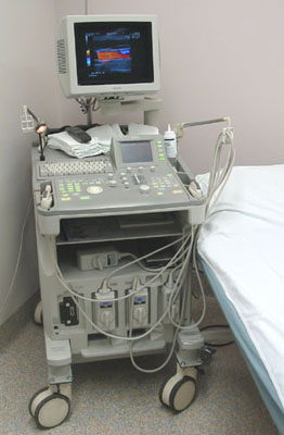
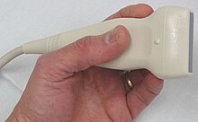
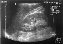
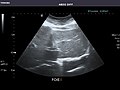
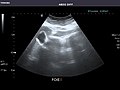
Medical ultrasound includes diagnostic techniques using ultrasound, as well as therapeutic applications of ultrasound. In diagnosis, it is used to create an image of internal body structures such as tendons, muscles, joints, blood vessels, and internal organs, to measure some characteristics or to generate an informative audible sound. The usage of ultrasound to produce visual images for medicine is called medical ultrasonography or simply sonography, or echography. The practice of examining pregnant women using ultrasound is called obstetric ultrasonography, and was an early development of clinical ultrasonography. The machine used is called an ultrasound machine, a sonograph or an echograph. The visual image formed using this technique is called an ultrasonogram, a sonogram or an echogram.

<span class="mw-page-title-main">Gallbladder</span> Organ in humans and other vertebrates

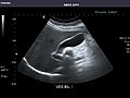
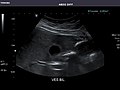
In vertebrates, the gallbladder, also known as the cholecyst, is a small hollow organ where bile is stored and concentrated before it is released into the small intestine. In humans, the pear-shaped gallbladder lies beneath the liver, although the structure and position of the gallbladder can vary significantly among animal species. It receives bile, produced by the liver, via the common hepatic duct, and stores it. The bile is then released via the common bile duct into the duodenum, where the bile helps in the digestion of fats.

<span class="mw-page-title-main">Cholecystitis</span> Inflammation of the gallbladder

Cholecystitis is inflammation of the gallbladder. Symptoms include right upper abdominal pain, pain in the right shoulder, nausea, vomiting, and occasionally fever. Often gallbladder attacks precede acute cholecystitis. The pain lasts longer in cholecystitis than in a typical gallbladder attack. Without appropriate treatment, recurrent episodes of cholecystitis are common. Complications of acute cholecystitis include gallstone pancreatitis, common bile duct stones, or inflammation of the common bile duct.

<span class="mw-page-title-main">Caroli disease</span> Medical condition

Caroli disease is a rare inherited disorder characterized by cystic dilatation of the bile ducts within the liver. There are two patterns of Caroli disease: focal or simple Caroli disease consists of abnormally widened bile ducts affecting an isolated portion of liver. The second form is more diffuse, and when associated with portal hypertension and congenital hepatic fibrosis, is often referred to as "Caroli syndrome". The underlying differences between the two types are not well understood. Caroli disease is also associated with liver failure and polycystic kidney disease. The disease affects about one in 1,000,000 people, with more reported cases of Caroli syndrome than of Caroli disease.

<span class="mw-page-title-main">Interventional radiology</span> Medical subspecialty

Interventional radiology (IR) is a medical specialty that performs various minimally-invasive procedures using medical imaging guidance, such as x-ray fluoroscopy, computed tomography, magnetic resonance imaging, or ultrasound. IR performs both diagnostic and therapeutic procedures through very small incisions or body orifices. Diagnostic IR procedures are those intended to help make a diagnosis or guide further medical treatment, and include image-guided biopsy of a tumor or injection of an imaging contrast agent into a hollow structure, such as a blood vessel or a duct. By contrast, therapeutic IR procedures provide direct treatment—they include catheter-based medicine delivery, medical device placement, and angioplasty of narrowed structures.

<span class="mw-page-title-main">Abdominal pain</span> Stomach aches

Abdominal pain, also known as a stomach ache, is a symptom associated with both non-serious and serious medical issues. Since the abdomen contains most of the body's vital organs, it can be an indicator of a wide variety of diseases. Given that, approaching the examination of a person and planning of a differential diagnosis is extremely important.

<span class="mw-page-title-main">Abdominal aorta</span> Largest artery in the abdomen

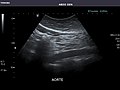
In human anatomy, the abdominal aorta is the largest artery in the abdominal cavity. As part of the aorta, it is a direct continuation of the descending aorta.

<span class="mw-page-title-main">Pancreaticoduodenectomy</span> Major surgical procedure involving the pancreas, duodenum, and other organs

A pancreaticoduodenectomy, also known as a Whipple procedure, is a major surgical operation most often performed to remove cancerous tumours from the head of the pancreas. It is also used for the treatment of pancreatic or duodenal trauma, or chronic pancreatitis. Due to the shared blood supply of organs in the proximal gastrointestinal system, surgical removal of the head of the pancreas also necessitates removal of the duodenum, proximal jejunum, gallbladder, and, occasionally, part of the stomach.

<span class="mw-page-title-main">Common bile duct stone</span> Medical condition

Common bile duct stone, also known as choledocholithiasis, is the presence of gallstones in the common bile duct (CBD). This condition can cause jaundice and liver cell damage. Treatments include choledocholithotomy and endoscopic retrograde cholangiopancreatography (ERCP).

<span class="mw-page-title-main">Renal artery</span> Vessel supplying blood to kidney

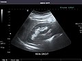
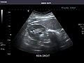
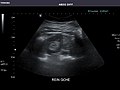
The renal arteries are paired arteries that supply the kidneys with blood. Each is directed across the crus of the diaphragm, so as to form nearly a right angle.

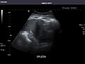
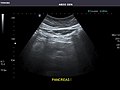
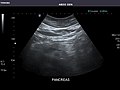
Organomegaly is the abnormal enlargement of organs. For example, cardiomegaly is enlargement of the heart. Visceromegaly is the enlargement of abdominal organs. Examples of visceromegaly are enlarged liver (hepatomegaly), spleen (splenomegaly), stomach, kidneys, and pancreas.

The Kocher manoeuvre is a surgical procedure to expose structures in the retroperitoneum behind the duodenum and pancreas. In vascular surgery, it is described as a method to expose the abdominal aorta. It usually has been in contrast to midline laparotomy and right retroperitoneal space dissection. These two procedures have been used for diverse cases, but have approximately equivalent outcomes.

<span class="mw-page-title-main">Endoscopic ultrasound</span> Medical imaging procedure

Endoscopic ultrasound (EUS) or echo-endoscopy is a medical procedure in which endoscopy is combined with ultrasound to obtain images of the internal organs in the chest, abdomen and colon. It can be used to visualize the walls of these organs, or to look at adjacent structures. Combined with Doppler imaging, nearby blood vessels can also be evaluated.

<span class="mw-page-title-main">Cholescintigraphy</span> Medical imaging of hepatobiliary tract using radiotracers

Cholescintigraphy or hepatobiliary scintigraphy is scintigraphy of the hepatobiliary tract, including the gallbladder and bile ducts. The image produced by this type of medical imaging, called a cholescintigram, is also known by other names depending on which radiotracer is used, such as HIDA scan, PIPIDA scan, DISIDA scan, or BrIDA scan. Cholescintigraphic scanning is a nuclear medicine procedure to evaluate the health and function of the gallbladder and biliary system. A radioactive tracer is injected through any accessible vein and then allowed to circulate to the liver, where it is excreted into the bile ducts and stored by the gallbladder until released into the duodenum.

<span class="mw-page-title-main">Aortic rupture</span> Rupture or breakage of the aorta, the largest artery in the body

Aortic rupture is the rupture or breakage of the aorta, the largest artery in the body. Aortic rupture is a rare, extremely dangerous condition. The most common cause is an abdominal aortic aneurysm that has ruptured spontaneously. Aortic rupture is distinct from aortic dissection, which is a tear through the inner wall of the aorta that can block the flow of blood through the aorta to the heart or abdominal organs.

<span class="mw-page-title-main">Abdominal trauma</span> Medical condition

Abdominal trauma is an injury to the abdomen. Signs and symptoms include abdominal pain, tenderness, rigidity, and bruising of the external abdomen. Complications may include blood loss and infection.

Biliary dyskinesia is a disorder of some component of biliary part of the digestive system in which bile cannot physically move in the proper direction through the tubular biliary tract. It most commonly involves abnormal biliary tract peristalsis muscular coordination within the gallbladder in response to dietary stimulation of that organ to squirt the liquid bile through the common bile duct into the duodenum. Ineffective peristaltic contraction of that structure produces postprandial right upper abdominal pain (cholecystodynia) and almost no other problem. When the dyskinesia is localized at the biliary outlet into the duodenum just as increased tonus of that outlet sphincter of Oddi, the backed-up bile can cause pancreatic injury with abdominal pain more toward the upper left side. In general, biliary dyskinesia is the disturbance in the coordination of peristaltic contraction of the biliary ducts, and/or reduction in the speed of emptying of the biliary tree into the duodenum.

<span class="mw-page-title-main">Sphincter of Oddi dysfunction</span> Medical condition

Sphincter of Oddi dysfunction refers to a group of functional disorders leading to abdominal pain due to dysfunction of the Sphincter of Oddi: functional biliary sphincter of Oddi and functional pancreatic sphincter of Oddi disorder. The sphincter of Oddi is a sphincter muscle, a circular band of muscle at the bottom of the biliary tree which controls the flow of pancreatic juices and bile into the second part of the duodenum. The pathogenesis of this condition is recognized to encompass stenosis or dyskinesia of the sphincter of Oddi ; consequently the terms biliary dyskinesia, papillary stenosis, and postcholecystectomy syndrome have all been used to describe this condition. Both stenosis and dyskinesia can obstruct flow through the sphincter of Oddi and can therefore cause retention of bile in the biliary tree and pancreatic juice in the pancreatic duct.

Cholecystostomy or (cholecystotomy) is a medical procedure used to drain the gallbladder through either a percutaneous or endoscopic approach. The procedure involves creating a stoma in the gallbladder, which can facilitate placement of a tube or stent for drainage, first performed by American surgeon, Dr. John Stough Bobbs, in 1867. It is sometimes used in cases of cholecystitis or other gallbladder disease where the person is ill, and there is a need to delay or defer cholecystectomy. The first endoscopic cholecystostomy was performed by Drs. Todd Baron and Mark Topazian in 2007 using ultrasound guidance to puncture the stomach wall and place a plastic biliary catheter for gallbladder drainage.

Canine gallbladder mucocele (GBM) is an emerging biliary disease in dogs described as the excessive and abnormal accumulation of thick, gelatinous mucus in the lumen, which results in an enlarged gallbladder. GBMs have been diagnosed more frequently in comparison to prior to the 2000s when it was considered rare. The mucus is usually pale yellow to dark green in appearance.

References

  1. Bisset RA, Khan AN, Sabih D (1 January 2008). Differential Diagnosis in Abdominal Ultrasound (3rd ed.). Elsevier India. p. 257. ISBN   978-81-312-1574-6 . Retrieved 10 April 2011.
  2. Whitworth M, Bricker L, Mullan C (July 2015). "Ultrasound for fetal assessment in early pregnancy". The Cochrane Database of Systematic Reviews. 2015 (7): CD007058. doi:10.1002/14651858.CD007058.pub3. PMC   4084925 . PMID   26171896.
  3. Salomon LJ, Alfirevic Z, Bilardo CM, Chalouhi GE, Ghi T, Kagan KO, et al. (January 2013). "ISUOG practice guidelines: performance of first-trimester fetal ultrasound scan". Ultrasound in Obstetrics & Gynecology. 41 (1): 102–113. doi: 10.1002/uog.12342 . PMID   23280739.
  4. 1 2 Jang T (2017-08-28). "Bedside Ultrasonography Evaluation of Abdominal Aortic Aneurysm - Technique". Medscape .
  5. 1 2 American Medical Society for Sports Medicine (24 April 2014), "Five Things Physicians and Patients Should Question", Choosing Wisely: an initiative of the ABIM Foundation , American Medical Society for Sports Medicine, retrieved 29 July 2014, which cites
    • Putukian M, O'Connor FG, Stricker P, McGrew C, Hosey RG, Gordon SM, et al. (July 2008). "Mononucleosis and athletic participation: an evidence-based subject review". Clinical Journal of Sport Medicine. 18 (4): 309–315. doi:10.1097/JSM.0b013e31817e34f8. PMID   18614881. S2CID   23780443.
    • Spielmann AL, DeLong DM, Kliewer MA (January 2005). "Sonographic evaluation of spleen size in tall healthy athletes". AJR. American Journal of Roentgenology. 184 (1): 45–49. doi:10.2214/ajr.184.1.01840045. PMID   15615949.
  6. Dietrich CF, Tuma J, Badea R (2010-07-28). "Ultrasound of the liver - EFSUMB – European Course Book" (PDF). European federation of societies for ultrasound in medicine and biology (EFSUMB). Archived from the original (PDF) on 2017-08-12. Retrieved 2017-12-22.
  7. Content initially copied from: Hansen KL, Nielsen MB, Ewertsen C (December 2015). "Ultrasonography of the Kidney: A Pictorial Review". Diagnostics. 6 (1): 2. doi: 10.3390/diagnostics6010002 . PMC   4808817 . PMID   26838799. (CC-BY 4.0)
  8. Macnaught F, Campbell-Rogers N (August 2009). "The liver: how we do it". Australasian Journal of Ultrasound in Medicine. 12 (3): 44–47. doi:10.1002/j.2205-0140.2009.tb00061.x. PMC   5024840 . PMID   28191063.