Gallbladder

Last updated

Gallbladder
Gallbladder (organ).png
2425 Gallbladder.jpg
The gallbladder sits beneath the liver
Details
Precursor Foregut
System Digestive system
Artery Cystic artery
Vein Cystic vein
Nerve Celiac ganglia, vagus nerve [1]
Identifiers
Latin vesica biliaris, vesica fellea
MeSH D005704
TA98 A05.8.02.001
TA2 3081
FMA 7202
Anatomical terminology

In vertebrates, the gallbladder, also known as the cholecyst, is a small hollow organ where bile is stored and concentrated before it is released into the small intestine. In humans, the pear-shaped gallbladder lies beneath the liver, although the structure and position of the gallbladder can vary significantly among animal species. It receives bile, produced by the liver, via the common hepatic duct, and stores it. The bile is then released via the common bile duct into the duodenum, where the bile helps in the digestion of fats.

Contents

The gallbladder can be affected by gallstones, formed by material that cannot be dissolved – usually cholesterol or bilirubin, a product of hemoglobin breakdown. These may cause significant pain, particularly in the upper-right corner of the abdomen, and are often treated with removal of the gallbladder (called a cholecystectomy). Cholecystitis, inflammation of the gallbladder, has a wide range of causes, including result from the impaction of gallstones, infection, and autoimmune disease.

Structure

The gallbladder is a hollow grey-blue organ that sits in a shallow depression below the right lobe of the liver. [2] In adults, the gallbladder measures approximately 7 to 10 centimetres (2.8 to 3.9 inches) in length and 4 centimetres (1.6 in) in diameter when fully distended. [3] The gallbladder has a capacity of about 50 millilitres (1.8 imperial fluid ounces). [2]

The gallbladder is shaped like a pear, with its tip opening into the cystic duct. [4] The gallbladder is divided into three sections: the fundus, body, and neck. The fundus is the rounded base, angled so that it faces the abdominal wall. The body lies in a depression in the surface of the lower liver. The neck tapers and is continuous with the cystic duct, part of the biliary tree. [2] The gallbladder fossa, against which the fundus and body of the gallbladder lie, is found beneath the junction of hepatic segments IVB and V. [5] The cystic duct unites with the common hepatic duct to become the common bile duct. At the junction of the neck of the gallbladder and the cystic duct, there is an out-pouching of the gallbladder wall forming a mucosal fold known as "Hartmann's pouch". [2]

Lymphatic drainage of the gallbladder follows the cystic node, which is located between the cystic duct and the common hepatic duct. Lymphatics from the lower part of the organ drain into lower hepatic lymph nodes. All the lymph finally drains into celiac lymph nodes.

Microanatomy

Micrograph of a normal gallbladder wall. H&E stain. Gallbladder - intermed mag.jpg
Micrograph of a normal gallbladder wall. H&E stain.

The gallbladder wall is composed of a number of layers. The innermost surface of the gallbladder wall is lined by a single layer of columnar cells with a brush border of microvilli, very similar to intestinal absorptive cells. [2] Underneath the epithelium is an underlying lamina propria, a muscular layer, an outer perimuscular layer and serosa. Unlike elsewhere in the intestinal tract, the gallbladder does not have a muscularis mucosae, and the muscular fibres are not arranged in distinct layers. [6]

The mucosa, the inner portion of the gallbladder wall, consists of a lining of a single layer of columnar cells, with cells possessing small hair-like attachments called microvilli. [2] This sits on a thin layer of connective tissue, the lamina propria. [6] The mucosa is curved and collected into tiny outpouchings called rugae. [2]

A muscular layer sits beneath the mucosa. This is formed by smooth muscle, with fibres that lie in longitudinal, oblique and transverse directions, and are not arranged in separate layers. The muscle fibres here contract to expel bile from the gallbladder. [6] A distinctive feature of the gallbladder is the presence of Rokitansky–Aschoff sinuses, deep outpouchings of the mucosa that can extend through the muscular layer, and which indicate adenomyomatosis. [7] The muscular layer is surrounded by a layer of connective and fat tissue. [2]

The outer layer of the fundus of gallbladder, and the surfaces not in contact with the liver, are covered by a thick serosa, which is exposed to the peritoneum. [2] The serosa contains blood vessels and lymphatics. [6] The surfaces in contact with the liver are covered in connective tissue. [2]

Variation

Abdominal ultrasonography showing gallbladder and common bile duct Gallbladder and common bile duct ultrasound.jpg
Abdominal ultrasonography showing gallbladder and common bile duct

The gallbladder varies in size, shape, and position among different people. [2] Rarely, two or even three gallbladders may coexist, either as separate bladders draining into the cystic duct, or sharing a common branch that drains into the cystic duct. Additionally, the gallbladder may fail to form at all. Gallbladders with two lobes separated by a septum may also exist. These abnormalities are not likely to affect function and are generally asymptomatic. [8]

The location of the gallbladder in relation to the liver may also vary, with documented variants including gallbladders found within, [9] above, on the left side of, behind, and detached or suspended from the liver. Such variants are very rare: from 1886 to 1998, only 110 cases of left-lying liver, or less than one per year, were reported in scientific literature. [10] [11] [2]

An anatomical variation can occur, known as a Phrygian cap, which is an innocuous fold in the fundus, named after its resemblance to the Phrygian cap. [12]

Development

The gallbladder develops from an endodermal outpouching of the embryonic gut tube. [13] Early in development, the human embryo has three germ layers and abuts an embryonic yolk sac. During the second week of embryogenesis, as the embryo grows, it begins to surround and envelop portions of this sac. The enveloped portions form the basis for the adult gastrointestinal tract. Sections of this foregut begin to differentiate into the organs of the gastrointestinal tract, such as the esophagus, stomach, and intestines. [13]

During the fourth week of embryological development, the stomach rotates. The stomach, originally lying in the midline of the embryo, rotates so that its body is on the left. This rotation also affects the part of the gastrointestinal tube immediately below the stomach, which will go on to become the duodenum. By the end of the fourth week, the developing duodenum begins to spout a small outpouching on its right side, the hepatic diverticulum, which will go on to become the biliary tree. Just below this is a second outpouching, known as the cystic diverticulum, that will eventually develop into the gallbladder. [13]

Function

1. Bile ducts: 2. Intrahepatic bile ducts, 3. Left and right hepatic ducts, 4. Common hepatic duct, 5. Cystic duct, 6. Common bile duct, 7. Ampulla of Vater, 8. Major duodenal papilla
9. Gallbladder.
10-11. Right and left lobes of liver.
12. Spleen.
13. Esophagus.
14. Stomach.
15. Pancreas: 16. Accessory pancreatic duct, 17. Pancreatic duct.
18. Small intestine: 19. Duodenum, 20. Jejunum
21-22. Right and left kidneys.
The front border of the liver has been lifted up (brown arrow). Biliary system multilingual.svg
1. Bile ducts : 2. Intrahepatic bile ducts, 3. Left and right hepatic ducts, 4. Common hepatic duct, 5. Cystic duct, 6. Common bile duct, 7. Ampulla of Vater, 8. Major duodenal papilla
9. Gallbladder.
10–11. Right and left lobes of liver.
12. Spleen.
13. Esophagus.
14. Stomach.
15. Pancreas : 16. Accessory pancreatic duct, 17. Pancreatic duct.
18. Small intestine : 19. Duodenum, 20. Jejunum
21–22. Right and left kidneys.
The front border of the liver has been lifted up (brown arrow).

The main functions of the gallbladder are to store and concentrate bile, also called gall, needed for the digestion of fats in food. Produced by the liver, bile flows through small vessels into the larger hepatic ducts and ultimately through the cystic duct (parts of the biliary tree) into the gallbladder, where it is stored. At any one time, 30 to 60 millilitres (1.0 to 2.0 US fl oz) of bile is stored within the gallbladder. [15]

When food containing fat enters the digestive tract, it stimulates the secretion of cholecystokinin (CCK) from I cells of the duodenum and jejunum. In response to cholecystokinin, the gallbladder rhythmically contracts and releases its contents into the common bile duct, eventually draining into the duodenum. The bile emulsifies fats in partly digested food, thereby assisting their absorption. Bile consists primarily of water and bile salts, and also acts as a means of eliminating bilirubin, a product of hemoglobin metabolism, from the body. [15]

The bile that is secreted by the liver and stored in the gallbladder is not the same as the bile that is secreted by the gallbladder. During gallbladder storage of bile, it is concentrated 3-10 fold [16] by removal of some water and electrolytes. This is through the active transport of sodium and chloride ions [17] across the epithelium of the gallbladder, which creates an osmotic pressure that also causes water and other electrolytes to be reabsorbed. [15]

A function of the gallbladder appears to be protection against carcinogenesis as indicated by observations that removal of the gallbladder (cholecystectomy) increases subsequent cancer risk. For instance, a systematic review and meta analysis of eighteen studies concluded that cholecystecomy has a harmful effect on the risk of right-sided colon cancer. [18] Another recent study reported a significantly increased total cancer risk, including increased risk of several different types of cancer, after cholecystectomy. [19]

Clinical significance

Gallstones

3D still showing gallstones Gallbladder stones.jpg
3D still showing gallstones

Gallstones form when the bile is saturated, usually with either cholesterol or bilirubin. [20] Most gallstones do not cause symptoms, with stones either remaining in the gallbladder or passed along the biliary system. [21] When symptoms occur, severe "colicky" pain in the upper right part of the abdomen is often felt. [20] If the stone blocks the gallbladder, inflammation known as cholecystitis may result. If the stone lodges in the biliary system, jaundice may occur; if the stone blocks the pancreatic duct, pancreatitis may occur. [21] Gallstones are diagnosed using ultrasound. [20] When a symptomatic gallstone occurs, it is often managed by waiting for it to be passed naturally. [21] Given the likelihood of recurrent gallstones, surgery to remove the gallbladder is often considered. [21] Some medication, such as ursodeoxycholic acid, may be used; lithotripsy, a non-invasive mechanical procedure used to break down the stones, may also be used. [21]

Inflammation

Known as cholecystitis, inflammation of the gallbladder is commonly caused by obstruction of the duct with gallstones, which is known as cholelithiasis. Blocked bile accumulates, and pressure on the gallbladder wall may lead to the release of substances that cause inflammation, such as phospholipase. There is also the risk of bacterial infection. An inflamed gallbladder is likely to cause sharp and localised pain, fever, and tenderness in the upper, right corner of the abdomen, and may have a positive Murphy's sign. Cholecystitis is often managed with rest and antibiotics, particularly cephalosporins and, in severe cases, metronidazole. Additionally the gallbladder may need to be removed surgically if inflammation has progressed far enough. [21]

Gallbladder removal

A cholecystectomy is a procedure in which the gallbladder is removed. It may be removed because of recurrent gallstones and is considered an elective procedure. A cholecystectomy may be an open procedure, or a laparoscopic one. In the surgery, the gallbladder is removed from the neck to the fundus, [22] and so bile will drain directly from the liver into the biliary tree. About 30 percent of patients may experience some degree of indigestion following the procedure, although severe complications are much rarer. [21] About 10 percent of surgeries lead to a chronic condition of postcholecystectomy syndrome. [23]

Complication

Biliary injury (bile duct injury) is the traumatic damage of the bile ducts. It is most commonly an iatrogenic complication of cholecystectomy — surgical removal of gall bladder, but can also be caused by other operations or by major trauma. The risk of biliary injury is more during laparoscopic cholecystectomy than during open cholecystectomy. Biliary injury may lead to several complications and may even cause death if not diagnosed in time and managed properly. Ideally biliary injury should be managed at a center with facilities and expertise in endoscopy, radiology and surgery. [24]

Biloma is collection of bile within the abdominal cavity. It happens when there is a bile leak, for example after surgery for removing the gallbladder (laparoscopic cholecystectomy), with an incidence of 0.3–2%. Other causes are biliary surgery, liver biopsy, abdominal trauma, and, rarely, spontaneous perforation. [25]

Cancer

Cancer of the gallbladder is uncommon and mostly occurs in later life. When cancer occurs, it is mostly of the glands lining the surface of the gallbladder (adenocarcinoma). [21] Gallstones are thought to be linked to the formation of cancer. Other risk factors include large (>1 cm) gallbladder polyps and having a highly calcified "porcelain" gallbladder. [21]

Cancer of the gallbladder can cause attacks of biliary pain, yellowing of the skin (jaundice), and weight loss. A large gallbladder may be able to be felt in the abdomen. Liver function tests may be elevated, particularly involving GGT and ALP, with ultrasound and CT scans being considered medical imaging investigations of choice. [21] Cancer of the gallbladder is managed by removing the gallbladder, however, as of 2010, the prognosis remains poor. [21]

Cancer of the gallbladder may also be found incidentally after surgical removal of the gallbladder, with 1–3% of cancers identified in this way. Gallbladder polyps are mostly benign growths or lesions resembling growths that form in the gallbladder wall, [26] and are only associated with cancer when they are larger in size (>1 cm). [21] Cholesterol polyps, often associated with cholesterolosis ("strawberry gallbladder", a change in the gallbladder wall due to excess cholesterol [27] ), often cause no symptoms and are thus often detected in this way. [21]

Tests

Abdominal ultrasonography showing biliary sludge and gallstones Ultrasonography of sludge and gallstones, annotated.jpg
Abdominal ultrasonography showing biliary sludge and gallstones

Tests used to investigate for gallbladder disease include blood tests and medical imaging. A full blood count may reveal an increased white cell count suggestive of inflammation or infection. Tests such as bilirubin and liver function tests may reveal if there is inflammation linked to the biliary tree or gallbladder, and whether this is associated with inflammation of the liver, and a lipase or amylase may be elevated if there is pancreatitis. Bilirubin may rise when there is obstruction of the flow of bile. A CA 19-9 level may be taken to investigate for cholangiocarcinoma. [21]

An ultrasound is often the first medical imaging test performed when gallbladder disease such as gallstones are suspected. [21] An abdominal X-ray or CT scan is another form of imaging that may be used to examine the gallbladder and surrounding organs. [21] Other imaging options include MRCP (magnetic resonance cholangiopancreatography), ERCP and percutaneous or intraoperative cholangiography. [21] A cholescintigraphy scan is a nuclear imaging procedure used to assess the condition of the gallbladder. [28]

Other animals

Most vertebrates have gallbladders, but the form and arrangement of the bile ducts may vary considerably. In many species, for example, there are several separate ducts running to the intestine, rather than the single common bile duct found in humans. Several species of mammals (including horses, deer, rats, and laminoids), [29] [30] several species of birds (such as pigeons and some psittacine species), lampreys and all invertebrates do not have a gallbladder. [31] [32]

The bile from several species of bears is used in traditional Chinese medicine; bile bears are kept alive in captivity while their bile is extracted, in an industry characterized by animal cruelty. [33] [34]

History

Depictions of the gallbladder and biliary tree are found in Babylonian models found from 2000 BCE, and in ancient Etruscan model from 200 BCE, with models associated with divine worship. [35]

Diseases of the gallbladder are known to have existed in humans since antiquity, with gallstones found in the mummy of Princess Amenen of Thebes dating to 1500 BCE. [35] [36] Some historians believe the death of Alexander the Great may have been associated with an acute episode of cholecystitis. [35] The existence of the gallbladder has been noted since the 5th century, but it is only relatively recently that the function and the diseases of the gallbladder has been documented, [36] particularly in the last two centuries. [35]

The first descriptions of gallstones appear to have been in the Renaissance, perhaps because of the low incidence of gallstones in earlier times owing to a diet with more cereals and vegetables and less meat. [37] Anthonius Benevinius in 1506 was the first to draw a connection between symptoms and the presence of gallstones. [37] Ludwig Georg Courvoisier, after examining a number of cases in 1890 that gave rise to the eponymous Courvoisier's law, stated that in an enlarged, nontender gallbladder, the cause of jaundice is unlikely to be gallstones. [35]

The first surgical removal of a gallstone (cholecystolithotomy) was in 1676 by physician Joenisius, who removed the stones from a spontaneously occurring biliary fistula. [35] Stough Hobbs in 1867 performed the first recorded cholecystotomy, [37] although such an operation was in fact described earlier by French surgeon Jean Louis Petit in the mid eighteenth century. [35] German surgeon Carl Langenbuch performed the first cholecystectomy in 1882 for a sufferer of cholelithiasis. [36] Before this, surgery had focused on creating a fistula for drainage of gallstones. [35] Langenbuch reasoned that given several other species of mammal have no gallbladder, humans could survive without one. [35]

The debate whether surgical removal of the gallbladder or simply gallstones was preferred was settled in the 1920s, with the consensus that removal of the gallbladder was preferred. [36] It was only in the mid and late parts of the twentieth century that medical imaging techniques such as use of contrast medium and CT scans were used to view the gallbladder. [35] The first laparoscopic cholecystectomy performed by Erich Mühe of Germany in 1985, although French surgeons Phillipe Mouret and Francois Dubois are often credited for their operations in 1987 and 1988 respectively. [38]

Society and culture

To have "gall" is associated with bold, belligerent behaviour, whereas to have "bile" is associated with sourness. [39]

In the Chinese medicine, the gallbladder ( ) is associated with the Wuxing element of wood, in excess its emotion is belligerence and in deficiency cowardice and judgement, in the Chinese language it is related to a myriad of idioms, including using terms such as "a body completely [of] gall" (渾身是膽) to describe a forward person, and "single, alone gallbladder hero" (孤膽英雄) to describe a lone hero, or "they have a lot of gall to talk like that". [40]

In the Zangfu theory of Chinese medicine it is an extraordinary Fu or yang organ, as it holds bile. The gallbladder not only has a digestive role, but is seen as the seat of decision-making and judgement. [40]

See also

Related Research Articles

<span class="mw-page-title-main">Bile</span> Dark greenish-brown fluid aiding in the digestion of fats

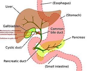
Bile, or gall, is a yellow-green fluid produced by the liver of most vertebrates that aids the digestion of lipids in the small intestine. In humans, bile is primarily composed of water, produced continuously by the liver, and stored and concentrated in the gallbladder. After a human eats, this stored bile is discharged into the first section of their small intestine.

<span class="mw-page-title-main">Bile duct</span> Type of organ

A bile duct is any of a number of long tube-like structures that carry bile, and is present in most vertebrates. The bile duct is separated into three main parts: the fundus (superior), the body (middle), and the neck (inferior).

<span class="mw-page-title-main">Gallstone</span> Disease where stones form in the gallbladder

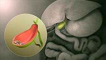
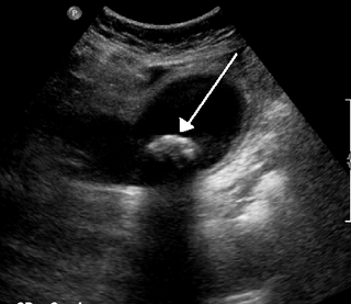
A gallstone is a stone formed within the gallbladder from precipitated bile components. The term cholelithiasis may refer to the presence of gallstones or to any disease caused by gallstones, and choledocholithiasis refers to the presence of migrated gallstones within bile ducts.

<span class="mw-page-title-main">Cholecystitis</span> Inflammation of the gallbladder

Cholecystitis is inflammation of the gallbladder. Symptoms include right upper abdominal pain, pain in the right shoulder, nausea, vomiting, and occasionally fever. Often gallbladder attacks precede acute cholecystitis. The pain lasts longer in cholecystitis than in a typical gallbladder attack. Without appropriate treatment, recurrent episodes of cholecystitis are common. Complications of acute cholecystitis include gallstone pancreatitis, common bile duct stones, or inflammation of the common bile duct.

<span class="mw-page-title-main">Cholecystectomy</span> Surgical removal of the gallbladder

Cholecystectomy is the surgical removal of the gallbladder. Cholecystectomy is a common treatment of symptomatic gallstones and other gallbladder conditions. In 2011, cholecystectomy was the eighth most common operating room procedure performed in hospitals in the United States. Cholecystectomy can be performed either laparoscopically, or via an open surgical technique.

<span class="mw-page-title-main">Mirizzi's syndrome</span> Medical condition

Mirizzi's syndrome is a rare complication in which a gallstone becomes impacted in the cystic duct or neck of the gallbladder causing compression of the common hepatic duct, resulting in obstruction and jaundice. The obstructive jaundice can be caused by direct extrinsic compression by the stone or from fibrosis caused by chronic cholecystitis (inflammation). A cholecystocholedochal fistula can occur.

Courvoisier's principle states that a painless palpably enlarged gallbladder accompanied with mild jaundice is unlikely to be caused by gallstones. Usually, the term is used to describe the physical examination finding of the right-upper quadrant of the abdomen. This sign implicates possible malignancy of the gallbladder or pancreas and the swelling is unlikely due to gallstones.

<span class="mw-page-title-main">Gastrointestinal disease</span> Medical condition

Gastrointestinal diseases refer to diseases involving the gastrointestinal tract, namely the esophagus, stomach, small intestine, large intestine and rectum, and the accessory organs of digestion, the liver, gallbladder, and pancreas.

<span class="mw-page-title-main">Common bile duct stone</span> Medical condition

Common bile duct stone, also known as choledocholithiasis, is the presence of gallstones in the common bile duct (CBD). This condition can cause jaundice and liver cell damage. Treatments include choledocholithotomy and endoscopic retrograde cholangiopancreatography (ERCP).

<span class="mw-page-title-main">Common hepatic duct</span> Exocrine duct

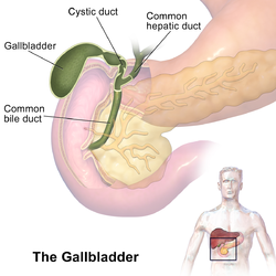
The common hepatic duct is the first part of the biliary tract. It joins the cystic duct coming from the gallbladder to form the common bile duct.

<span class="mw-page-title-main">Gallbladder cancer</span> Medical condition

Gallbladder cancer is a relatively uncommon cancer, with an incidence of fewer than 2 cases per 100,000 people per year in the United States. It is particularly common in central and South America, central and eastern Europe, Japan and northern India; it is also common in certain ethnic groups e.g. Native American Indians and Hispanics. If it is diagnosed early enough, it can be cured by removing the gallbladder, part of the liver and associated lymph nodes. Most often it is found after symptoms such as abdominal pain, jaundice and vomiting occur, and it has spread to other organs such as the liver.

<span class="mw-page-title-main">Ascending cholangitis</span> Medical condition

Ascending cholangitis, also known as acute cholangitis or simply cholangitis, is inflammation of the bile duct, usually caused by bacteria ascending from its junction with the duodenum. It tends to occur if the bile duct is already partially obstructed by gallstones.

<span class="mw-page-title-main">Biliary colic</span> Medical condition in which gallstones cause acute pain

Biliary colic, also known as symptomatic cholelithiasis, a gallbladder attack or gallstone attack, is when a colic occurs due to a gallstone temporarily blocking the cystic duct. Typically, the pain is in the right upper part of the abdomen, and can be severe. Pain usually lasts from 15 minutes to a few hours. Often, it occurs after eating a heavy meal, or during the night. Repeated attacks are common. Cholecystokinin - a gastrointestinal hormone - plays a role in the colic, as following the consumption of fatty meals, the hormone triggers the gallbladder to contract, which may expel stones into the duct and temporarily block it until being successfully passed.

<span class="mw-page-title-main">Biliary tract</span> Organ system

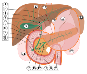
The biliary tract refers to the liver, gallbladder and bile ducts, and how they work together to make, store and secrete bile. Bile consists of water, electrolytes, bile acids, cholesterol, phospholipids and conjugated bilirubin. Some components are synthesized by hepatocytes ; the rest are extracted from the blood by the liver.

<span class="mw-page-title-main">Gallbladder disease</span> Medical condition

Gallbladder diseases are diseases involving the gallbladder and is closely linked to biliary disease, with the most common cause being gallstones (cholelithiasis).

<span class="mw-page-title-main">Choledochal cysts</span> Medical condition

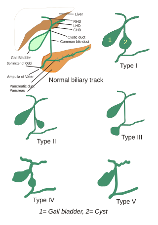
Choledochal cysts are congenital conditions involving cystic dilatation of bile ducts. They are uncommon in western countries but not as rare in East Asian nations like Japan and China.

Biliary injury is the traumatic damage of the bile ducts. It is most commonly an iatrogenic complication of cholecystectomy, but can also be caused by other operations or by major trauma. The risk of biliary injury is higher during laparoscopic cholecystectomy than during open cholecystectomy. Biliary injury may lead to several complications and may even cause death if not diagnosed in time and managed properly. Ideally biliary injury should be managed at a center with facilities and expertise in endoscopy, radiology and surgery.

The cystic node is the sentinel lymph node of the gall bladder. It is located within the cystohepatic triangle.

Cholecystostomy or (cholecystotomy) is a medical procedure used to drain the gallbladder through either a percutaneous or endoscopic approach. The procedure involves creating a stoma in the gallbladder, which can facilitate placement of a tube or stent for drainage, first performed by American surgeon, Dr. John Stough Bobbs, in 1867. It is sometimes used in cases of cholecystitis or other gallbladder disease where the person is ill, and there is a need to delay or defer cholecystectomy. The first endoscopic cholecystostomy was performed by Drs. Todd Baron and Mark Topazian in 2007 using ultrasound guidance to puncture the stomach wall and place a plastic biliary catheter for gallbladder drainage.

<span class="mw-page-title-main">Choledochoduodenostomy</span>

Choledochoduodenostomy (CDD) is a surgical procedure to create an anastomosis, a surgical connection, between the common bile duct (CBD) and an alternative portion of the duodenum. In healthy individuals, the CBD meets the pancreatic duct at the ampulla of Vater, which drains via the major duodenal papilla to the second part of duodenum. In cases of benign conditions such as narrowing of the distal CBD or recurrent CBD stones, performing a CDD provides the diseased patient with CBD drainage and decompression. A side-to-side anastomosis is usually performed.

References

  1. Ginsburg, Ph.D., J.N. (August 22, 2005). "Control of Gastrointestinal Function". In Thomas M. Nosek, Ph.D. (ed.). Gastrointestinal Physiology. Essentials of Human Physiology. Augusta, Georgia, United States: Medical College of Georgia. pp.  p. 30. Archived from the original on April 1, 2008. Retrieved June 29, 2007.
  2. 1 2 3 4 5 6 7 8 9 10 11 12 Gray's Anatomy 2008, p. 1187-81.
  3. Jon W. Meilstrup (1994). Imaging Atlas of the Normal Gallbladder and Its Variants. Boca Raton: CRC Press. p. 4. ISBN   978-0-8493-4788-7.
  4. Nagral, Sanjay (2005). "Anatomy relevant to cholecystectomy". Journal of Minimal Access Surgery. 1 (2): 53–8. doi: 10.4103/0972-9941.16527 . PMC   3004105 . PMID   21206646.
  5. Shakelford's Surgery of Alimentary Tract, ed.7. 2013
  6. 1 2 3 4 Young, Barbara; et al. (2006). Wheater's functional histology: a text and colour atlas (5th ed.). [Edinburgh?]: Churchill Livingstone/Elsevier. p.  298. ISBN   978-0-443-06850-8.
  7. Ross, M.; Pawlina, W. (2011). Histology: A Text and Atlas (6th ed.). Lippincott Williams & Wilkins. p.  646. ISBN   978-0-7817-7200-6.
  8. Leeuw, Th.G.; Verbeek, P.C.M.; Rauws, E.A.J.; Gouma, D.J. (September 1995). "A double or bilobar gallbladder as a cause of severe complications after (laparoscopic) cholecystectomy". Surgical Endoscopy. 9 (9): 998–1000. doi:10.1007/BF00188459. PMID   7482221. S2CID   2581053.
  9. Segura-Sampedro, JJ; Navarro-Sánchez, A; Ashrafian, H; Martínez-Isla, A (February 2015). "Laparoscopic approach to the intrahepatic gallbladder. A case report". Revista Espanola de Enfermedades Digestivas. 107 (2): 122–3. PMID   25659400. Archived from the original on March 4, 2016.
  10. Dhulkotia, A; Kumar, S; Kabra, V; Shukla, HS (March 1, 2002). "Aberrant gallbladder situated beneath the left lobe of liver". HPB. 4 (1): 39–42. doi:10.1080/136518202753598726. PMC   2023911 . PMID   18333151.
  11. Naganuma, S.; Ishida, H.; Konno, K.; Hamashima, Y.; Hoshino, T.; Naganuma, H.; Komatsuda, T.; Ohyama, Y.; Yamada, N.; Ishida, J.; Masamune, O. (March 6, 2014). "Sonographic findings of anomalous position of the gallbladder". Abdominal Imaging. 23 (1): 67–72. doi:10.1007/s002619900287. PMID   9437066. S2CID   30176379.
  12. Meilstrup JW; Hopper KD; Thieme GA (December 1991). "Imaging of gallbladder variants". AJR Am J Roentgenol. 157 (6): 1205–8. doi:10.2214/ajr.157.6.1950867. PMID   1950867.
  13. 1 2 3 Gary C. Schoenwolf; et al. (2009). Larsen's human embryology (Thoroughly rev. and updated 4th ed.). Philadelphia: Churchill Livingstone/Elsevier. pp. Development of the Gastrointestinal Tract. ISBN   978-0-443-06811-9.
  14. Standring S, Borley NR, eds. (2008). Gray's anatomy : the anatomical basis of clinical practice. Brown JL, Moore LA (40th ed.). London: Churchill Livingstone. pp. 1163, 1177, 1185–6. ISBN   978-0-8089-2371-8.
  15. 1 2 3 Hall, Arthur C. Guyton, John E. (2005). Textbook of medical physiology (11th ed.). Philadelphia: W.B. Saunders. pp. 802–804. ISBN   978-0-7216-0240-0.{{cite book}}: CS1 maint: multiple names: authors list (link)
  16. KO, CYNTHIA (2005). "Biliary Sludge Is Formed by Modification of Hepatic Bile by the Gallbladder Mucosa". Clinical Gastroenterology and Hepatology. 3 (7): 672–8. doi:10.1016/s1542-3565(05)00369-1. PMID   16206500. S2CID   27488720.
  17. Meyer, G.; Guizzardi, F.; Rodighiero, S.; Manfredi, R.; Saino, S.; Sironi, C.; Garavaglia, M. L.; Bazzini, C.; Bottà, G. (June 2005). "Ion transport across the gallbladder epithelium". Current Drug Targets. Immune, Endocrine and Metabolic Disorders. 5 (2): 143–151. doi:10.2174/1568008054064805. ISSN   1568-0088. PMID   16089346.
  18. Mu L, Li W, Ren W, Hu D, Song Y. The association between cholecystectomy and the risk of colorectal cancer: an updated systematic review and meta-analysis of cohort studies. Transl Cancer Res. 2023 Jun 30;12(6):1452-1465. doi: 10.21037/tcr-22-2049. Epub 2023 May 22. PMID: 37434692; PMCID: PMC10331452
  19. Choi YJ, Jin EH, Lim JH, Shin CM, Kim N, Han K, Lee DH. Increased Risk of Cancer after Cholecystectomy: A Nationwide Cohort Study in Korea including 123,295 Patients. Gut Liver. 2022 May 15;16(3):465-473. doi: 10.5009/gnl210009. PMID: 35502586; PMCID: PMC9099388
  20. 1 2 3 "Cholelithiasis - Hepatic and Biliary Disorders - MSD Manual Professional Edition". MSD Manual Professional Edition. Retrieved October 18, 2017.
  21. 1 2 3 4 5 6 7 8 9 10 11 12 13 14 15 16 17 Nicki R. Colledge; Brian R. Walker; Stuart H. Ralston, eds. (2010). Davidson's principles and practice of medicine (21st ed.). Edinburgh: Churchill Livingstone/Elsevier. pp. 977–984. ISBN   978-0-7020-3085-7.
  22. Neri V; Ambrosi A; Fersini A; Tartaglia N; Valentino TP (2007). "Antegrade dissection in laparoscopic cholecystectomy". Journal of the Society of Laparoendoscopic Surgeons. 11 (2): 225–8. PMC   3015719 . PMID   17761085.
  23. nhs.uk, Complications of a gallbladder removal
  24. VK Kapoor (2007), "Bile duct injury repair: when? what? who?", Journal of Hepato-Biliary-Pancreatic Surgery, 14 (5): 476–479, doi:10.1007/s00534-007-1220-y, PMID   17909716
  25. Norton J. Greenberger; et al. (2009), "Endoscopic Management of Acute Biliary & Pancreatic Conditions", Gastroenterology, Hepatology, and Endoscopy, Current Medical Diagnosis and Treatment, pp. 355–356
  26. "Gallbladder Polyps". MayoClinic. Retrieved March 19, 2015.
  27. Strawberry gallbladder – cancerweb.ncl.ac.uk.
  28. "HIDA scan - Overview". Mayo Clinic. Retrieved October 18, 2017.
  29. C. Michael Hogan. 2008. Guanaco: Lama guanicoe, GlobalTwitcher.com, ed. N. Strömberg Archived March 4, 2011, at the Wayback Machine
  30. Higashiyama, H; Sumitomo, H; Ozawa, A; Igarashi, H; Tsunekawa, N; Kurohmaru, M; Kanai, Y (2016). "Anatomy of the Murine Hepatobiliary System: A Whole-Organ-Level Analysis Using a Transparency Method". The Anatomical Record. 299 (2): 161–172. doi: 10.1002/ar.23287 . PMID   26559382.
  31. Romer, Alfred Sherwood; Parsons, Thomas S. (1977). The Vertebrate Body. Philadelphia, PA: Holt-Saunders International. p. 355. ISBN   978-0-03-910284-5.
  32. Hagey, L. R.; Vidal, N.; Hofmann, A. F.; Krasowski, M. D. (2010). "Complex Evolution of Bile Salts in Birds". The Auk. 127 (4): 820–831. doi: 10.1525/auk.2010.09155 . PMC   2990222 . PMID   21113274.
  33. Actman, Jani (May 5, 2016). "Inside the Disturbing World of Bear-Bile Farming". National Geographic. Archived from the original on May 5, 2016. Retrieved October 23, 2017.
  34. Hance, Jeremy (April 9, 2015). "Is the end of 'house of horror' bear bile factories in sight?". The Guardian. Retrieved October 23, 2017.
  35. 1 2 3 4 5 6 7 8 9 10 Eachempati, Soumitra R.; II, R. Lawrence Reed (2015). Acute Cholecystitis. Springer. pp. 1–16. ISBN   9783319148243.
  36. 1 2 3 4 Jarnagin, William R. (2012). Blumgart's Surgery of the Liver, Pancreas and Biliary Tract E-Book: Expert Consult - Online. Elsevier Health Sciences. p. 511. ISBN   978-1455746064.
  37. 1 2 3 Bateson, M. C. (2012). Gallstone Disease and its Management. Springer Science & Business Media. pp. 1–2. ISBN   9789400941731.
  38. Reynolds, Walker (January–March 2001). "The First Laparoscopic Cholecystectomy". Journal of the Society of Laparoendoscopic Surgeons. 5 (1): 89–94. PMC   3015420 . PMID   11304004.
  39. Lifang, Qu (2020). Chinese Medicine Psychology: A Clinical Guide to Mental and Emotional Wellness. Jessica Kingsley Publishers. ISBN   9781787752764.
  40. 1 2 Yu, Ning (January 1, 2003). "Metaphor, Body, and Culture: The Chinese Understanding of Gallbladder and Courage". Metaphor and Symbol. 18 (1): 13–31. doi:10.1207/S15327868MS1801_2. S2CID   143595915.
Books