| Punctate epithelial erosions | |
|---|---|
| Other names | Punctate erosive keratopathy or Superficial punctate keratitis |
 | |
| This condition affects the cornea | |
| Specialty | Ophthalmology |
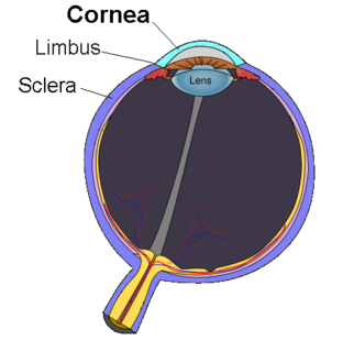
Punctate epithelial erosions are a pathology affecting the cornea.
| Punctate epithelial erosions | |
|---|---|
| Other names | Punctate erosive keratopathy or Superficial punctate keratitis |
| | |
| This condition affects the cornea | |
| Specialty | Ophthalmology |
Punctate epithelial erosions are a pathology affecting the cornea.
It is a characterized by a breakdown or damage of the epithelium of the cornea in a pinpoint pattern, which can be seen with examination with a slit-lamp. Patients may present with non-specific symptoms such as red eye, tearing, foreign body sensation, photophobia and burning.
Punctate epithelial erosions may be seen with different disorders:
| | This section is empty. You can help by adding to it. (September 2017) |
Slit lamp examination
Due to the different underlying causes, proper diagnosis, treatment, and prognosis can only be determined by an eye care professional. Punctate epithelial erosions may be treated with artificial tears. In some disorders, topical antibiotic is added to the treatment. Patients should discontinue contact lens wear until recovery.

Keratoconus (KC) is a disorder of the eye that results in progressive thinning of the cornea. This may result in blurry vision, double vision, nearsightedness, irregular astigmatism, and light sensitivity leading to poor quality-of-life. Usually both eyes are affected. In more severe cases a scarring or a circle may be seen within the cornea.

Keratitis is a condition in which the eye's cornea, the clear dome on the front surface of the eye, becomes inflamed. The condition is often marked by moderate to intense pain and usually involves any of the following symptoms: pain, impaired eyesight, photophobia, red eye and a 'gritty' sensation.

Dry eye syndrome (DES), also known as keratoconjunctivitis sicca (KCS), is the condition of having dry eyes. Other associated symptoms include irritation, redness, discharge, and easily fatigued eyes. Blurred vision may also occur. Symptoms range from mild and occasional to severe and continuous. Scarring of the cornea may occur in untreated cases.

Thygeson's superficial punctate keratopathy (TSPK) is a disease of the eyes. The causes of TSPK are not currently known, but details of the disease were first published in the Journal of the American Medical Association in 1950 by renowned American ophthalmologist Phillips Thygeson (1903–2002), after whom it is named.

A slit lamp is an instrument consisting of a high-intensity light source that can be focused to shine a thin sheet of light into the eye. It is used in conjunction with a biomicroscope. The lamp facilitates an examination of the anterior segment and posterior segment of the human eye, which includes the eyelid, sclera, conjunctiva, iris, natural crystalline lens, and cornea. The binocular slit-lamp examination provides a stereoscopic magnified view of the eye structures in detail, enabling anatomical diagnoses to be made for a variety of eye conditions. A second, hand-held lens is used to examine the retina.
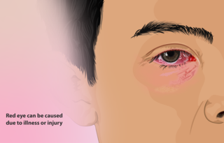
A red eye is an eye that appears red due to illness or injury. It is usually injection and prominence of the superficial blood vessels of the conjunctiva, which may be caused by disorders of these or adjacent structures. Conjunctivitis and subconjunctival hemorrhage are two of the less serious but more common causes.
Fuchs dystrophy, also referred to as Fuchs endothelial corneal dystrophy (FECD) and Fuchs endothelial dystrophy (FED), is a slowly progressing corneal dystrophy that usually affects both eyes and is slightly more common in women than in men. Although early signs of Fuchs dystrophy are sometimes seen in people in their 30s and 40s, the disease rarely affects vision until people reach their 50s and 60s.
Recurrent corneal erosion is a disorder of the eyes characterized by the failure of the cornea's outermost layer of epithelial cells to attach to the underlying basement membrane. The condition is excruciatingly painful because the loss of these cells results in the exposure of sensitive corneal nerves. This condition can often leave patients with temporary blindness due to extreme light sensitivity (photophobia).

Corneal abrasion is a scratch to the surface of the cornea of the eye. Symptoms include pain, redness, light sensitivity, and a feeling like a foreign body is in the eye. Most people recover completely within three days.

Corneal dystrophy is a group of rare hereditary disorders characterised by bilateral abnormal deposition of substances in the transparent front part of the eye called the cornea.

Acanthamoeba keratitis (AK) is a rare disease in which amoebae of the genus Acanthamoeba invade the clear portion of the front (cornea) of the eye. It affects roughly 100 people in the United States each year. Acanthamoeba are protozoa found nearly ubiquitously in soil and water and can cause infections of the skin, eyes, and central nervous system.

A pinguecula is a common type of conjunctival stromal degeneration in the eye. It appear as an elevated yellow-white plaque in the bulbar conjunctiva near to limbus. Calcification may also seen occasionally.

Corneal ulcer is an inflammatory or, more seriously, infective condition of the cornea involving disruption of its epithelial layer with involvement of the corneal stroma. It is a common condition in humans particularly in the tropics and the agrarian societies. In developing countries, children afflicted by Vitamin A deficiency are at high risk for corneal ulcer and may become blind in both eyes, which may persist lifelong. In ophthalmology, a corneal ulcer usually refers to having an infectious cause while the term corneal abrasion refers more to physical abrasions.

Band keratopathy is a corneal disease derived from the appearance of calcium on the central cornea. This is an example of metastatic calcification, which by definition, occurs in the presence of hypercalcemia.

Meesmann corneal dystrophy (MECD) is a rare hereditary autosomal dominant disease that is characterized as a type of corneal dystrophy and a keratin disease. MECD is characterized by the formation of microcysts in the outermost layer of the cornea, known as the anterior corneal epithelium. The anterior corneal epithelium also becomes fragile. This usually affects both eyes rather than a single eye and worsens over time. There are two phenotypes, Meesmann corneal dystrophy 1 (MECD1) and Meesmann corneal dystrophy 2 (MECD2), which affect the genes KRT3 and KRT12, respectively. A heterozygous mutation in either of these genes will lead to a single phenotype. Many with Meesmann corneal dystrophy are asymptomatic or experience mild symptoms.

Conjunctivochalasis is a common eye surface condition characterized by the presence of excess folds of the conjunctiva located between the globe of the eye and the eyelid margin.

Herpetic simplex keratitis is a form of keratitis caused by recurrent herpes simplex virus (HSV) infection in the cornea.
Neurotrophic keratitis (NK) is a degenerative disease of the cornea caused by damage of the trigeminal nerve, which results in impairment of corneal sensitivity, spontaneous corneal epithelium breakdown, poor corneal healing and development of corneal ulceration, melting and perforation. This is because, in addition to the primary sensory role, the nerve also plays a role maintaining the integrity of the cornea by supplying it with trophic factors and regulating tissue metabolism.
Tear break-up time (TBUT) also known as tear film break-up time (TFBUT) is the time taken to appear first dry spot on cornea after a complete blinking. TFBUT measurement is an easy and fast method used to assess the stability of tear film. It is a standard diagnostic procedure in the dry eye clinics. The volume of tear in the eye depends on two factors, drainage through the lacrimal passages and evaporation. Factors like decreased tear production, increased evaporation rate, tearfilm instability, tear hyperosmolarity, inflammations, ocular surface damages etc. can cause dryness to the eyes.
Exposure keratopathy is medical condition affecting the cornea of eyes. It can lead to corneal ulceration and permanent loss of vision due to corneal opacity.