Vascular surgery

Last updated

Vascular surgery
SurgicalAAA 01 Base 275.jpg
Open infrarenal aortic repair model
ICD-9-CM 38-39
MeSH D014656
OPS-301 code 5-38...5-39

Vascular surgery is a surgical subspecialty in which vascular diseases involving the arteries, veins, or lymphatic vessels, are managed by medical therapy, minimally-invasive catheter procedures and surgical reconstruction. The specialty evolved from general and cardiovascular surgery where it refined the management of just the vessels, no longer treating the heart or other organs. Modern vascular surgery includes open surgery techniques, endovascular (minimally invasive) techniques and medical management of vascular diseases - unlike the parent specialities. The vascular surgeon is trained in the diagnosis and management of diseases affecting all parts of the vascular system excluding the coronaries and intracranial vasculature. Vascular surgeons also are called to assist other physicians to carry out surgery near vessels, or to salvage vascular injuries that include hemorrhage control, dissection, occlusion or simply for safe exposure of vascular structures. [1]

Contents

History

Early leaders of the field included Russian surgeon Nikolai Korotkov, noted for developing early surgical techniques, American interventional radiologist Charles Theodore Dotter who is credited with inventing minimally invasive angioplasty (1964), and Australian Robert Paton, who helped the field achieve recognition as a specialty. Edwin Wylie of San Francisco was one of the early American pioneers who developed and fostered advanced training in vascular surgery and pushed for its recognition as a specialty in the United States in the 1970s. The most notable historic figure in vascular surgery is the 1912 Nobel Prize winning surgeon, Alexis Carrel for his techniques used to suture vessels.

Evolution

Medical science has advanced significantly since 1507, when Leonardo da Vinci drew this diagram of the internal organs and vascular systems of a woman. DaVinciCardiovascularWoman.jpg
Medical science has advanced significantly since 1507, when Leonardo da Vinci drew this diagram of the internal organs and vascular systems of a woman.

The specialty continues to be based on operative arterial and venous surgery but since the early 1990s has evolved greatly. There is now considerable emphasis on minimally invasive alternatives to surgery. The field was originally pioneered by interventional radiologists like Dr. Charles Dotter, who invented angioplasty using serial dilatation of vessels.

The surgeon Dr. Thomas J. Fogarty invented a balloon catheter, designed to remove clots from occluded vessels, which was used as the eventual model to do endovascular angioplasty. Further development of the field has occurred via joint efforts between interventional radiology, vascular surgery, and interventional cardiology. This area of vascular surgery is called Endovascular Surgery or Interventional Vascular Radiology, a term that some in the specialty append to their primary qualification as Vascular Surgeon. Endovascular and endovenous procedures (e.g., EVAR) can now form the bulk of a vascular surgeon's practice.

The treatment of the aorta, the body's largest artery, dates back to Greek surgeon Antyllus, who first performed surgeries for various aneurysms in the second century AD. Modern treatment of aortic diseases stems from development and advancements from Michael DeBakey and Denton Cooley. In 1955, DeBakey and Cooley performed the first replacement of a thoracic aneurysm with a homograft. In 1958, they began using the Dacron graft, resulting in a revolution for surgeons in the repair of aortic aneurysms. He also was first to perform cardiopulmonary bypass to repair the ascending aorta, using antegrade perfusion of the brachiocephalic artery.

Dr. Ted Diethrich, one of Dr. DeBakey's associates, went on to pioneer many of the minimally invasive techniques that later became hallmarks of endovascular surgery. [2] Dietrich later founded the Arizona Heart Hospital in 1998 and served as its medical director from 1998 to 2010. In 2000, Diethrich performed the first endovascular aneurysm repair (EVAR) for ruptured abdominal aortic aneurysm. Dietrich trained several future leaders in the field of endovascular surgery at the Arizona Heart Hospital including Venkatesh Ramaiah, MD [3] who served as medical director of the institution following Dietrich's death in 2017. [4]

The development of endovascular surgery has been accompanied by a gradual separation of vascular surgery from its origin in general surgery. Most vascular surgeons would now confine their practice to vascular surgery and, similarly, general surgeons would not be trained or practise the larger vascular surgery operations or most endovascular procedures. More recently, professional vascular surgery societies and their training program have formally separated vascular surgery into a separate specialty with its own training program, meetings and accreditation. Notable societies are Society for Vascular Surgery (SVS), USA; Australia and New Zealand Society of Vascular Surgeons (ANZSVS). Local societies also exist (e.g., New South Wales Vascular and Melbourne Vascular Surgical Association (MVSA)). Larger societies of surgery actively separate and encourage specialty surgical societies under their umbrella (e.g., Royal Australasian College of Surgeons (RACS)).

Currently

Arterial and venous disease treatment by angiography, stenting, and non-operative varicose vein treatment sclerotherapy, endovenous laser treatment have largely replaced major surgery in many first world countries. These procedures provide reasonable outcomes that are comparable to surgery with the advantage of short hospital stay (day or overnight for most cases) with lower morbidity and mortality rates. Historically performed by interventional radiologists, vascular surgeons have become increasingly proficient with endovascular methods. [5] The durability of endovascular arterial procedures is generally good, especially when viewed in the context of their common clinical usage i.e. arterial disease occurring in elderly patients and usually associated with concurrent significant patient comorbidities especially ischemic heart disease. The cost savings from shorter hospital stays and less morbidity are considerable but are somewhat balanced by the high cost of imaging equipment, construction and staffing of dedicated procedural suites, and of the implant devices themselves. The benefits for younger patients and in venous disease are less persuasive but there are strong trends towards nonoperative treatment options driven by patient preference, health insurance company costs, trial demonstrating comparable efficacy at least in the medium term.

A recent trend in the United States is the stand-alone day angiography facility associated with a private vascular surgery clinic, thus allowing treatment of most arterial endovascular cases conveniently and possibly with lesser overall community cost.[ citation needed ] Similar non-hospital treatment facilities for non-operative vein treatment have existed for some years and are now widespread in many countries.

NHS England conducted a review of all 70 vascular surgery sites across England in 2018 as part of its Getting It Right First Time programme. The review specified that vascular hubs should perform at least 60 abdominal aortic aneurysm procedures and 40 carotid endarterectomies a year. 12 trusts missed both targets and many more missed one of them. A programme of concentrating vascular surgery in fewer centres is proceeding. [6]

Vascular surgery encompasses surgery of the aorta, carotid arteries, and lower extremities, including the iliac, femoral, vascular trauma and tibial arteries. Vascular surgery also involves surgery of veins, for conditions such as May–Thurner syndrome and for varicose veins. In some regions, vascular surgery also includes dialysis access surgery and transplant surgery.

Management of arterial diseases

Infrarenal aortic occlusion imaged with magnetic resonance angiography (MRA). Leriche Syndrom 76w - MRA.jpg
Infrarenal aortic occlusion imaged with magnetic resonance angiography (MRA).

The management of arterial pathology excluding coronary and intracranial disease is within the scope of vascular surgeons. Disease states generally arise from narrowing of the arterial system known as stenosis or abnormal dilation referred to as an aneurysm. There are multiple mechanisms by which the arterial lumen can narrow, the most common of which is atherosclerosis. [7] Symptomatic stenosis may also result from a complication of arterial dissection. Other less common causes of stenosis include fibromuscular dysplasia, radiation induced fibrosis or cystic adventitial disease. Dilation of an artery which retains histologic layers is called an aneurysm. An aneurysms can be fusiform (concentric dilation), saccular (outpouching) or a combination of the two. Arterial dilation which does not contain three histologic layers is considered a pseudoaneurysm. Additionally, there are a number of congenital vascular anomalies which lead to symptomatic disease that are managed by the vascular surgeon, a few of which include aberrant subclavian artery, popliteal artery entrapment syndrome or persistent sciatic artery. [8] Vascular surgeons treat arterial diseases with a range of therapies including lifestyle modification, medications, endovascular therapy and surgery.

Aneurysms

Aortic aneurysms

The aorta is the largest artery in the body and the descending aorta has both a thoracic and an abdominal component. A thoracic aortic aneurysm is located in the chest, and an abdominal aortic aneurysm is located in the abdomen. Not pictured here are aneurysms which span both cavities and are referred to as thoracoabdominal aortic aneurysms. Aortic aneurysm.jpg
The aorta is the largest artery in the body and the descending aorta has both a thoracic and an abdominal component. A thoracic aortic aneurysm is located in the chest, and an abdominal aortic aneurysm is located in the abdomen. Not pictured here are aneurysms which span both cavities and are referred to as thoracoabdominal aortic aneurysms.
Abdominal

An abdominal aortic aneurysm (AAA) refers to aneurysmal dilation of the aorta confined to the abdominal cavity. Most commonly, aneurysms are asymptomatic and located in the infrarenal position. Often, they are discovered incidentally or on screening exams in patients with risk factors such as a history of smoking. Patients with aneurysms which have a diameter less than 5 cm are at <1% rupture risk per year. When the aneurysm meets size criteria it can be treated with aortic replacement or EVAR.

Thoracic

Thoracic aortic aneurysms are contained in the chest. Aneurysms of the descending aorta can often be treated with thoracic endovascular aortic repair or TEVAR. Treating aneurysms which involve the ascending aorta are generally within the scope of cardiac surgeons, but upcoming endovascular technology may allow for a more minimally invasive approach in some patients.

Thoracoabdominal

Thoroacoabdominal aneurysms are those which span the chest and abdominal cavities. The Crawford classification was developed and describes five types of thoracoabdominal aneurysms. [9]

Other arterial aneurysms

In addition to treating aneurysms which arise from the aorta, vascular surgeons also treat aneurysms elsewhere in the body.

Visceral arteries

Visceral artery aneurysms include those isolated to the renal artery, splenic artery, celiac artery, and hepatic artery. Of these, data shows that splenic artery aneurysms are the most common. [10]

Indications for repair differ slightly between arteries. For instance, current guidelines recommend repair of renal and splenic artery aneurysms greater than 3 cm, and those of any size in women of childbearing age; whereas celiac and hepatic artery aneurysms are indicated for repair when their size is greater than 2 cm. This is in contrast to superior mesenteric artery aneurysms which should be repaired regardless of size when they are discovered. [11]

Popliteal artery

A popliteal artery aneurysm is an arterial aneurysm localized in the popliteal artery which courses behind the knee. Unlike aneurysms located in the abdomen, popliteal artery aneurysm rarely present with rupture but rather with symptoms of acute limb ischemia due to embolization of thrombus. Thus, when a patient presents with an asymptomatic popliteal aneurysm that is greater than 2 cm in diameter a vascular surgeon are able to offer vascular bypass or endovascular exclusion depending on several factors. [12]

Arterial dissections

Early classification schemes of aortic dissection. Stanford type A are those which originate in the ascending aorta whereas Stanford type B originate distal to the left subclavian artery (descending aorta). The Debakey classification describes where the original tear is and the extent of the dissection. Disekce klasifikace DeBakey a Stanford.png
Early classification schemes of aortic dissection. Stanford type A are those which originate in the ascending aorta whereas Stanford type B originate distal to the left subclavian artery (descending aorta). The Debakey classification describes where the original tear is and the extent of the dissection.

The artery wall is composed of three concentric layers: the intima, media and adventitia. In general, an arterial dissection is a tear in the innermost layer of the arterial wall that makes a separation which allows blood to flow, and collect, between the layers. Arterial dissections include: an aortic dissection (aorta), a coronary artery dissection (coronary artery), two types of cervical artery dissection involving one of the arteries in the neck – a carotid artery dissection (carotid artery), and a vertebral artery dissection (vertebral artery), a pulmonary artery dissection is an extremely rare condition as a complication of chronic pulmonary hypertension. [13]

Whereas cardiac surgeons are usually in charge of managing type A dissections, type B dissections are typically managed by vascular surgeons. The most common risk factor for type B aortic dissection is hypertension. The first line treatment for type B aortic dissection is aimed at reducing both heart rate and blood pressure and is referred to as anti-impulse therapy.

A thoracic aortic stent graft, seen on chest X-ray which was placed during a TEVAR procedure. TAstent.PNG
A thoracic aortic stent graft, seen on chest X-ray which was placed during a TEVAR procedure.

Should initial medical management fail or there is the involvement of a major branch of the aorta, vascular surgery may be needed for these type B dissections. Treatment may include thoracic endovascular aortic repair (TEVAR) with or without extra-anatomic bypass such as carotid-carotid bypass, carotid-subclavian bypass, or subclavian-carotid transposition. [14]

Visceral artery dissection

Visceral artery dissections are arterial dissections involving the superior mesenteric artery, celiac artery, renal arteries, hepatic artery and others. When they are an extension of an aortic dissection, this condition is managed simultaneously with aortic treatment. In isolation, visceral artery dissections are discovered incidentally in up to a third of patients and in these cases may be managed medically by a vascular surgeon. In cases where the dissection results in organ damage it is generally accepted by vascular surgeons that surgery is necessary. Surgical management strategies depend on the associated complications, surgical ability and patient preference. [15] [16]

Mesenteric ischemia

Mesenteric ischemia results from the acute or chronic obstruction of the superior mesenteric artery (SMA). The SMA arises from the abdominal aorta and usually supplies blood from the distal duodenum through two-thirds of the transverse colon and the pancreas.

Chronic mesenteric ischemia

The symptoms of chronic mesenteric ischemia can be classified as abdominal angina [17] which is abdominal pain which occurs a fixed period of time after eating. Due to this, patient's may avoid eating, resulting in unintended weight loss. The first surgical treatment is thought to be performed by R.S. Shaw and described in the New England Journal of Medicine in 1958. The procedure Shaw described is referred to as mesenteric endarterectomy. [18] Since then, many advances in treatment have been made in minimally invasive, endovascular techniques including angioplasty and stenting.

Acute mesenteric ischemia

Acute mesenteric ischemia (AMI) results from the sudden occlusion of the superior mesenteric artery.

Renovascular hypertension

The renal arteries supply oxygenated blood to the kidneys. The kidneys serve to filter the flood and control blood pressure through the renin-angiotensin system. One cause of resistant hypertension is atherosclerotic disease in the renal arteries and is generally referred to as renovascular hypertension. If renovascular hypertension is diagnosed and maximal medical fails to control high blood pressure, the vascular surgeon may offer surgical treatment, either endovascular or open surgical reconstruction.

Cerebrovascular disease

Carotid ultrasound. Carotid ultrasound.jpg
Carotid ultrasound.
Carotid endarterectomy. Cad endarterectomy.jpg
Carotid endarterectomy.

Vascular surgeons are responsible for treating extracranial cerebrovascular disease as well as the interpretation of non-invasive vascular imaging relating to extracranial and intracranial circulation such as carotid ultrasonography and transcranial doppler. The most common of cerebrovascular conditions treated by vascular surgeons is carotid artery stenosis which is a narrowing of the carotid arteries and may be either clinically symptomatic or asymptomatic (silent). Carotid artery stenosis is caused by atherosclerosis whereby the buildup of atheromatous plaque inside the artery causes narrowing.

Symptoms of carotid artery stenosis can include transient ischemic attack or stroke. Both symptomatic and asymptomatic carotid stenosis can be diagnosed with the aid of carotid duplex ultrasound which allows for the estimation of severity of narrowing as well as characterize the plaque. Treatment can include medical therapy, carotid endarterectomy or carotid stenting.

The Society for Vascular Surgery publishes clinical practice guidelines for the management of extracranial cerebrovascular disease. [19] Less common diseases involving cerebral circulation treated by vascular surgeons include vertebrobasilar insufficiency, subclavian steal syndrome, carotid artery dissection, vertebral artery dissection, carotid body tumor and carotid artery aneurysm among others.

Peripheral arterial disease

Peripheral artery disease PAD is the abnormal narrowing of the arteries which supply the limbs. Patients with this condition can present with intermittent claudication which is pain mainly in the calves and thighs while walking. If there is progression, a patient may also present with chronic limb threatening ischemia which encompasses pain at rest and non-healing wounds. Vascular surgeons are experts in the diagnosis, medical management, endovascular and open surgical treatment of PAD. [20]

A vascular surgeon may diagnose PAD using a combination of history, physical exam and medical imaging. Medical imaging may include ankle-brachial index, doppler ultrasonography and computed tomography angiography, among others. Treatments are individualized and may include medical therapy, endovascular intervention or open surgical options including angioplasty, stenting, atherectomy, endarterectomy and vascular bypass, among others.

Management of venous diseases

Chronic venous disease

Chronic venous insufficiency is the abnormal pooling of blood in the lower extremity venous system which can lead to reticular veins, varicose veins, chronic edema and inflammation among other things. Population data suggests that chronic venous insufficiency affects up to 40% of females and 17% of males. [21] When chronic insufficiency leads to pain, swelling and skin changes it is referred to as chronic venous disease. Chronic venous insufficiency (CVI) is distinguished from post-thrombotic syndrome (PTS) in that CVI is primarily an issue of valvular incompetence of the superficial or deep veins whereas PTS may occur as a long-term complication of deep venous thrombosis. [22]

The vascular surgeon has several modalities to treat lower extremity venous disease which including medical, interventional and surgical procedures. For instance, venous ulceration may be treated with Unna's boots, superficial venous reflux with radiofrequency, laser ablation or vein stripping if indicated. When indicated, insufficiency in the deep veins may be treated with reconstruction of the venous valves with internal or external valvuloplasty. [23]

Varicose veins

A medical illustration of lower extremity varicose veins. Varicose veins.jpg
A medical illustration of lower extremity varicose veins.

Lower extremity varicose veins is the condition in which the superficial veins become tortuous (snakelike) and dilated (enlarged) to greater than 3 mm (0.12 in) in the upright position. [24] Incompetent or faulty valves are often present in these veins when investigated with duplex ultrasonography. Vascular treatments can include compression stockings, venous ablation or vein stripping, depending on specific patient presentation, severity of disease, among other things.

Nonthrombotic iliac vein lesions

Nonthrombotic iliac vein lesions (NIVL) include May-Thurner Syndrome (MTS) whereby there is compression of the left iliac venous outflow usually by the right iliac artery leading to left leg discomfort, pain, swelling and varicose veins. NIVL encompasses compression of the iliac veins on either the right or left side. [25] Vascular surgeons may offer different treatment modalities depending on the patient presentation. Minimally invasive diagnostic and therapeutic options might include intravascular ultrasound, venography and iliac vein stenting whereas surgical management may be offered in refractory cases. [26] Surgical management strategies involve reconstruction or bypass of the affected segment such as cross-pubic venous bypass, also known as the Palma procedure. [27] [28]

Deep vein thrombosis

Deep vein thrombosis (DVT) is the formation of thrombus in a deep vein. DVT is more likely to occur in the lower extremity than the upper extremity or jugular vein. When a DVT involves the pelvic and lower extremity veins it can sometimes be classified as an iliofemoral DVT. Some evidence to suggests that performing an intervention in these cases may be beneficial whereas other evidence does not. [29] Overall, the data shows that there may be a reduction in the incidence in post-thrombotic syndrome in patients who undergo certain procedures for iliofemoral DVT but it is not without risks. [30] A vascular surgeon may offer venogram, endovascular suction or mechanical thrombectomy and in some cases pharmacomechanical thrombectomy.[ citation needed ] Some lower extremity DVT can be severe enough to cause a condition called phlegmasia cerulea dolens or phlegmasia alba dolens and can be limb-threatening events. When phlegmasia is present, intervention is often warranted and may include venous thrombectomy.

Post-thrombotic syndrome

Post-thrombotic syndrome (PTS) is a medical condition that sometimes occurs as a long-term complication of DVT and is characterized by long term edema and skin changes following DVT. Presenting symptoms may include itchiness, pain, cramps and paresthesia. It is estimated that between 20% and 50% of patients will experience some degree of PTS. [31] A treatment strategy for PTS may involve the use of compression stockings.

Pulmonary embolism

Surgical management of an acute pulmonary embolism (pulmonary thrombectomy) is uncommon and has largely been abandoned because of poor long-term outcomes. However, recently, it has gone through a resurgence with the revision of the surgical technique and is thought to benefit certain people. [32] Chronic pulmonary embolism leading to pulmonary hypertension (known as chronic thromboembolic hypertension) is treated with a surgical procedure known as a pulmonary thromboendarterectomy. [33]

Compressive venopathies

Compression of large veins by adjacent structures or masses may lead to distinct clinical syndromes including May–Thurner syndrome (MTS), nutcracker syndrome and superior vena cava syndrome to name a few. Treatment modalities include venography, intravascular ultrasound and venous stenting as well as more invasive open venous reconstruction and bypass.

Management of hemodialysis access

Patients with chronic kidney disease may have progression of disease which requires renal replacement therapy to filter their blood. One strategy for this therapy is hemodialysis, which is a procedure that involves filtering a patient's blood to remove waste products and returning their blood back to them. One method which avoids repeated arterial trauma is to create an arteriovenous fistula (AVF). The first procedure described for this purpose is named the Cimino fistula, after one of the surgeons who first had success with it. Vascular surgeons may create an AVF for a patient as well as undertake minimally invasive procedures to ensure the fistula remains patent.

Management of vascular trauma

One way that vascular trauma may be understood is by categorizing vascular injury by three criteria: mechanism of injury, anatomical site of injury and contextual circumstances. Mechanism of injury refers to etiology, e.g. iatrogenic, blunt, penetrating, blast injury, etc. Anatomical site functionally refers to whether there is compressible versus non-compressible hemorrhage, while contextual circumstances refers to injuries sustained in the civilian or military realm. Each context can be further broken down: military into combatant vs. noncombatant and civil into urban vs rural trauma. [34] This categorization scheme is of both epidemiologic and clinical significance. For instance, arterial injury in military combatants currently occurs predominantly in males in their twenties who are exposed to improvised explosive devices or gunshot wounds; whereas in the civilian realm, one study conducted in the United States showed the most common mechanisms to include motor vehicle collisions, firearm injuries, stab wounds and falls from heights. [35]

Blunt thoracic aortic injury

Advances in vascular surgery, specifically endovascular technologies, have led to a dramatic change in the operative approach to blunt thoracic aortic injury (BTAI). BTAI results from a high speed insult to the thorax such as a motor vehicle collision or a fall from a height. One widely-used classification scheme is based on the extent of injury to the anatomic layers of the aorta as seen with computed tomography angiography or intravascular ultrasound. Grade 1 BTAI are those which tear the aortic intima; grade 2 injuries refer to intramural hematoma; grade 3 injuries are pseudoaneurysm and are only contained by adventitial tissue; and grade 4 refer to free rupture of blood into the chest and surrounding tissue. [36] When indicated, first line intervention involves TEVAR.

Investigations

Major trials

NameNumber of
patients
Description
Netherlands Vascular Study [37]
Multicentre Aneurysm Screening Study (MASS)Found reduced mortality after screening for abdominal aortic aneurysms in the UK. [38]
UK Small Aneurysm Trial1090AAA 4–5.5 cm; Immediate surgery vs. ultrasound surveillance (and treatment for rapid expansion or AAA >5.5); 30-day mortality after elective AAA repair is 5.8%. No difference in survival. [39]
ADAM VA Cooperative Group Trial73451Patients in VA screened with no known history of aneurysm; Age 50–79; AAA 4.0-5.4 cm; similar conclusion to UK Small Aneurysm Trial. [40]
Joint Vascular Research Group Trial284Studied the relationship between intraoperative intravenous heparinization, blood loss during surgery and thrombotic complications. Conclusion: Intraoperative heparin, given before aortic cross-clamping, is an important prophylactic against perioperative MI[ clarification needed ] in aortic aneurysm surgery. [41]
North American Symptomatic Carotid Endarterectomy Trial (NASCET)1415Showed carotid endarterectomy was beneficial in symptomatic patients. Two year stroke rate in patients with > 70% carotid stenosis decreased from 26% to 9%. Two year stroke rate in patients with > 50% decreased from 15% to 9%. [42] [43]
Asymptomatic Carotid Atherosclerosis Study (ACAS)1662Demonstrated benefit in asymptomatic patients with >60% stenosis. Five year stroke rate reduced from 11% to 5.1% with carotid endarterectomy. [44] [45]

Training

Previously considered a field within general surgery, it is now considered a specialty in its own right. As a result, there are two pathways for training in the United States. Traditionally, a five-year general surgery residency is followed by a 1-2 year (typically 2 years) vascular surgery fellowship. An alternative path is to perform a five or six year vascular surgery residency. In many countries, Vascular surgeons can opt for additional training in cardiac surgery as well as post-residency.

Programs of training vary slightly between different regions of the world.

CountryStandards bodiesProfessional representationMinimum length of training (post intern)
Australia and New Zealand Royal Australasian College of Surgeons Australian & New Zealand Society of Vascular Surgery (ANZSVS)6 years
Canada Royal College of Surgeons of Canada Canadian Society for Vascular Surgery5 years
Iran Iran National Board of Vascular Surgery Iranian College of Vascular Surgeons7 years (4 years of general surgery + 3 years of vascular surgery)
Italy5 years
United Kingdom Royal College of Surgeons of England
Royal College of Surgeons of Edinburgh
Vascular Society of Great Britain and Ireland8 years (2 years of core surgery + 6 years of vascular surgery)
United States American Board of Surgery
American Osteopathic Board of Surgery
Society for Vascular Surgery
American College of Surgeons
5 years (4 via 5-year integrated Vascular Surgery Residency).
7 if completing as a 2-year fellowship following general surgery [46]

See also

Related Research Articles

<span class="mw-page-title-main">Aortic dissection</span> Injury to the innermost layer of the aorta

Aortic dissection (AD) occurs when an injury to the innermost layer of the aorta allows blood to flow between the layers of the aortic wall, forcing the layers apart. In most cases, this is associated with a sudden onset of severe chest or back pain, often described as "tearing" in character. Vomiting, sweating, and lightheadedness may also occur. Damage to other organs may result from the decreased blood supply, such as stroke, lower extremity ischemia, or mesenteric ischemia. Aortic dissection can quickly lead to death from insufficient blood flow to the heart or complete rupture of the aorta.

<span class="mw-page-title-main">Aneurysm</span> Bulge in the wall of a blood vessel

An aneurysm is an outward bulging, likened to a bubble or balloon, caused by a localized, abnormal, weak spot on a blood vessel wall. Aneurysms may be a result of a hereditary condition or an acquired disease. Aneurysms can also be a nidus for clot formation (thrombosis) and embolization. As an aneurysm increases in size, the risk of rupture, which leads to uncontrolled bleeding, increases. Although they may occur in any blood vessel, particularly lethal examples include aneurysms of the circle of Willis in the brain, aortic aneurysms affecting the thoracic aorta, and abdominal aortic aneurysms. Aneurysms can arise in the heart itself following a heart attack, including both ventricular and atrial septal aneurysms. There are congenital atrial septal aneurysms, a rare heart defect.

<span class="mw-page-title-main">Interventional radiology</span> Medical subspecialty

Interventional radiology (IR) is a medical specialty that performs various minimally-invasive procedures using medical imaging guidance, such as x-ray fluoroscopy, computed tomography, magnetic resonance imaging, or ultrasound. IR performs both diagnostic and therapeutic procedures through very small incisions or body orifices. Diagnostic IR procedures are those intended to help make a diagnosis or guide further medical treatment, and include image-guided biopsy of a tumor or injection of an imaging contrast agent into a hollow structure, such as a blood vessel or a duct. By contrast, therapeutic IR procedures provide direct treatment—they include catheter-based medicine delivery, medical device placement, and angioplasty of narrowed structures.

<span class="mw-page-title-main">Aortic aneurysm</span> Excessive enlargement of the human aorta

An aortic aneurysm is an enlargement (dilatation) of the aorta to greater than 1.5 times normal size. They usually cause no symptoms except when ruptured. Occasionally, there may be abdominal, back, or leg pain. The prevalence of abdominal aortic aneurysm ("AAA") has been reported to range from 2 to 12% and is found in about 8% of men more than 65 years of age. The mortality rate attributable to AAA is about 15,000 per year in the United States and 6,000 to 8,000 per year in the United Kingdom and Ireland. Between 2001 and 2006, there were approximately 230,000 AAA surgical repairs performed on Medicare patients in the United States.

<span class="mw-page-title-main">Thoracic aortic aneurysm</span> Medical condition

A thoracic aortic aneurysm is an aortic aneurysm that presents primarily in the thorax.

<span class="mw-page-title-main">Pseudoaneurysm</span> Collection of blood between outer artery layers

A pseudoaneurysm, also known as a false aneurysm, is a locally contained hematoma outside an artery or the heart due to damage to the vessel wall. The injury passes through all three layers of the arterial wall, causing a leak, which is contained by a new, weak "wall" formed by the products of the clotting cascade. A pseudoaneurysm does not contain any layer of the vessel wall.

<span class="mw-page-title-main">Celiac artery</span> First major branch of the abdominal aorta

The celiacartery, also known as the celiac trunk or truncus coeliacus, is the first major branch of the abdominal aorta. It is about 1.25 cm in length. Branching from the aorta at thoracic vertebra 12 (T12) in humans, it is one of three anterior/ midline branches of the abdominal aorta.

<span class="mw-page-title-main">Nutcracker syndrome</span> Medical condition

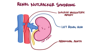
The nutcracker syndrome (NCS) results most commonly from the compression of the left renal vein (LRV) between the abdominal aorta (AA) and superior mesenteric artery (SMA), although other variants exist. The name derives from the fact that, in the sagittal plane and/or transverse plane, the SMA and AA appear to be a nutcracker crushing a nut.

<span class="mw-page-title-main">Vascular disease</span> Medical condition

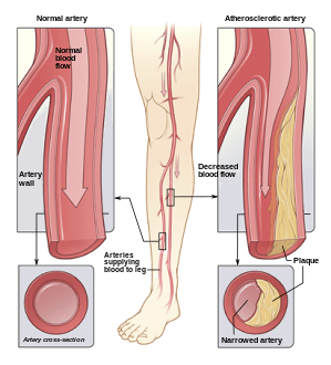
Vascular disease is a class of diseases of the vessels of the circulatory system in the body, including blood vessels – the arteries and veins, and the lymphatic vessels. Vascular disease is a subgroup of cardiovascular disease. Disorders in this vast network of blood and lymph vessels can cause a range of health problems that can sometimes become severe, and fatal. Coronary heart disease for example, is the leading cause of death for men and women in the United States.

<span class="mw-page-title-main">Traumatic aortic rupture</span> Medical condition

Traumatic aortic rupture, also called traumatic aortic disruption or transection, is a condition in which the aorta, the largest artery in the body, is torn or ruptured as a result of trauma to the body. The condition is frequently fatal due to the profuse bleeding that results from the rupture. Since the aorta branches directly from the heart to supply blood to the rest of the body, the pressure within it is very great, and blood may be pumped out of a tear in the blood vessel very rapidly. This can quickly result in shock and death. Thus traumatic aortic rupture is a common killer in automotive accidents and other traumas, with up to 18% of deaths that occur in automobile collisions being related to the injury. In fact, aortic disruption due to blunt chest trauma is the second leading cause of injury death behind traumatic brain injury.

<span class="mw-page-title-main">Endovascular aneurysm repair</span> Surgery used to treat abdominal aortic aneurysm

Endovascular aneurysm repair (EVAR) is a type of minimally-invasive endovascular surgery used to treat pathology of the aorta, most commonly an abdominal aortic aneurysm (AAA). When used to treat thoracic aortic disease, the procedure is then specifically termed TEVAR for "thoracic endovascular aortic/aneurysm repair." EVAR involves the placement of an expandable stent graft within the aorta to treat aortic disease without operating directly on the aorta. In 2003, EVAR surpassed open aortic surgery as the most common technique for repair of AAA, and in 2010, EVAR accounted for 78% of all intact AAA repair in the United States.

<span class="mw-page-title-main">Intestinal ischemia</span> Restriction of blood flow to the small intestine resulting in injury

Intestinal ischemia is a medical condition in which injury to the large or small intestine occurs due to not enough blood supply. It can come on suddenly, known as acute intestinal ischemia, or gradually, known as chronic intestinal ischemia. The acute form of the disease often presents with sudden severe abdominal pain and is associated with a high risk of death. The chronic form typically presents more gradually with abdominal pain after eating, unintentional weight loss, vomiting, and fear of eating.

<span class="mw-page-title-main">Peter H. Lin</span>

Peter Lin is an American vascular surgeon, medical researcher, specializing in minimally invasive endovascular treatment of vascular disease. He has published extensively in the area of vascular surgery and endovascular surgery.

Surgical Outcomes Analysis & Research, SOAR, is a research laboratory of the Department of Surgery at Boston University School of Medicine and Boston Medical Center with expertise in outcomes research. SOAR investigates surgical diseases and perioperative outcomes. The group focuses on pancreatic cancer, other gastrointestinal and hepatobiliary malignancies, vascular disease, and transplant surgery. SOAR's goal is to examine quality, delivery, and financing of care in order to have an immediate impact on patient care and system improvements. The group members utilize national health services and administrative databases, as well as institutional databases, to investigate and to address factors contributing to disease outcomes and healthcare disparities.

<span class="mw-page-title-main">Open aortic surgery</span> Surgical technique

Open aortic surgery (OAS), also known as open aortic repair (OAR), describes a technique whereby an abdominal, thoracic or retroperitoneal surgical incision is used to visualize and control the aorta for purposes of treatment, usually by the replacement of the affected segment with a prosthetic graft. OAS is used to treat aneurysms of the abdominal and thoracic aorta, aortic dissection, acute aortic syndrome, and aortic ruptures. Aortobifemoral bypass is also used to treat atherosclerotic disease of the abdominal aorta below the level of the renal arteries. In 2003, OAS was surpassed by endovascular aneurysm repair (EVAR) as the most common technique for repairing abdominal aortic aneurysms in the United States.

Juan Carlos Parodi is an Argentinian vascular surgeon who introduced the minimally invasive endovascular aneurysm repair (EVAR) to the world and performed the first successful endovascular repair of an abdominal aortic aneurysm on 7 September 1990 in Buenos Aires. In 1992 he was the first in the United States to perform minimally invasive aortic aneurysm surgery together with Drs. Frank Veith, Michael L. Marin and Claudio J. Schonholz. He continues to develop new techniques, including seat belt and air bag technique for cerebral protection during carotid stenting. He is recognized as a renowned pioneer in the specialty of endovascular repairs of the aorta.

Endovascular and hybrid trauma and bleeding management is a new and rapidly evolving concept within medical healthcare and endovascular resuscitation. It involves early multidisciplinary evaluation and management of hemodynamically unstable patients with traumatic injuries as well as being a bridge to definitive treatment. It has recently been shown that the EVTM concept may also be applied to non-traumatic hemodynamically unstable patients.

<span class="mw-page-title-main">Nicolai L. Volodos</span> Ukrainian surgeon (b. 1934 d. 2016)

Nicolai Leontievich Volodos, was a Soviet/Ukrainian cardiovascular surgeon and scientist. An innovator, Volodos developed and introduced into clinical practice the world's first endovascular stent graft for the treatment of stenotic and aneurysmal diseases of arterial system. Volodos was described by his colleagues as ”a pioneer innovator and a giant in vascular and endovascular surgery” and ”a giant of historic proportions in the vascular and endovascular specialties, and the father of endovascular grafting”.

Gustavo S. Oderich is a Brazilian American vascular and endovascular surgeon who serves as a professor and chief of vascular and endovascular surgery, and is the director of the Advanced Endovascular Aortic Program at McGovern Medical School at The University of Texas Health Science Center at Houston and Memorial Hermann Health System. He previously served as chair of vascular and endovascular division at the Mayo Clinic in Rochester, Minnesota. Oderich is recognized for his work in minimally invasive endovascular surgery and research in fenestrated and branched stent-graft technology to treat complex aortic aneurysms and dissections.

<span class="mw-page-title-main">Isolated superior mesenteric artery dissection</span> Medical condition

Isolated superior mesenteric artery dissection (ISMAD) is a rare but potentially life-threatening condition that causes acute abdominal pain. It refers to a dissection that occurs solely in the superior mesenteric artery (SMA), typically spontaneously, and does not involve the aorta. Although aortic dissection can frequently extend into its peripheral territories, it is rare for these branches to have dissection without main aortic trunk involvement. The SMA is the most common site of dissection among visceral arteries compared to other gastrointestinal arteries.

References

  1. Hemingway, Jake F.; Desikan, Sarasijhaa; Dasari, Mohini; Tran, Cuong; Hoffman, Rachel; Gobble, Alexandra; Spurlock, Aaron; Singh, Niten; Quiroga, Elina; Tran, Nam; Starnes, Benjamin W. (2021). "Intraoperative consultation of vascular surgeons is increasing at a major American trauma center". Journal of Vascular Surgery. 74 (5): 1581–1587. doi: 10.1016/j.jvs.2021.04.065 . PMID   34022381. S2CID   235126667.
  2. Coselli, Joseph S.; Preventza, Ourania (2017-06-01). "In Memoriam: Edward B. Diethrich, MD (1935–2017)". Texas Heart Institute Journal. 44 (3): 164–166. doi:10.14503/THIJ-17-6354. ISSN   0730-2347. PMC   5505391 .
  3. Dr. Venkatesh Ramaiah: "Dr. Ted Diethrich lessons carried forward" - The Antegrade Flow Show , retrieved 2021-10-28
  4. "Dr. Ted Diethrich, founder of Arizona Heart Institute, dies at 81". www.beckershospitalreview.com. Retrieved 2021-10-28.
  5. Suckow BD, Goodney PP, Columbo JA, Kang R, Stone DH, Sedrakyan A, Cronenwett JL, Fillinger MF (Jun 2018). "National Trends in Open Surgical, Endovascular and Branched/Fenestrated Endovascular Aortic Aneurysm Repair in Medicare Patients". Journal of Vascular Surgery. 67 (6): 1690–1697.e1. doi: 10.1016/j.jvs.2017.09.046 . PMC   5970963 . PMID   29290495 . Retrieved 23 Sep 2020.
  6. "Trusts reveal plans to centralise services after GIRFT review". Health Service Journal. 19 March 2018. Retrieved 13 May 2018.
  7. Ho, Karen J (2023). Rutherford's Vascular Surgery and Endovascular Therapy. Chapter 4 - Atherosclerosis: Elsevier Health. pp. 41–50. ISBN   978-0-323-77557-1.{{cite book}}: CS1 maint: location (link)
  8. Van Hooft, I. M.; Zeebregts, C. J.; Van Sterkenburg, S. M.; De Vries, W. R.; Reijnen, M. M. (2009). "The persistent sciatic artery". European Journal of Vascular and Endovascular Surgery. 37 (5): 585–591. doi: 10.1016/j.ejvs.2009.01.014 . PMID   19231248.
  9. "Crawford classification" . Retrieved 13 February 2023.
  10. Kassem, M. M.; Gonzalez, L. (2022). "Splenic Artery Aneurysm". StatPearls. PMID   28613599.
  11. Chaer, R. A.; Abularrage, C. J.; Coleman, D. M.; Eslami, M. H.; Kashyap, V. S.; Rockman, C.; Murad, M. H. (2020). "The Society for Vascular Surgery clinical practice guidelines on the management of visceral aneurysms". Journal of Vascular Surgery. 72 (1S): 3S–39S. doi: 10.1016/j.jvs.2020.01.039 . PMID   32201007. S2CID   214617284.
  12. Farber, A.; Angle, N.; Avgerinos, E.; Dubois, L.; Eslami, M.; Geraghty, P.; Haurani, M.; Jim, J.; Ketteler, E.; Pulli, R.; Siracuse, J. J.; Murad, M. H. (2022). "The Society for Vascular Surgery clinical practice guidelines on popliteal artery aneurysms". Journal of Vascular Surgery. 75 (1S): 109S–120S. doi: 10.1016/j.jvs.2021.04.040 . PMID   34023430. S2CID   235170081.
  13. Khattar, R. S.; Fox, D. J.; Alty, J. E.; Arora, A. (1 February 2005). "Pulmonary artery dissection: an emerging cardiovascular complication in surviving patients with chronic pulmonary hypertension". Heart. 91 (2): 142–145. doi: 10.1136/hrt.2004.045799 . PMC   1768672 . PMID   15657218. S2CID   11516764 . Retrieved 13 February 2023.
  14. Lombardi, J. V.; Hughes, G. C.; Appoo, J. J.; Bavaria, J. E.; Beck, A. W.; Cambria, R. P.; Charlton-Ouw, K.; Eslami, M. H.; Kim, K. M.; Leshnower, B. G.; Maldonado, T.; Reece, T. B.; Wang, G. J. (2020). "Society for Vascular Surgery (SVS) and Society of Thoracic Surgeons (STS) reporting standards for type B aortic dissections". Journal of Vascular Surgery. 71 (3): 723–747. doi: 10.1016/j.jvs.2019.11.013 . PMID   32001058. S2CID   210984324.
  15. Acosta, S.; Gonçalves, F. B. (2021). "Management of Spontaneous Isolated Mesenteric Artery Dissection: A Systematic Review". Scandinavian Journal of Surgery. 110 (2): 130–138. doi:10.1177/14574969211000546. PMC   8258720 . PMID   33724090.
  16. Gobble, Ryan M.; Brill, Eliott R.; Rockman, Caron B.; Hecht, Elizabeth M.; Lamparello, Patrick J.; Jacobowitz, Glenn R.; Maldonado, Thomas S. (2009). "Endovascular treatment of spontaneous dissections of the superior mesenteric artery". Journal of Vascular Surgery. 50 (6): 1326–1332. doi: 10.1016/j.jvs.2009.07.019 . PMID   19782510.
  17. Goodman, G.H. (1918). "Angina Abdominus". The American Journal of the Medical Sciences. 155 (4): 524–528. doi:10.1097/00000441-191804000-00003. S2CID   72885362.
  18. SHAW RS; MAYNARD EP 3rd (1958). "Acute and chronic thrombosis of the mesenteric arteries associated with malabsorption; a report of two cases successfully treated by thromboendarterectomy". The New England Journal of Medicine. 258 (18): 874–878. doi:10.1056/NEJM195805012581803. PMID   13541677.{{cite journal}}: CS1 maint: numeric names: authors list (link)
  19. Aburahma, A. F.; Avgerinos, E. D.; Chang, R. W.; Darling Rc, 3rd; Duncan, A. A.; Forbes, T. L.; Malas, M. B.; Murad, M. H.; Perler, B. A.; Powell, R. J.; Rockman, C. B.; Zhou, W. (2022). "Society for Vascular Surgery clinical practice guidelines for management of extracranial cerebrovascular disease". Journal of Vascular Surgery. 75 (1S): 4S–22S. doi: 10.1016/j.jvs.2021.04.073 . PMID   34153348. S2CID   235596620.{{cite journal}}: CS1 maint: numeric names: authors list (link)
  20. "Specialty at a Glance | Society for Vascular Surgery".
  21. Patel, S. K.; Surowiec, S. M. (2022). "Venous Insufficiency". StatPearls. PMID   28613694.
  22. Waheed, S. M.; Kudaravalli, P.; Hotwagner, D. T. (2022). "Deep Vein Thrombosis". StatPearls. PMID   29939530.
  23. Dalsing, Michael C (2023). Rutherford's Vascular Surgery and Endovascular Therapy. Chapter 159 - Chronic Venous Insufficiency: Deep Vein Valve Reconstruction: Elsevier Health. pp. 2098–2111. ISBN   978-0-323-77557-1.{{cite book}}: CS1 maint: location (link)
  24. Gloviczki, Peter (26 December 2008). Handbook of Venous Disorders: Guidelines of the American Venous Forum Third Edition. CRC Press. ISBN   978-0-340-93880-5.
  25. Joh, M.; Desai, K. R. (2021). "Treatment of Nonthrombotic Iliac Vein Lesions". Seminars in Interventional Radiology. 38 (2): 155–159. doi:10.1055/s-0041-1727101. PMC   8175115 . PMID   34108800.
  26. Jayaraj, Arjun (2023). Rutherford's Vascular Surgery and Endovascular Therapy. Chapter 160- Iliocaval Venous Obstruction: Surgical Treatment: Elsevier Health. pp. 2132–2147. ISBN   978-0-323-77557-1.{{cite book}}: CS1 maint: location (link)
  27. Alimi, Yves S Sr. (2023). Rutherford's Vascular Surgery and Endovascular Therapy. Chapter 161- Iliocaval Venous Obstruction: Endovascular Treatment: Elsevier Health. pp. 2112–2131. ISBN   978-0-323-77557-1.{{cite book}}: CS1 maint: location (link)
  28. Power, Adam H.; Gloviczki, Peter (2014). "Left iliac vein occlusion treated with a Palma procedure". Journal of Vascular Surgery: Venous and Lymphatic Disorders. 2 (2): 204–205. doi:10.1016/j.jvsv.2012.10.053. PMID   26993190.
  29. Stevens, S. M.; Woller, S. C.; Baumann Kreuziger, L.; Bounameaux, H.; Doerschug, K.; Geersing, G. J.; Huisman, M. V.; Kearon, C.; King, C. S.; Knighton, A. J.; Lake, E.; Murin, S.; Vintch JRE; Wells, P. S.; Moores, L. K. (2021). "Executive Summary: Antithrombotic Therapy for VTE Disease: Second Update of the Chest Guideline and Expert Panel Report". Chest. 160 (6): 2247–2259. doi:10.1016/j.chest.2021.07.056. PMID   34352279. S2CID   236933535.
  30. Broderick, Cathryn; Watson, Lorna; Armon, Matthew P. (2021). "Thrombolytic strategies versus standard anticoagulation for acute deep vein thrombosis of the lower limb". Cochrane Database of Systematic Reviews. 2021 (1): CD002783. doi:10.1002/14651858.CD002783.pub5. PMC   8094969 . PMID   33464575.
  31. Azirar, Sara; Appelen, Diebrecht; Prins, Martin H.; Neumann, Martino HAM; De Feiter, Adriaan NP; Kolbach, Dinanda N. (2019). "Compression therapy for treating post-thrombotic syndrome". Cochrane Database of Systematic Reviews. 2019 (9): CD004177. doi:10.1002/14651858.CD004177.pub2. PMC   6749555 . PMID   31531971.
  32. Augustinos P, Ouriel K (August 2004). "Invasive approaches to treatment of venous thromboembolism". Circulation. 110 (9 Suppl 1): I27–34. doi: 10.1161/01.CIR.0000140900.64198.f4 . PMID   15339878.
  33. Madani, Michael M. (2016). "50. Pulmonary Thromboendarterectomy". In Peacock, Andrew J.; Naeije, Robert; Rubin, Lewis J. (eds.). Pulmonary Circulation: Diseases and Their Treatment, Fourth Edition. CRC Press. p. 541. ISBN   978-1-4987-1991-9.
  34. Gogalniceanu, Peter (2021). Rich's Vascular Trauma (4th ed.). Chapter 2 - Epidemiology of Vascular Trauma: Elsevier Medical. pp. 23–33. ISBN   978-0-323-69766-8.{{cite book}}: CS1 maint: location (link)
  35. Barmparas, G.; Inaba, K.; Talving, P.; David, J. S.; Lam, L.; Plurad, D.; Green, D.; Demetriades, D. (2010). "Pediatric vs adult vascular trauma: A National Trauma Databank review". Journal of Pediatric Surgery. 45 (7): 1404–1412. doi:10.1016/j.jpedsurg.2009.09.017. PMID   20638516.
  36. Arbabi, Cassra N (2023). Rutherford's Vascular Surgery and Endovascular Therapy (10th ed.). Chapter 181 - Thoracic Vascular Trauma: Elsevier Medical. pp. 2397–2410. ISBN   978-0-323-77559-5.{{cite book}}: CS1 maint: location (link)
  37. Hooi JD; Kester AD; Stoffers HE; Overdijk MM; van Ree JW; Knottnerus JA (April 2001). "Incidence of and risk factors for asymptomatic peripheral arterial occlusive disease: a longitudinal study". Am J Epidemiol. 153 (7): 666–72. doi: 10.1093/aje/153.7.666 . PMID   11282794.
  38. Ashton HA; Buxton MJ; Day NE; et al. (November 2002). "The Multicentre Aneurysm Screening Study (MASS) into the effect of abdominal aortic aneurysm screening on mortality in men: a randomised controlled trial". Lancet. 360 (9345): 1531–9. doi:10.1016/S0140-6736(02)11522-4. PMID   12443589. S2CID   21497118.
  39. "Mortality results for randomised controlled trial of early elective surgery or ultrasonographic surveillance for small abdominal aortic aneurysms. The UK Small Aneurysm Trial Participants". Lancet. 352 (9141): 1649–55. November 1998. doi:10.1016/S0140-6736(98)10137-X. PMID   9853436. S2CID   24733279.
  40. Lederle FA; Wilson SE; Johnson GR; et al. (August 1994). "Design of the abdominal aortic Aneurysm Detection and Management Study. ADAM VA Cooperative Study Group". J Vasc Surg. 20 (2): 296–303. doi:10.1016/0741-5214(94)90019-1. PMID   8040955.
  41. Thompson JF; Mullee MA; Bell PR; et al. (July 1996). "Intraoperative heparinisation, blood loss and myocardial infarction during aortic aneurysm surgery: a Joint Vascular Research Group study". Eur J Vasc Endovasc Surg. 12 (1): 86–90. doi: 10.1016/S1078-5884(96)80281-4 . PMID   8696904.
  42. Barnett, Henry J.M.; Taylor, D. Wayne; Eliasziw, Michael; Fox, Allan J.; Ferguson, Gary G.; Haynes, R. Brian; Rankin, Richard N.; Clagett, G. Patrick; Hachinski, Vladimir C.; Sackett, David L.; Thorpe, Kevin E. (1998-11-12). "Benefit of Carotid Endarterectomy in Patients with Symptomatic Moderate or Severe Stenosis". New England Journal of Medicine. 339 (20): 1415–1425. doi: 10.1056/NEJM199811123392002 . ISSN   0028-4793. PMID   9811916.
  43. LLC, Peripheral Brain. "NASCET - Wiki Journal Club". www.wikijournalclub.org. Retrieved 2021-02-11.
  44. "Table 1: The Single Nucleotide Polymorphisms in cathepsin B protein mined from literature (PMID: 16492714)". doi: 10.7717/peerj.7425/table-1 .{{cite journal}}: Cite journal requires |journal= (help)
  45. "ACAS - Wiki Journal Club". www.wikijournalclub.org. Retrieved 2021-02-11.
  46. VascularWeb: New Vascular Surgery Training Paradigms