Endovascular aneurysm repair

Last updated
Endovascular aneurysm repair
Aneurysm endovascular.jpg
Endovascular aneurysm repair
Other namesEndovascular aortic repair
ICD-9-CM 39.51, 39.52, 39.7

Endovascular aneurysm repair (EVAR) is a type of minimally-invasive endovascular surgery used to treat pathology of the aorta, most commonly an abdominal aortic aneurysm (AAA). When used to treat thoracic aortic disease, the procedure is then specifically termed TEVAR for "thoracic endovascular aortic/aneurysm repair." EVAR involves the placement of an expandable stent graft within the aorta to treat aortic disease without operating directly on the aorta. In 2003, EVAR surpassed open aortic surgery as the most common technique for repair of AAA, [1] and in 2010, EVAR accounted for 78% of all intact AAA repair in the United States. [2]

Contents

Medical uses

Standard EVAR is appropriate for aneurysms that begin below the renal arteries, where there exists an adequate length of normal aorta (the "proximal aortic neck") for reliable attachment of the endograft without leakage of blood around the device (" endoleak "). [3] If the proximal aortic neck is also involved with the aneurysm, the patient may be a candidate for complex visceral EVAR with a fenestrated or branched EVAR.

Patients with aneurysms require elective repair of their aneurysm when it reaches a diameter large enough (typically greater than 5.5 cm) such that the risk of rupture is greater than the risk of surgery. Repair is also warranted for aneurysms that rapidly enlarge or those that have been the source of emboli (debris from the aneurysm that dislodges and travel into other arteries). Lastly, the repair is also indicated for aneurysms that are the source of pain and tenderness, which may indicate impending rupture. The options for repair include traditional open aortic surgery or endovascular repair.[ citation needed ]

Endovascular procedures aim to reduce the morbidity and mortality of treating arterial disease in a patient population that is increasingly older and less fit than when major open repairs were developed and popularized. Even in the early days, significant risks were accepted in the understanding that the large open operation was the only option. That is not the case in most patients today.[ citation needed ]

Studies that assign aneurysm patients to treatment with EVAR or traditional open surgery have demonstrated fewer early complications with the minimally invasive approach. Some studies have also observed a lower mortality rate with EVAR. [4] [5] The reduction in death, however, does not persist long-term. After a few years, the survival after repair is similar to EVAR or open surgery. This observation may be the result of durability problems with early endograft, with a corresponding need for additional procedures to repair endoleaks and other device-related issues. Newer, improved technology may reduce the need for such secondary procedures. If so, the results of EVAR may improve to the point where long-term survival benefit becomes evident.[ citation needed ]

EVAR is also used for rupture of the abdominal and descending thoracic aorta, and in rare cases used to treat pathology of the ascending aorta. [6]

Aortic dissection

Endografts have been used in patients with aortic dissection, noting the extremely complex nature of open surgical repair in these patients. In uncomplicated aortic dissections, no benefit has been demonstrated over medical management alone. In uncomplicated type B aortic dissection, TEVAR does not seem either to improve or compromise 2-year survival and adverse event rates. [7] Its use in complicated aortic dissection is under investigation. In the Clinical Practice Guidelines of the European Society for Vascular Surgery, it is recommended that in patients with complicated acute type B aortic dissection, endovascular repair with thoracic endografting should be the first line intervention. [8]

Before people are deemed to be suitable candidates for this treatment, they have to go through a rigorous set of tests. These include a CT scan of the complete thorax/abdomen/pelvis and blood tests. The CT scan gives precise measurements of the aneurysm and the surrounding anatomy. In particular, the calibre/tortuosity of the iliac arteries and the relationship of the neck of the aneurysm to the renal arteries are important determinants of whether the aneurysm is amenable to endoluminal repair. In certain occasions where the renal arteries are too close to the aneurysm, the custom-made fenestrated graft stent is now an accepted alternative to doing open surgery.[ citation needed ]

Relative contraindications

A patient's anatomy can be unsuitable for EVAR in several ways. Most commonly, in an infrarenal aneurysm, a potential EVAR candidate lacks adequate length of the normal-diameter aorta between the aneurysm and the takeoff of the renal arteries, the "infra-renal neck". Another relative contraindications include prohibitively small iliac arteries, aneurysmal iliac arteries, prohibitively small femoral arteries, or circumferential calcification of the femoral or iliac arteries.[ citation needed ]

In addition to a short proximal aortic neck, the neck may be angulated, large in diameter, or shaped like a funnel (conical) where the neck diameter at the top is larger than the neck diameter at the bottom. Along with a short proximal aortic neck, necks with any of these characteristics are called "hostile necks" and endovascular repair can be either contraindicated or associated with early-late complications of endoleak, or endograft migration, or both.[ citation needed ]

Many of the advances in EVAR technique aim to adapt EVAR for these situations, and advanced techniques allow EVAR to be employed in patients who previously were not candidates.[ citation needed ]

Technique

Abdominal aortic endograft on a CT scan; original aneurysm marked in blue Endovasc.jpg
Abdominal aortic endograft on a CT scan; original aneurysm marked in blue

The procedure is carried out in a sterile environment under fluoroscopic guidance. It is usually carried out by a vascular surgeon, interventional radiologist or cardiac surgeon, and occasionally, general surgeon or interventional cardiologist. [9] [10] [11] [12] The procedure can be performed under general, regional (spinal or epidural) or even local anesthesia. [13]

Access to the patient's femoral arteries can be with surgical incisions or percutaneously in the groin on both sides. Vascular sheaths are introduced into the patient's femoral arteries, through which guidewires, catheters, and the endograft are passed.[ citation needed ]

Diagnostic angiography images are captured of the aorta to determine the location of the patient's renal arteries, so the stent-graft can be deployed without blocking these. Failure to achieve this will cause kidney failure. With most devices, the "main body" of the endograft is placed first, followed by the "limbs" which join the main body and extend to the iliac arteries, effectively protecting the aneurysm sac from blood pressure.[ citation needed ]

The abdominal aneurysm extends down to the common iliac arteries in about 25%-30% of patients. In such cases, the iliac limbs can be extended into the external iliac artery to bypass a common iliac aneurysm. Alternatively, a specially designed endograft, (an iliac branch device) can be used to preserve flow to the internal iliac arteries. The preservation of the hypogastric (internal iliac) arteries is important to prevent buttock claudication and impotence, and every effort should be made to preserve flow to at least one hypogastric artery.[ citation needed ]

The endograft acts as an artificial lumen for blood to flow through, protecting the surrounding aneurysm sac. This reduces the pressure in the aneurysm, which itself will usually thrombose and shrink in size over time. [14]

Staging such procedures is common, particularly to address aortic branch points near the diseased aortic segment. One example in the treatment of thoracic aortic disease is revascularization of the left common carotid artery and/or the left subclavian artery from the innominate artery or the right common carotid artery to allow treatment of a thoracic aortic aneurysm that encroaches proximally into the aortic arch. These "extra-anatomic bypasses" can be performed without an invasive thoracotomy. Another example in the abdominal aorta is the embolization of the internal iliac artery on one side prior to coverage by an iliac limb device. Continued improvement in stent-graft design, including branched endografts, will reduce but not eliminate multi-stage procedures.[ citation needed ]

Percutaneous EVAR

Standard EVAR involves a surgical cut-down on either the femoral or iliac arteries, with the creation of a 4–6 cm incision. Like many surgical procedures, EVAR has advanced to a more minimally invasive technique, by accessing the femoral arteries percutaneously In percutaneous EVAR (PEVAR), small, sub-centimeter incisions are made over the femoral artery, and endovascular techniques are used to place the device over a wire. Percutaneous EVAR has been systematically compared to the standard EVAR cut-down femoral artery approach. [15] Moderate quality evidence suggests that there are no differences in short-term mortality, aneurysm sealing, long and short-term complications, or infections at the wound site. [15] Higher quality evidence suggests that there are no differences in post-repair bleeding complications or haematoma between the two approaches. [15] The percutaneous approach may have reduced surgical time. [15]

Fenestrated EVAR

An example of a physician-modified aortic endograft with fenestrations added to accommodate the visceral branch arteries FenGraft.jpg
An example of a physician-modified aortic endograft with fenestrations added to accommodate the visceral branch arteries

Fenestrated endovascular aortic/aneurysm repair (FEVAR) is performed in the cases where the aneurysm extends near or involves the visceral vessels (such as juxta-renal, para-renal, thoraco-abdominal aortic aneurysms). A custom-made graft with fenestrations (holes on the graft body to maintain the patency of the visceral arteries) is used for the procedure. When the aneurysm involves the visceral arteries, standard EVAR is contraindicated due to the lack of suitable infra-aortic segment for the endograft attachment; FEVAR achieves seal between the stent graft and para-visceral segment and/or more proximal segment while preserving the flow of the visceral arteries. FEVAR has been in use in the United Kingdom for over a decade and early results were published in Jun 2012. [16]

Branched EVAR

Artist's rendering of a branched/fenestrated EVAR in the visceral segment of the aorta above an abdominal aortic aneurysm ArtistsFenBranchInSituCropped.jpg
Artist's rendering of a branched/fenestrated EVAR in the visceral segment of the aorta above an abdominal aortic aneurysm

Thoracoabdominal aortic aneurysms (TAAA) involve the aorta in the chest and abdomen. As such, major branch arteries to the head, arms, spinal cord, intestines, and kidneys may originate from the aneurysm. An endovascular repair of a TAAA is only possible if blood flow to these critical arteries is preserved. Hybrid procedures offer one option, but a more direct approach involves the use of a branched endograft. However, the complex anatomy associated with the supra-aortic vessels is particularly difficult to accommodate with branched endograft devices. [17] Dr. Timothy Chuter pioneered this approach, with a completely endovascular solution. After partial deployment of the main body of an endograft, separate endograft limbs are deployed from the main body to each major aortic branch. This procedure is long, technically difficult, and currently only performed in a few centers. When the aneurysm begins above the renal arteries, neither fenestrated endografts nor "EndoAnchoring" of an infrarenal endograft is useful (an open surgical repair may be necessary). Alternatively, a "branched" endograft may be used. A branched endograft has graft limbs that branch off of the main portion of the device to directly provide blood flow to the kidneys or the visceral arteries. [18] [19]

Hybrid procedures

On occasion, there is inadequate length or quality of the proximal or distal aortic neck. In these cases, a fully minimally invasive option is not possible. One solution, however, is a hybrid repair, which combines an open surgical bypass with EVAR or TEVAR. In hybrid procedures, the endograft is positioned over major aortic branches. While such a position would normally cause problems from disruption of blood flow to the covered branches (renal, visceral, or branches to the head or arms), the prior placement of bypass grafts to these critical vessels allowed the deployment of the endograft at a level that would otherwise not be possible.[ citation needed ]

If a patient has calcified or narrow femoral arteries that prohibit the introduction of the endograft transfemorally, an iliac conduit may be used. This is typically a piece of PTFE that is sewn directly to the iliac arteries, which are exposed via an open retroperitoneal approach. The endograft is then introduced into the aorta through the conduit.[ citation needed ]

In patients with thoracic aortic disease involving the arch and descending aorta, it is not always possible to perform a completely endovascular repair. This is because head vessels of the aortic arch supplying blood to the brain cannot be covered and for this reason, there is often an inadequate landing zone for stent-graft delivery. A hybrid repair strategy offers a reasonable choice for treating such patients. A commonly used hybrid repair procedure is the "frozen elephant trunk repair". [20] This technique involves midline sternotomy. The aortic arch is transected and the stent-graft device is delivered in an ante-grade fashion in the descending aorta. The aortic arch is subsequently reconstructed and the proximal portion of the stent-graft device is then directly sutured into the surgical graft. Patients with anomalies of the arch and some disease extension into the descending aorta are often ideal candidates. Studies have reported successful use of hybrid techniques for treating Kommerell diverticulum [21] and descending aneurysms in patients with previous coarctation repairs. [22] [23]

In addition, hybrid techniques combining both open and endovascular repair are also used in managing emergency complications in the aortic arch, such as retrograde ascending dissection and endoleaks from previous stent grafting of descending aorta. A "reverse frozen elephant trunk repair" is shown to be particularly effective. [24]

Adjunctive procedures

Risks

The complications of EVAR can be divided into those that are related to the repair procedure and those related to the endograft device. For example, a myocardial infarction that occurs immediately after the repair is normally related to the procedure and not the device. By contrast, the development of an endoleak from degeneration of endograft fabric would be a device-related complication. [19]

Durability and problems such as 'endoleaks' may require careful surveillance and adjuvant procedures to ensure the success of the EVAR or EVAR/hybrid procedure. CT angiography (CTA) imaging has, in particular, made a key contribution to planning, success, durability in this complex area of vascular surgery.[ citation needed ]

A major cause of complications in EVAR is the failure of the seal between the proximal, infra-renal aneurysm neck and the endovascular graft. [27] [28] [29] Risk of this form of failure is especially elevated in adverse or challenging proximal neck anatomies, where this seal could be compromised by unsuitable geometric fit between the graft and vessel wall, as well as instability of the anatomy. [30] [31] [32] New recent techniques have been introduced to address these risks by utilizing a segment of the supra-renal portion of the aorta to increase the sealing zone, such as with fenestrated EVAR, chimneys and snorkels. [33] These techniques may be suitable in certain patients with qualifying factors, e.g., configuration of renal arteries, renal function. However, these are more complex procedures than standard EVAR and may be subject to further complications. [34] [35] [36]

An approach that directly augments the fixation and sealing between the graft and aorta to mimic the stability of a surgical anastomosis is EndoAnchoring. [37] [38] EndoAnchors are small, helically shaped implants that directly lock the graft to the aortic wall with the goal to prevent complications of the seal, especially in adverse neck anatomies. [39] [40] These EndoAnchors may also be used to treat identified leaks between the graft and proximal neck. [41] [42] [43]

Arterial dissection, contrast-induced kidney failure, thromboembolizaton, ischemic colitis, groin hematoma, wound infection, type II endoleaks, myocardial infarction, congestive heart failure, cardiac arrhythmias, respiratory failure.[ citation needed ]

Endograft migration, aneurysm rupture, graft limb stenosis/kinking, type I/III/IV endoleaks, stent graft thrombosis, or infection. [44] Device infection occurs in 1-5% of aortic prosthesis placements and is a life-threatening complication. [45]

Endoleaks

An endoleak is a leak into the aneurysm sac after endovascular repair. Five types of endoleaks exist: [14]

Type I and III leaks are considered high-pressure leaks and are more concerning than other leak types. Depending on the aortic anatomy, they may require further intervention to treat. Type 2 leaks are common and often can be left untreated unless the aneurysm sac continues to expand after EVAR. [46]

Spinal cord injury

Spinal cord injury is a devastating complication after aortic surgery, specifically for thoracoabdominal aortic aneurysm repair; severe injury could lead to urine and fecal incontinence, paresthesia and even paraplegia. [47] The risk varies between studies with two metanalysis demonstrating a pooled incidence of spinal cord injury 2.2% [48] and 11%. [49] Predictive factors include increasing extent of coverage, hypogastric artery occlusion, prior aortic repair and perioperative hypotension. [50] Spinal cord injury related to aortic repair occurs due to impaired blood flow to the spine after coverage of blood vessels, important to the blood circulation of the spine, namely intercostal- and lumbar arteries. [51] A few methods exist for potentially reversing spinal cord injury, if it arises, elevated blood pressure, increased oxygenation, blood transfusion and cerebrospinal fluid drainage.

Cerebrospinal fluid drainage

Cerebrospinal fluid drainage is one of the adjunct methods used to reverse spinal cord injury. With increased drainage of spinal fluid, the intrathecal pressure decreases which allows for increase blood perfusion to the spine, possibly reversing the ischemic injury of the spinal tissue due to lessened blood supply. The benefits of this procedure have been established in open aortic repair [52] and suggested in endovascular aortic repair. [47]

Recovery after EVAR

Unlike traditional aortic repair, standard recovery after EVAR is remarkably straightforward. Patients who have undergone EVAR typically spend one night in the hospital to be monitored, although it has been suggested that EVAR can be performed as a same-day procedure. [53]

Patients are advised to slowly return to normal activity. There are no specific activity restrictions after EVAR, however, patients typically are seen by their surgeon within one month after EVAR to begin post-EVAR surveillance.[ citation needed ]

There is limited research looking at patients' experience of recovery after more complex and staged EVAR for thoracoabdominal aortic diseases. One qualitative study found that patients with complex aortic diseases struggle with physical and psychological setbacks, continuing years after their operations. [54]

History

Dr. Juan C. Parodi introduced the minimally-invasive endovascular aneurysm repair (EVAR) to the world and performed the first successful endovascular repair of an abdominal aortic aneurysm on 7 September 1990 in Buenos Aires on a friend of Carlos Menem, the then President of Argentina. The first device was simple, according to Parodi: "It was a graft I designed with expandable ends, the extra-large Palmaz stent, a Teflon sheath with a valve, a wire, and the valvuloplasty balloon, which I took from the cardiologists." Parodi's first patient lived for nine years after the procedure and died from pancreatic cancer. [55] [56] The first EVAR performed in the United States was in 1992 by Drs. Frank Veith, Michael Marin, Juan Parodi and Claudio Schonholz at Montefiore Medical Center affiliated with Albert Einstein College of Medicine.[ citation needed ]

The modern endovascular device used to repair abdominal aortic aneurysms, which is bifurcated and modular, was pioneered and first employed by Dr. Timothy Chuter while a fellow at the University of Rochester. [57] The first clinical series of his device was published from Nottingham in 1994. [58] The first endovascular repair of a ruptured abdominal aortic aneurysm was also reported from Nottingham in 1994. [59]

By 2003, four devices were on the market in the United States. [60] Each of these devices has since been either abandoned or further refined to improve its characteristics in vivo.

Special populations

Women

Women are known to have smaller aortas on average than men, so are potential candidates for AAA treatment at smaller maximum aneurysm diameters than men.[ citation needed ]

Transplant candidates

As immunosuppressive medications are known to increase the rate of aneurysm growth, transplant candidates are AAA repair candidates at smaller maximum aneurysm diameters than the general population.[ citation needed ]

Other animals

Due to the expense associated with EVAR stent-graft devices and their specificity to human aortic anatomy, EVAR is not used in other animals.[ citation needed ]

Videos

Related Research Articles

<span class="mw-page-title-main">Aortic dissection</span> Injury to the innermost layer of the aorta

Aortic dissection (AD) occurs when an injury to the innermost layer of the aorta allows blood to flow between the layers of the aortic wall, forcing the layers apart. In most cases, this is associated with a sudden onset of severe chest or back pain, often described as "tearing" in character. Vomiting, sweating, and lightheadedness may also occur. Damage to other organs may result from the decreased blood supply, such as stroke, lower extremity ischemia, or mesenteric ischemia. Aortic dissection can quickly lead to death from insufficient blood flow to the heart or complete rupture of the aorta.

<span class="mw-page-title-main">Aneurysm</span> Bulge in the wall of a blood vessel

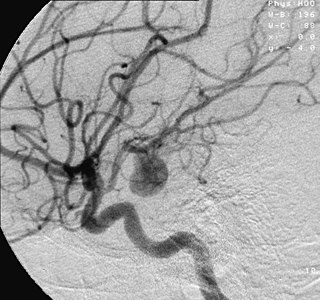
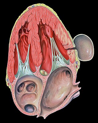
An aneurysm is an outward bulging, likened to a bubble or balloon, caused by a localized, abnormal, weak spot on a blood vessel wall. Aneurysms may be a result of a hereditary condition or an acquired disease. Aneurysms can also be a nidus for clot formation (thrombosis) and embolization. As an aneurysm increases in size, the risk of rupture, which leads to uncontrolled bleeding, increases. Although they may occur in any blood vessel, particularly lethal examples include aneurysms of the circle of Willis in the brain, aortic aneurysms affecting the thoracic aorta, and abdominal aortic aneurysms. Aneurysms can arise in the heart itself following a heart attack, including both ventricular and atrial septal aneurysms. There are congenital atrial septal aneurysms, a rare heart defect.

<span class="mw-page-title-main">Interventional radiology</span> Medical subspecialty

Interventional radiology (IR) is a medical specialty that performs various minimally-invasive procedures using medical imaging guidance, such as x-ray fluoroscopy, computed tomography, magnetic resonance imaging, or ultrasound. IR performs both diagnostic and therapeutic procedures through very small incisions or body orifices. Diagnostic IR procedures are those intended to help make a diagnosis or guide further medical treatment, and include image-guided biopsy of a tumor or injection of an imaging contrast agent into a hollow structure, such as a blood vessel or a duct. By contrast, therapeutic IR procedures provide direct treatment—they include catheter-based medicine delivery, medical device placement, and angioplasty of narrowed structures.

<span class="mw-page-title-main">Vascular surgery</span> Medical specialty, operative procedures for the treatment of vascular disorders

Vascular surgery is a surgical subspecialty in which vascular diseases involving the arteries, veins, or lymphatic vessels, are managed by medical therapy, minimally-invasive catheter procedures and surgical reconstruction. The specialty evolved from general and cardiovascular surgery where it refined the management of just the vessels, no longer treating the heart or other organs. Modern vascular surgery includes open surgery techniques, endovascular techniques and medical management of vascular diseases - unlike the parent specialities. The vascular surgeon is trained in the diagnosis and management of diseases affecting all parts of the vascular system excluding the coronaries and intracranial vasculature. Vascular surgeons also are called to assist other physicians to carry out surgery near vessels, or to salvage vascular injuries that include hemorrhage control, dissection, occlusion or simply for safe exposure of vascular structures.

<span class="mw-page-title-main">Aortic aneurysm</span> Excessive enlargement of the human aorta

An aortic aneurysm is an enlargement (dilatation) of the aorta to greater than 1.5 times normal size. They usually cause no symptoms except when ruptured. Occasionally, there may be abdominal, back, or leg pain. The prevalence of abdominal aortic aneurysm ("AAA") has been reported to range from 2 to 12% and is found in about 8% of men more than 65 years of age. The mortality rate attributable to AAA is about 15,000 per year in the United States and 6,000 to 8,000 per year in the United Kingdom and Ireland. Between 2001 and 2006, there were approximately 230,000 AAA surgical repairs performed on Medicare patients in the United States.

<span class="mw-page-title-main">Abdominal aortic aneurysm</span> Medical condition

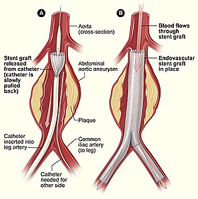
Abdominal aortic aneurysm (AAA) is a localized enlargement of the abdominal aorta such that the diameter is greater than 3 cm or more than 50% larger than normal. An AAA usually causes no symptoms, except during rupture. Occasionally, abdominal, back, or leg pain may occur. Large aneurysms can sometimes be felt by pushing on the abdomen. Rupture may result in pain in the abdomen or back, low blood pressure, or loss of consciousness, and often results in death.

<span class="mw-page-title-main">Thoracic aortic aneurysm</span> Medical condition

A thoracic aortic aneurysm is an aortic aneurysm that presents primarily in the thorax.

<span class="mw-page-title-main">Pseudoaneurysm</span> Collection of blood between outer artery layers

A pseudoaneurysm, also known as a false aneurysm, is a locally contained hematoma outside an artery or the heart due to damage to the vessel wall. The injury passes through all three layers of the arterial wall, causing a leak, which is contained by a new, weak "wall" formed by the products of the clotting cascade. A pseudoaneurysm does not contain any layer of the vessel wall.

<span class="mw-page-title-main">Aortoiliac occlusive disease</span> Medical condition

In medicine, aortoiliac occlusive disease is a form of central artery disease involving the blockage of the abdominal aorta as it transitions into the common iliac arteries.

<span class="mw-page-title-main">Traumatic aortic rupture</span> Medical condition

Traumatic aortic rupture, also called traumatic aortic disruption or transection, is a condition in which the aorta, the largest artery in the body, is torn or ruptured as a result of trauma to the body. The condition is frequently fatal due to the profuse bleeding that results from the rupture. Since the aorta branches directly from the heart to supply blood to the rest of the body, the pressure within it is very great, and blood may be pumped out of a tear in the blood vessel very rapidly. This can quickly result in shock and death. Thus traumatic aortic rupture is a common killer in automotive accidents and other traumas, with up to 18% of deaths that occur in automobile collisions being related to the injury. In fact, aortic disruption due to blunt chest trauma is the second leading cause of injury death behind traumatic brain injury.

Michael L. Marin is an American vascular surgeon. Together with Drs. Frank Veith, Juan C. Parodi and Claudio J. Schonholz, he was the first in the United States to perform minimally invasive aortic aneurysm surgery. In 2004, he was the first doctor to implant an intravascular telemetric monitor -- a device that alerts to physicians any leakage in aortic stent-grafts.

Surgical Outcomes Analysis & Research, SOAR, is a research laboratory of the Department of Surgery at Boston University School of Medicine and Boston Medical Center with expertise in outcomes research. SOAR investigates surgical diseases and perioperative outcomes. The group focuses on pancreatic cancer, other gastrointestinal and hepatobiliary malignancies, vascular disease, and transplant surgery. SOAR's goal is to examine quality, delivery, and financing of care in order to have an immediate impact on patient care and system improvements. The group members utilize national health services and administrative databases, as well as institutional databases, to investigate and to address factors contributing to disease outcomes and healthcare disparities.

<span class="mw-page-title-main">Hybrid cardiac surgery</span>

A hybrid cardiac surgical procedure in a narrow sense is defined as a procedure that combines a conventional, more invasive surgical part with an interventional part, using some sort of catheter-based procedure guided by fluoroscopy imaging in a hybrid operating room (OR) without interruption. The hybrid technique has a reduced risk of surgical complications and has shown decreased recovery time. It can be used to treat numerous heart diseases and conditions and with the increasing complexity of each case, the hybrid surgical technique is becoming more common.

<span class="mw-page-title-main">Open aortic surgery</span> Surgical technique

Open aortic surgery (OAS), also known as open aortic repair (OAR), describes a technique whereby an abdominal, thoracic or retroperitoneal surgical incision is used to visualize and control the aorta for purposes of treatment, usually by the replacement of the affected segment with a prosthetic graft. OAS is used to treat aneurysms of the abdominal and thoracic aorta, aortic dissection, acute aortic syndrome, and aortic ruptures. Aortobifemoral bypass is also used to treat atherosclerotic disease of the abdominal aorta below the level of the renal arteries. In 2003, OAS was surpassed by endovascular aneurysm repair (EVAR) as the most common technique for repairing abdominal aortic aneurysms in the United States.

Juan Carlos Parodi is an Argentinian vascular surgeon who introduced the minimally invasive endovascular aneurysm repair (EVAR) to the world and performed the first successful endovascular repair of an abdominal aortic aneurysm on 7 September 1990 in Buenos Aires. In 1992 he was the first in the United States to perform minimally invasive aortic aneurysm surgery together with Drs. Frank Veith, Michael L. Marin and Claudio J. Schonholz. He continues to develop new techniques, including seat belt and air bag technique for cerebral protection during carotid stenting. He is recognized as a renowned pioneer in the specialty of endovascular repairs of the aorta.

Endovascular and hybrid trauma and bleeding management is a new and rapidly evolving concept within medical healthcare and endovascular resuscitation. It involves early multidisciplinary evaluation and management of hemodynamically unstable patients with traumatic injuries as well as being a bridge to definitive treatment. It has recently been shown that the EVTM concept may also be applied to non-traumatic hemodynamically unstable patients.

<span class="mw-page-title-main">Nicolai L. Volodos</span> Ukrainian surgeon (b. 1934 d. 2016)

Nicolai Leontievich Volodos, was a Soviet/Ukrainian cardiovascular surgeon and scientist. An innovator, Volodos developed and introduced into clinical practice the world's first endovascular stent graft for the treatment of stenotic and aneurysmal diseases of arterial system. Volodos was described by his colleagues as ”a pioneer innovator and a giant in vascular and endovascular surgery” and ”a giant of historic proportions in the vascular and endovascular specialties, and the father of endovascular grafting”.

<span class="mw-page-title-main">Hazim J. Safi</span> Physician

Hazim J. Safi, MD, FACS, is a physician and surgeon who is well known for his research in the surgical treatment of aortic disease. Safi and his colleagues at Baylor College of Medicine were the first to identify variables associated with early death and postoperative complications in patients undergoing thoracoabdominal aortic operations. Safi now serves as professor of cardiothoracic surgery, and founding chair at McGovern Medical School at The University of Texas Health Science Center in Houston, TX.

Gustavo S. Oderich is a Brazilian American vascular and endovascular surgeon who serves as a professor and chief of vascular and endovascular surgery, and is the director of the Advanced Endovascular Aortic Program at McGovern Medical School at The University of Texas Health Science Center at Houston and Memorial Hermann Health System. He previously served as chair of vascular and endovascular division at the Mayo Clinic in Rochester, Minnesota. Oderich is recognized for his work in minimally invasive endovascular surgery and research in fenestrated and branched stent-graft technology to treat complex aortic aneurysms and dissections.

<span class="mw-page-title-main">Benjamin Starnes</span> American physician and vascular surgeon

Benjamin Starnes is a vascular surgeon and medical researcher. He holds the Alexander Whitehill Clowes Endowed Chair in Vascular surgery at the University of Washington. He served as a U.S. Army surgeon for 15 years, doing three tours of duty, including in the last M.A.S.H. unit. On the day of the September 11 attacks he was at the Pentagon rendering medical aid to victims, and his experience was later recounted in the book American Phoenix: Heroes of the Pentagon on 9/11. He is among the primary authors of the official guidelines for diagnosis and management of aortic disease adopted by the American College of Cardiology and the American Heart Association.

References

  1. Sethi RK, Henry AJ, Hevelone ND, Lipsitz SR, Belkin M, Nguyen LL (September 2013). "Impact of hospital market competition on endovascular aneurysm repair adoption and outcomes". J. Vasc. Surg. 58 (3): 596–606. doi: 10.1016/j.jvs.2013.02.014 . PMID   23684424.
  2. Dua, A (2014). "Epidemiology of aortic aneurysm repair in the United States from 2000 to 2010". J Vasc Surg. 59 (6): 1512–7. doi: 10.1016/j.jvs.2014.01.007 . PMID   24560865.
  3. Zarkowsky, DS; Sorber, R; Ramirez, JL; Goodney, PP; Iannuzzi, JC; Wohlauer, M; Hicks, CW (August 2021). "Aortic Neck IFU Violations During EVAR for Ruptured Infrarenal Aortic Aneurysms are Associated with Increased In-Hospital Mortality". Annals of Vascular Surgery. 75: 12–21. doi:10.1016/j.avsg.2021.04.019. PMC   9843606 . PMID   33951521. S2CID   233869750.
  4. Jackson, Rubie (Apr 2012). "Comparison of Long-term Survival After Open vs Endovascular Repair of Intact Abdominal Aortic Aneurysm Among Medicare Beneficiaries". JAMA. 307 (15): 1621–8. doi:10.1001/jama.2012.453. PMID   22511690.
  5. Schermerhorn, Marc (January 2008). "Endovascular vs. open repair of abdominal aortic aneurysms in the Medicare population". New England Journal of Medicine. 358 (5): 464–74. doi: 10.1056/NEJMoa0707348 . PMID   18234751. S2CID   205089378.
  6. McCallum, JC; Limmer, KK; Perricone, A; Bandyk, D; Kansal, N (July 2013). "Total endovascular repair of acute ascending aortic rupture: a case report and review of the literature". Vascular and Endovascular Surgery. 47 (5): 374–8. doi:10.1177/1538574413486838. PMID   23697343. S2CID   115272001.
  7. Nienaber CA, Rousseau H, Eggebrecht H, et al. (December 2009). "Randomized comparison of strategies for type B aortic dissection: the INvestigation of STEnt Grafts in Aortic Dissection (INSTEAD) trial". Circulation. 120 (25): 2519–28. doi: 10.1161/CIRCULATIONAHA.109.886408 . PMID   19996018.
  8. Riambau V, Böckler D, Brunkwall J, et al. (January 2017). "Editor's choice - Management of descending thoracic aorta diseases: Clinical practice guidelines of the European Society of Vascular Surgery (ESVS)". European Journal of Vascular and Endovascular Surgery. 53 (1): 4–52. doi: 10.1016/j.ejvs.2016.06.005 . hdl: 1983/6273178d-f3ec-42f0-a26c-fd8ffdb5e82f . PMID   28081802.
  9. McCutcheon BA, Talamini MA, Chang DC, Rose JA, Bandyk DF, Wilson SE (September 2013). "The comparative effectiveness of surgeons over interventionalists in endovascular repairs of abdominal aortic aneurysm". Ann. Surg. 258 (3): 476–82. doi:10.1097/SLA.0b013e3182a196b5. PMID   24022440. S2CID   20701190.
  10. "National Vascular Registry - 2018 Annual Report" (PDF), VSQIP (PDF), retrieved 2019-12-09
  11. Andrew England & Richard Mc Williams (2013). "Endovascular aortic aneurysm repair (EVAR)". The Ulster Medical Journal. 82 (1): 3–10. PMC   3632841 . PMID   23620623.
  12. Koncar I, Tolić M, Ilić N, Cvetković S, Dragas M, Cinara I, Kostić D, Davidović L (2012). "Endovascular aortic repair: first twenty years". Srp Arh Celok Lek. 140 (11–12): 792–9. doi: 10.2298/SARH1212792K . PMID   23350259.
  13. Noh, M; Choi, BM; Kwon, H; Han, Y; Ko, GY; Kwon, TW; Noh, GJ; Cho, YP (August 2018). "General anesthesia versus local anesthesia for endovascular aortic aneurysm repair". Medicine. 97 (32): e11789. doi:10.1097/MD.0000000000011789. PMC   6133456 . PMID   30095638.
  14. 1 2 Greenhalgh RM, Powell JT (2008). "Endovascular repair of abdominal aortic aneurysm". N. Engl. J. Med. 358 (5): 494–501. doi:10.1056/NEJMct0707524. PMID   18234753. S2CID   21462830.
  15. 1 2 3 4 Wang, Qi; Wu, Jing; Ma, Yanfang; Zhu, Ying; Song, Xiaoyang; Xie, Shitong; Liang, Fuxiang; Gimzewska, Madelaine; Li, Meixuan; Yao, Liang (2023-01-11). "Totally percutaneous versus surgical cut-down femoral artery access for elective bifurcated abdominal endovascular aneurysm repair". The Cochrane Database of Systematic Reviews. 1 (1): CD010185. doi:10.1002/14651858.CD010185.pub4. ISSN   1469-493X. PMC   9832535 . PMID   36629152.
  16. British Society for Endovascular Therapy and the Global Collaborators on Advanced Stent-Graft Techniques for Aneurysm Repair (GLOBALSTAR) Registry. Early results of fenestrated endovascular repair of juxtarenal aortic aneurysms in the United Kingdom. Circulation. 2012 Jun 5;125(22):2707-15. doi: 10.1161/CIRCULATIONAHA.111.070334. Correspondence to S R Vallabhaneni.
  17. Smorenburg SP, Montesano M, Hoogteijling TJ, Truijers M, Symersky P, Jansen EK, Zandbergen HR, Wisselink W, van Schaik TG, Yeung KK (October 2020). "Anatomic Suitability for Branched Thoracic Endovascular Repair in Patients with Aortic Arch Pathological Features". J. Am. Heart Assoc. 9 (20): e016695. doi:10.1161/JAHA.120.016695. PMC   7763389 . PMID   33012240.
  18. "Abdominal Aortic Aneurysms". The Lecturio Medical Concept Library. 16 October 2020. Retrieved 2021-06-25.
  19. 1 2 "Background". The use of fenestrated and branched endovascular aneurysm repair for juxtarenal and thoracoabdominal aneurysms: a systematic review and cost-effectiveness analysis. NIHR Journals Library. December 2014. Retrieved 2021-06-25 via National Center for Biotechnology Information, U.S. National Library of Medicine.
  20. Lima, B., Roselli, E. E., Soltesz, E. G., Johnston, D. R., Pujara, A. C., Idrees, J., & Svensson, L. G. (2012). Modified and "reverse" frozen elephant trunk repairs for extensive disease and complications after stent grafting. The Annals of Thoracic Surgery, 93(1), 103-109.
  21. Idrees J, Keshavamurthy S, Subramanian S, Clair DG, Svensson LG, Roselli EE (2014). "Hybrid repair of Kommerell diverticulum". The Journal of Thoracic and Cardiovascular Surgery. 147 (3): 973–976. doi: 10.1016/j.jtcvs.2013.02.063 . PMID   23535153.
  22. Idrees J, Arafat A, Svensson LG, Clair D, Roselli EE (2014). "Hybrid repair of aortic aneurysm in patients with previous coarctation". The Journal of Thoracic and Cardiovascular Surgery. 148 (1): 60–64. doi:10.1016/j.jtcvs.2013.03.045. PMID   23988283.
  23. Roselli, E. E., Qureshi, A., Idrees, J., Lima, B., Greenberg, R. K., Svensson, L. G., & Pettersson, G. (2012). Open, hybrid, and endovascular treatment for aortic coarctation and post-repair aneurysm in adolescents and adults. The Annals of Thoracic Surgery, 94(3), 751-758.
  24. Idrees J, Arafat A, Johnston DR, Svensson LG, Roselli EE (2014). "Repair of retrograde ascending dissection after descending stent grafting". The Journal of Thoracic and Cardiovascular Surgery. 147 (1): 151–154. doi: 10.1016/j.jtcvs.2013.08.075 . PMID   24139893.
  25. Eberhardt KM, Sadeghi-Azandaryani M, Worlicek S, Koeppel T, Reiser MF, Treitl M (2014). "Treatment of type I endoleaks using transcatheter embolization with onyx". J Endovasc Ther. 21 (1): 162–71. doi:10.1583/13-4349MR2.1. PMID   24502498. S2CID   12765777.
  26. Deaton DH (2012). "Improving proximal fixation and seal with the HeliFx Aortic EndoAnchor". Semin Vasc Surg. 25 (4): 187–92. doi:10.1053/j.semvascsurg.2012.09.002. PMID   23206564.
  27. Greenhalgh RM, Brown LC, Powell JT, Thompson SG, Epstein D, Sculpher MJ (May 2010). "Endovascular versus open repair of abdominal aortic aneurysm. United Kingdom EVAR Trial Investigators". N Engl J Med. 362 (20): 1863–71. doi:10.1056/nejmoa0909305. PMC   3150002 . PMID   21977143.
  28. De Bruin JL, et al. (2010). "Long-term outcome of open or endovascular repair of abdominal aortic aneurysm". N Engl J Med. 362 (20): 1881–9. doi:10.1056/nejmoa0909499. hdl: 2066/87282 . PMID   20484396. S2CID   23955432.
  29. Becquemin JP, et al. (May 2011). "A randomized controlled trial of endovascular aneurysm repair versus open surgery for abdominal aortic aneurysms in low- to moderate-risk patients". J Vasc Surg. 53 (5): 1167–1173. doi: 10.1016/j.jvs.2010.10.124 . PMID   21276681.
  30. Leurs LJ, et al. (2006). "Influence of infrarenal neck length on outcome of endovascular abdominal aortic aneurysm repair". J Endovasc Ther. 13 (5): 640–8. doi:10.1583/06-1882.1. PMID   17042668. S2CID   20282634.
  31. Aburahma AF, et al. (2009). "The correlation of aortic neck length to early and late outcomes in endovascular aneurysm repair patients". J Vasc Surg. 50 (4): 738–48. doi: 10.1016/j.jvs.2009.04.061 . PMID   19595545.
  32. Sternbergh WC, et al. (2002). "Aortic neck angulation predicts adverse outcome with endovascular abdominal aortic aneurysm repair". J Vasc Surg. 35 (3): 482–6. doi: 10.1067/mva.2002.119506 . PMID   11877695.
  33. Deaton DH (Jun 2009). "Future technologies to address the failed endoprosthesis". Semin Vasc Surg. 22 (2): 111–8. doi:10.1053/j.semvascsurg.2009.04.010. PMID   19573751.
  34. Katsargyris A, Oikonomou K, Klonaris C, Töpel I, Verhoeven EL (Apr 2013). "Comparison of outcomes with open, fenestrated, and chimney graft repair of juxtarenal aneurysms: are we ready for a paradigm shift?". J Endovasc Ther. 20 (2): 159–69. doi:10.1583/1545-1550-20.2.159. PMID   23581756. S2CID   207187756.
  35. Coscas R, Kobeiter H, Desgranges P, Becquemin JP (Jun 2011). "Technical aspects, current indications, and results of chimney grafts for juxtarenal aortic aneurysms". J Vasc Surg. 53 (6): 1520–7. doi: 10.1016/j.jvs.2011.01.067 . PMID   21514774.
  36. Cross J, Gurusamy K, Gadhvi V, Simring D, Harris P, Ivancev K, Richards T (Feb 2012). "Fenestrated endovascular aneurysm repair". Br J Surg. 99 (2): 152–9. doi: 10.1002/bjs.7804 . PMID   22183704. S2CID   205512667.
  37. Melas; et al. (2012). "Helical Endostaples Enhance Endograft Fixation in an Experimental Model Using Human Cadaveric Aortas". J Vasc Surg. 55 (6): 1726–1733. doi: 10.1016/j.jvs.2011.11.048 . PMID   22322119.
  38. Deaton DH (Dec 2012). "Improving proximal fixation and seal with the HeliFx Aortic EndoAnchor". Semin Vasc Surg. 25 (4): 187–92. doi:10.1053/j.semvascsurg.2012.09.002. PMID   23206564.
  39. De Vries JP, et al. (Feb 2014). "Rationale of EndoAnchors in abdominal aortic aneurysms with short or angulated necks". J Cardiovasc Surg (Torino). 55 (1): 103–7.
  40. Perdikides et al "Primary EndoAnchoring in the Endovascular Repair of Abdominal Aortic Aneurysms With an Unfavorable Neck" J Endovasc Ther 2012; 19.
  41. Successful Treatment of a Proximal Type I Endoleak with HeliFX EndoAnchors. Hogendoorn W et al. Ann Vasc Surg 2013.
  42. Kasprzak P, et al. (2013). "EndoAnchor Placement in Thoracic and Thoracoabdominal Stent-Grafts to Repair Complications of Nonalignment". J Endovasc Ther. 20 (4): 471–480. doi:10.1583/12-4125.1. PMID   23914854. S2CID   8953331.
  43. de Vries; et al. (2011). "Use of endostaples to secure migrated endografts and proximal cuffs after failed endovascular abdominal aortic aneurysm repair". J Vasc Surg. 54 (6): 1792–1794. doi: 10.1016/j.jvs.2011.05.099 . PMID   21784606.
  44. Becquemin, J. P.; Qvarfordt, P.; Kron, J.; Cavillon, A.; Desgranges, P.; Allaire, E.; Melliere, D. (December 1997). "Aortic graft infection: is there a place for partial graft removal?". European Journal of Vascular and Endovascular Surgery. 14 Suppl A: 53–58. doi: 10.1016/s1078-5884(97)80155-4 . ISSN   1078-5884. PMID   9467616.
  45. Antonello, Roberta Maria; D'Oria, Mario; Cavallaro, Marco; Dore, Franca; Cova, Maria Assunta; Ricciardi, Maria Chiara; Comar, Manola; Campisciano, Giuseppina; Lepidi, Sandro; De Martino, Randall R.; Chiarandini, Stefano; Luzzati, Roberto; Di Bella, Stefano (2019). "Management of abdominal aortic prosthetic graft and endograft infections. A multidisciplinary update". Journal of Infection and Chemotherapy. 25 (9): 669–680. doi:10.1016/j.jiac.2019.05.013. PMID   31182331. S2CID   184485045.
  46. Kumar, L; Cowled, P; Boult, M; Howell, S; Fitridge, R (October 2017). "Type II Endoleak after Endovascular Aneurysm Repair: Natural History and Treatment Outcomes". Annals of Vascular Surgery. 44: 94–102. doi:10.1016/j.avsg.2017.04.029. hdl: 2440/106018 . PMID   28483626.
  47. 1 2 Cheung, Albert T.; Pochettino, Alberto; McGarvey, Michael L.; Appoo, Jehangir J.; Fairman, Ronald M.; Carpenter, Jeffrey P.; Moser, William G.; Woo, Edward Y.; Bavaria, Joseph E. (October 2005). "Strategies to Manage Paraplegia Risk After Endovascular Stent Repair of Descending Thoracic Aortic Aneurysms". The Annals of Thoracic Surgery. 80 (4): 1280–1289. doi: 10.1016/j.athoracsur.2005.04.027 . PMID   16181855.
  48. Dijkstra, Martijn L.; Vainas, Tryfon; Zeebregts, Clark J.; Hooft, Lotty; Van Der Laan, Maarten J. (2018). "Editor's Choice – Spinal Cord Ischaemia in Endovascular Thoracic and Thoraco-abdominal Aortic Repair: Review of Preventive Strategies". European Journal of Vascular and Endovascular Surgery. 55 (6): 829–841. doi: 10.1016/j.ejvs.2018.02.002 . PMID   29525741. S2CID   3887120.
  49. Pini, Rodolfo; Faggioli, Gianluca; Paraskevas, Kosmas I.; Alaidroos, Moad; Palermo, Sergio; Gallitto, Enrico; Gargiulo, Mauro (2022). "A systematic review and meta-analysis of the occurrence of spinal cord ischemia after endovascular repair of thoracoabdominal aortic aneurysms". Journal of Vascular Surgery. 75 (4): 1466–1477.e8. doi:10.1016/j.jvs.2021.10.015. PMID   34736999. S2CID   240443129.
  50. Tenorio, Emanuel R.; Eagleton, Matthew J.; Kärkkäinen, Jussi M.; Oderich, Gustavo S. (February 2019). "Prevention of spinal cord injury during endovascular thoracoabdominal repair". The Journal of Cardiovascular Surgery. 60 (1): 54–65. doi:10.23736/S0021-9509.18.10739-7. ISSN   1827-191X. PMID   30260147. S2CID   52842416.
  51. Etz, Christian D.; Weigang, Ernst; Hartert, Marc; Lonn, Lars; Mestres, Carlos A.; Di Bartolomeo, Roberto; Bachet, Jean E.; Carrel, Thierry P.; Grabenwöger, Martin; Schepens, Marc A.A.M.; Czerny, Martin (2015). "Contemporary spinal cord protection during thoracic and thoracoabdominal aortic surgery and endovascular aortic repair: A position paper of the vascular domain of the European Association for Cardio-Thoracic Surgery". European Journal of Cardio-Thoracic Surgery. 47 (6): 943–957. doi: 10.1093/ejcts/ezv142 . PMID   25991554.
  52. Coselli, Joseph S.; LeMaire, Scott A.; Köksoy, Cüneyt; Schmittling, Zachary C.; Curling, Patrick E. (April 2002). "Cerebrospinal fluid drainage reduces paraplegia after thoracoabdominal aortic aneurysm repair: Results of a randomized clinical trial". Journal of Vascular Surgery. 35 (4): 631–639. doi: 10.1067/mva.2002.122024 . PMID   11932655.
  53. Lachat ML, Pecoraro F, Mayer D, et al. (November 2013). "Outpatient endovascular aortic aneurysm repair: experience in 100 consecutive patients". Ann. Surg. 258 (5): 754–8, discussion 758–9. doi:10.1097/SLA.0b013e3182a617f1. PMID   24045449. S2CID   29499221.
  54. Haakseth, Linda; Wanhainen, Anders; Björck, Martin; Jangland, Eva (2019). "Understanding patients' experiences of recovery after staged complex aortic repair: A phenomenological study". Journal of Advanced Nursing. 75 (11): 2834–2844. doi:10.1111/jan.14103. ISSN   1365-2648. PMID   31222821. S2CID   195192470.
  55. Parodi, JC (February 1997). "Endoluminal treatment of arterial diseases using a stent-graft combination: reflections 20 years after the initial concept". Journal of Endovascular Surgery. 4 (1): 3–4. doi:10.1177/152660289700400102. PMID   9034912. S2CID   208500761.
  56. "Endovascular revolution in the aorta: 25 years of a landmark case". vascularnews.com. Society for Vascular Surgery. 26 January 2016. Retrieved 20 July 2020. On 7 September 1990, a 70-year-old man with severe chronic obstructive pulmonary disease and severe back pain caused by a 6cm abdominal aortic aneurysm underwent the first successful endovascular aneurysm repair (EVAR) in the Western world. The case by Juan C Parodi and Julio Palmaz became the first widely known endovascular repair of the aorta and was described the following year in the Annals of Vascular Surgery. On the 25th anniversary of that landmark case, we dedicate these pages to some of the EVAR pioneers.
  57. Chuter TA, Green RM, Ouriel K, Fiore WM, DeWeese JA (August 1993). "Transfemoral endovascular aortic graft placement". J. Vasc. Surg. 18 (2): 185–95, discussion 195–7. doi:10.1016/0741-5214(93)90598-G. PMID   8350427.
  58. SW Yusuf, DM Baker, TAM Chuter, SC Whitaker, PW Wenham, BR Hopkinson TRANSFEMORAL ENDOLUMINAL REPAIR OF ABDOMINAL AORTIC ANEURYSM WITH BIFURCATED GRAFT Lancet 1994; 344: 650-651.
  59. S W Yusuf, S C Whitaker, TAM Chuter, PW Wenham, BR Hopkinson EMERGENCY ENDOVASCULAR ABDOMINAL AORTIC ANEURYSM REPAIR (letter) Lancet 1994; 344: 1645
  60. Rutherford, RB (March 2012). "Open versus endovascular stent graft repair for abdominal aortic aneurysms: an historical view". Seminars in Vascular Surgery. 25 (1): 39–48. doi:10.1053/j.semvascsurg.2012.03.005. PMID   22595481.