The history of anatomy extends from the earliest examinations of sacrificial victims to the sophisticated analyses of the body performed by modern anatomists and scientists. Written descriptions of human organs and parts can be traced back thousands of years to ancient Egyptian papyri, where attention to the body was necessitated by their highly elaborate burial practices.
An autopsy is a surgical procedure that consists of a thorough examination of a corpse by dissection to determine the cause, mode, and manner of death; or the exam may be performed to evaluate any disease or injury that may be present for research or educational purposes. For animals, the term necropsy is generally reserved.

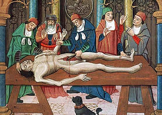
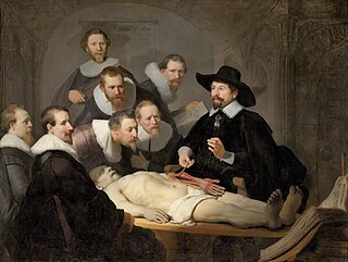
Dissection is the dismembering of the body of a deceased animal or plant to study its anatomical structure. Autopsy is used in pathology and forensic medicine to determine the cause of death in humans. Less extensive dissection of plants and smaller animals preserved in a formaldehyde solution is typically carried out or demonstrated in biology and natural science classes in middle school and high school, while extensive dissections of cadavers of adults and children, both fresh and preserved are carried out by medical students in medical schools as a part of the teaching in subjects such as anatomy, pathology and forensic medicine. Consequently, dissection is typically conducted in a morgue or in an anatomy lab.
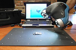
3D scanning is the process of analyzing a real-world object or environment to collect three dimensional data of its shape and possibly its appearance. The collected data can then be used to construct digital 3D models.

Materialise Mimics is an image processing software for 3D design and modeling, developed by Materialise NV, a Belgian company specialized in additive manufacturing software and technology for medical, dental and additive manufacturing industries. Materialise Mimics is used to create 3D surface models from stacks of 2D image data. These 3D models can then be used for a variety of engineering applications. Mimics is an acronym for Materialise Interactive Medical Image Control System. It is developed in an ISO environment with CE and FDA 510k premarket clearance. Materialise Mimics is commercially available as part of the Materialise Mimics Innovation Suite, which also contains Materialise3-matic, a design and meshing software for anatomical data. The current version is 24.0(released in 2021), and it supports Windows 10, Windows 7, Vista and XP in x64.
A tissue bank is an establishment that collects and recovers human cadaver tissue for the purposes of medical research, education and allograft transplantation. A tissue bank may also refer to a location where biomedical tissue is stored under cryogenic conditions and is generally used in a more clinical sense.
A cadaver or corpse is a dead human body. Cadavers are used by medical students, physicians and other scientists to study anatomy, identify disease sites, determine causes of death, and provide tissue to repair a defect in a living human being. Students in medical school study and dissect cadavers as a part of their education. Others who study cadavers include archaeologists and arts students. In addition, a cadaver may be used in the development and evaluation of surgical instruments.
Virtopsy is a virtual alternative to a traditional autopsy, conducted with scanning and imaging technology. The name is a portmanteau of "virtual" and "autopsy" and is a trademark registered to Richard Dirnhofer (de), the former head of the Institute of Forensic Medicine of the University of Bern, Switzerland.
Professor Paul Gerard McMenamin is an Australian academic and researcher specialising in the structure and immunology of the eye.
3D Indiana is a commercial Educational software for teaching and research on the human anatomy. The name is an acronym for Three-Dimensional Interactive Digital Anatomy. This software is based on the principles of volumetric anatomy which uses three intersecting coordinate planes to locate the organs of the human body based on mathematical calculations. This is in contrast to the traditional method of describing the location of the organs in relation to one another. The Gall bladder is traditionally described thus: 'It is situated on the right side of the body closely in contact with the inferior surface of the liver, along the right edge of the quadrate lobe in a shallow fossa extending from the right edge of the porta hepatis to the inferior lobe of the liver'.
VOXEL-MAN is the name of a set of a computer programs for creation and visualization of three-dimensional digital models of the human body derived from cross-sectional images of computer tomography, magnetic resonance tomography or photography. It was developed at the University Medical Center Hamburg-Eppendorf. Applications include diagnostic imaging, digital anatomical atlases and surgery simulators. The 3D interactive atlases of anatomy and radiology for brain/skull and inner organs are available for free download. The name Voxel-Man is derived from the term voxel, the elementary cuboid component of a digital representation of a three-dimensional object. Occasionally the name Voxel-Man is also used as a general term for a digital representation of the human body.

GIMIAS is a workflow-oriented environment focused on biomedical image computing and simulation. The open-source framework is extensible through plug-ins and is focused on building research and clinical software prototypes. Gimias has been used to develop clinical prototypes in the fields of cardiac imaging and simulation, angiography imaging and simulation, and neurology

In 3D computer graphics, 3D modeling is the process of developing a mathematical coordinate-based representation of any surface of an object in three dimensions via specialized software by manipulating edges, vertices, and polygons in a simulated 3D space.

Computer-generated imagery (CGI) is a specific-technology or application of computer graphics for creating or improving images in art, printed media, simulators, videos and video games. These images are either static or dynamic. CGI both refers to 2D computer graphics and 3D computer graphics with the purpose of designing characters, virtual worlds, or scenes and special effects. The application of CGI for creating/improving animations is called computer animation, or CGI animation.
Computational human phantoms are models of the human body used in computerized analysis. Since the 1960s, the radiological science community has developed and applied these models for ionizing radiation dosimetry studies. These models have become increasingly accurate with respect to the internal structure of the human body.
A medical animation is a short educational film, usually based around a physiological or surgical topic, that is rendered using 3D computer graphics. While it may be intended for an array of audiences, the medical animation is most commonly utilized as an instructional tool for medical professionals or their patients.
Alexander Tsiaras is an American photographer, entrepreneur, technology innovator, and journalist whose work has appeared on the cover of over 150 magazines. He is the founder, CEO, and editor-in-chief of TheVisualMD and StoryMD.
Cultural property imaging is a necessary part of long term preservation of cultural heritage. While the physical conditions of objects will change over time, imaging serves as a way to document and represent heritage in a moment in time of the life of the item. Different methods of imaging produce results that are applicable in various circumstances. Not every method is appropriate for every object, and not every object needs to be imaged by multiple methods. In addition to preservation and conservation-related concerns, imaging can also serve to enhance research and study of cultural heritage.

Susan Christina Potter was a cancer survivor, a disability rights activist and a body donor for the Visible Human Project. During the 15 years between signing on to the project in 2000 and her death by pneumonia in 2015 at the age of 87, Potter became a public figure and an outspoken advocate for medical education, mentoring medical students at the University of Colorado.
The Visible Embryo Project (VEP) is a multi-institutional, multidisciplinary research project originally created in the early 1990s as a collaboration between the Developmental Anatomy Center at the National Museum of Health and Medicine and the Biomedical Visualization Laboratory (BVL) at the University of Illinois at Chicago, "to develop software strategies for the development of distributed biostructural databases using cutting-edge technologies for high-performance computing and communications (HPCC), and to implement these tools in the creation of a large-scale digital archive of multidimensional data on normal and abnormal human development." This project related to BVL's other research in the areas of health informatics, educational multimedia, and biomedical imaging science. Over the following decades, the list of VEP collaborators grew to include over a dozen universities, national laboratories, and companies around the world.

