| Enamel pearl | |
|---|---|
 | |
| Recently pulled wisdom tooth with enamel pearl | |
| Specialty | Dentistry |
Enamel pearls are developmental variations of teeth that present as beads or nodules of enamel in places where they are not normally observed.
| Enamel pearl | |
|---|---|
| | |
| Recently pulled wisdom tooth with enamel pearl | |
| Specialty | Dentistry |
Enamel pearls are developmental variations of teeth that present as beads or nodules of enamel in places where they are not normally observed.
Enamel pearls most commonly present as spheroid in shape, but can also be conical, cylindrical, oval, teardrop, and irregularly shaped. They vary in size with an average diameter of 1.7mm [1] .
Enamel pearls are most frequently found in the root furcation of molars, and are not commonly seen in teeth with a single root, such as incisors and canines [1] .
Several theories of enamel pearl development exist, although no definitive mechanism has been established. Enamel pearls are composed primarily of enamel, but most also have a core of dentin within them.
The most widely accepted theory suggests enamel pearls are formed from remnants of Hertwig’s epithelial root sheath (HERS), which adhere to the tooth surface after root development. [1] During normal tooth development, after dentin formation is initiated on the root surface, the root sheath disintegrates and moves away from the root allowing the cells of the dental sac to contact predentin. This begins the differentiation of cementoblasts which in turn deposit cementum. However, when HERS remains adherent to the root surface, these fragments have the potential to develop into functional ameloblasts, which then deposit enamel onto the root surface, thereby forming enamel pearls. [1] This theory is considered inconclusive as it does not account for the dentin component seen in some enamel pearls.
Alternative theories propose that enamel pearl formation is akin to the development of supernumerary tubercles or cusps, thus pearl formation must occur during initial dentin formation. [1] Intradental enamel pearls may form during tooth formation, when ameloblasts are invaginated inside the developing dentin. [1] Inner enamel epithelium cells may similarly invade connective tissue of the dental papilla, resulting in an internal enamel pearl. [1]
Enamel pearls can be composed of different dental tissues (enamel, dentin, etc.) and can thus be classified based on this composition. Enamel-dentin pearls make up the largest proportion pearls and consist of a core of tubular dentin surrounded by enamel. Large enamel-dentin pearls may contain pulp within and are termed enamel-dentin-pulp pearls. Pearls that are composed exclusively of enamel are termed true enamel pearls or simple enamel pearls [1] .
Enamel pearls are estimated to occur in 1.1-9.7% of permanent molars, although higher rates are found when pearl detection is performed histologically instead of clinically [1] . The highest prevalence of enamel pearls is found in the maxillary third molar, with an incidence of approximately 75% [1] . Other common sites of occurrence include the mandibular third molar, and the maxillary second molar. The presence of two enamel pearls on the same tooth occurs in 8.7% of affected teeth, and up to four enamel pearls on the same tooth have been documented [1] .

The human teeth function to mechanically break down items of food by cutting and crushing them in preparation for swallowing and digesting. Humans have four types of teeth: incisors, canines, premolars, and molars, which each have a specific function. The incisors cut the food, the canines tear the food and the molars and premolars crush the food. The roots of teeth are embedded in the maxilla or the mandible and are covered by gums. Teeth are made of multiple tissues of varying density and hardness.

Cementum is a specialized calcified substance covering the root of a tooth. The cementum is the part of the periodontium that attaches the teeth to the alveolar bone by anchoring the periodontal ligament.

Tooth decay, also known as cavities or caries, is the breakdown of teeth due to acids produced by bacteria. The cavities may be a number of different colors from yellow to black. Symptoms may include pain and difficulty with eating. Complications may include inflammation of the tissue around the tooth, tooth loss and infection or abscess formation.

Dentin or dentine is a calcified tissue of the body and, along with enamel, cementum, and pulp, is one of the four major components of teeth. It is usually covered by enamel on the crown and cementum on the root and surrounds the entire pulp. By volume, 45% of dentin consists of the mineral hydroxyapatite, 33% is organic material, and 22% is water. Yellow in appearance, it greatly affects the color of a tooth due to the translucency of enamel. Dentin, which is less mineralized and less brittle than enamel, is necessary for the support of enamel. Dentin rates approximately 3 on the Mohs scale of mineral hardness. There are two main characteristics which distinguish dentin from enamel: firstly, dentin forms throughout life; secondly, dentin is sensitive and can become hypersensitive to changes in temperature due to the sensory function of odontoblasts, especially when enamel recedes and dentin channels become exposed.

The pulp is the part in the centre of a tooth made up of living connective tissues and odontoblasts. The pulp is a part of the dentin–pulp complex (endodontium). The vitality of the dentin-pulp complex, both during health and after injury, depends on pulp cell activity and the signalling processes that regulate the cell's behaviour.

Ameloblasts are cells present only during tooth development that deposit tooth enamel, which is the hard outermost layer of the tooth forming the surface of the crown.
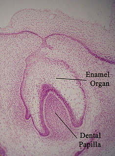
The enamel organ, also known as the dental organ, is a cellular aggregation seen in a developing tooth and it lies above the dental papilla. The enamel organ is responsible for the formation of enamel, initiation of dentine formation, establishment of the shape of a tooth's crown, and establishment of the dentoenamel junction.

Tooth development or odontogenesis is the complex process by which teeth form from embryonic cells, grow, and erupt into the mouth. For human teeth to have a healthy oral environment, all parts of the tooth must develop during appropriate stages of fetal development. Primary (baby) teeth start to form between the sixth and eighth week of prenatal development, and permanent teeth begin to form in the twentieth week. If teeth do not start to develop at or near these times, they will not develop at all, resulting in hypodontia or anodontia.
In vertebrates, an odontoblast is a cell of neural crest origin that is part of the outer surface of the dental pulp, and whose biological function is dentinogenesis, which is the formation of dentin, the substance beneath the tooth enamel on the crown and the cementum on the root.
Dentinogenesis is the formation of dentin, a substance that forms the majority of teeth. Dentinogenesis is performed by odontoblasts, which are a special type of biological cell on the outer wall of dental pulps, and it begins at the late bell stage of a tooth development. The different stages of dentin formation after differentiation of the cell result in different types of dentin: mantle dentin, primary dentin, secondary dentin, and tertiary dentin.

The Hertwig epithelial root sheath (HERS) or epithelial root sheath is a proliferation of epithelial cells located at the cervical loop of the enamel organ in a developing tooth. Hertwig epithelial root sheath initiates the formation of dentin in the root of a tooth by causing the differentiation of odontoblasts from the dental papilla. The root sheath eventually disintegrates with the periodontal ligament, but residual pieces that do not completely disappear are seen as epithelial cell rests of Malassez (ERM). These rests can become cystic, presenting future periodontal infections.
Cementogenesis is the formation of cementum, one of the three mineralized substances of a tooth. Cementum covers the roots of teeth and serves to anchor gingival and periodontal fibers of the periodontal ligament by the fibers to the alveolar bone.

Dentinogenesis imperfecta (DI) is a genetic disorder of tooth development. It is inherited in an autosomal dominant pattern, as a result of mutations on chromosome 4q21, in the dentine sialophosphoprotein gene (DSPP). It is one of the most frequently occurring autosomal dominant features in humans. Dentinogenesis imperfecta affects an estimated 1 in 6,000-8,000 people.
Dentin hypersensitivity is dental pain which is sharp in character and of short duration, arising from exposed dentin surfaces in response to stimuli, typically thermal, evaporative, tactile, osmotic, chemical or electrical; and which cannot be ascribed to any other dental disease.
Dens invaginatus (DI), also known as tooth within a tooth, is a rare dental malformation where there is an infolding of enamel into dentine. The prevalence of condition is 0.3 - 10%, affecting more males than females. The condition is presented in two forms, coronal and radicular, with the coronal form being more common.
Dental anatomy is a field of anatomy dedicated to the study of human tooth structures. The development, appearance, and classification of teeth fall within its purview. Tooth formation begins before birth, and the teeth's eventual morphology is dictated during this time. Dental anatomy is also a taxonomical science: it is concerned with the naming of teeth and the structures of which they are made, this information serving a practical purpose in dental treatment.
Dental pertains to the teeth, including dentistry. Topics related to the dentistry, the human mouth and teeth include:

Enamel hypoplasia is a defect of the teeth in which the enamel is deficient in quantity, caused by defective enamel matrix formation during enamel development, as a result of inherited and acquired systemic condition(s). It can be identified as missing tooth structure and may manifest as pits or grooves in the crown of the affected teeth, and in extreme cases, some portions of the crown of the tooth may have no enamel, exposing the dentin. It may be generalized across the dentition or localized to a few teeth. Defects are categorized by shape or location. Common categories are pit-form, plane-form, linear-form, and localised enamel hypoplasia. Hypoplastic lesions are found in areas of the teeth where the enamel was being actively formed during a systemic or local disturbance. Since the formation of enamel extends over a long period of time, defects may be confined to one well-defined area of the affected teeth. Knowledge of chronological development of deciduous and permanent teeth makes it possible to determine the approximate time at which the developmental disturbance occurred. Enamel hypoplasia varies substantially among populations and can be used to infer health and behavioural impacts from the past. Defects have also been found in a variety of non-human animals.

Pulp stones are nodular, calcified masses appearing in either or both the coronal and root portion of the pulp organ in teeth. Pulp stones are not painful unless they impinge on nerves.

Tooth discoloration is abnormal tooth color, hue or translucency. External discoloration is accumulation of stains on the tooth surface. Internal discoloration is due to absorption of pigment particles into tooth structure. Sometimes there are several different co-existent factors responsible for discoloration.