Related Research Articles

Human teeth function to mechanically break down items of food by cutting and crushing them in preparation for swallowing and digesting. As such, they are considered part of the human digestive system. Humans have four types of teeth: incisors, canines, premolars, and molars, which each have a specific function. The incisors cut the food, the canines tear the food and the molars and premolars crush the food. The roots of teeth are embedded in the maxilla or the mandible and are covered by gums. Teeth are made of multiple tissues of varying density and hardness.
Hyperdontia is the condition of having supernumerary teeth, or teeth that appear in addition to the regular number of teeth. They can appear in any area of the dental arch and can affect any dental organ. The opposite of hyperdontia is hypodontia, where there is a congenital lack of teeth, which is a condition seen more commonly than hyperdontia. The scientific definition of hyperdontia is "any tooth or odontogenic structure that is formed from tooth germ in excess of usual number for any given region of the dental arch." The additional teeth, which may be few or many, can occur on any place in the dental arch. Their arrangement may be symmetrical or non-symmetrical.

Dental surgery is any of a number of medical procedures that involve artificially modifying dentition; in other words, surgery of the teeth, gums and jaw bones.
Hypodontia is defined as the developmental absence of one or more teeth excluding the third molars. It is one of the most common dental anomalies, and can have a negative impact on function, and also appearance. It rarely occurs in primary teeth and the most commonly affected are the adult second premolars and the upper lateral incisors. It usually occurs as part of a syndrome that involves other abnormalities and requires multidisciplinary treatment.

Deciduous teeth or primary teeth, also informally known as baby teeth, milk teeth, or temporary teeth, are the first set of teeth in the growth and development of humans and other diphyodonts, which include most mammals but not elephants, kangaroos, or manatees, which are polyphyodonts. Deciduous teeth develop during the embryonic stage of development and erupt during infancy. They are usually lost and replaced by permanent teeth, but in the absence of their permanent replacements, they can remain functional for many years into adulthood.

Tooth development or odontogenesis is the complex process by which teeth form from embryonic cells, grow, and erupt into the mouth. For human teeth to have a healthy oral environment, all parts of the tooth must develop during appropriate stages of fetal development. Primary (baby) teeth start to form between the sixth and eighth week of prenatal development, and permanent teeth begin to form in the twentieth week. If teeth do not start to develop at or near these times, they will not develop at all, resulting in hypodontia or anodontia.

The maxillary central incisor is a human tooth in the front upper jaw, or maxilla, and is usually the most visible of all teeth in the mouth. It is located mesial to the maxillary lateral incisor. As with all incisors, their function is for shearing or cutting food during mastication (chewing). There is typically a single cusp on each tooth, called an incisal ridge or incisal edge. Formation of these teeth begins at 14 weeks in utero for the deciduous (baby) set and 3–4 months of age for the permanent set.

Dentinogenesis imperfecta (DI) is a genetic disorder of tooth development. It is inherited in an autosomal dominant pattern, as a result of mutations on chromosome 4q21, in the dentine sialophosphoprotein gene (DSPP). It is one of the most frequently occurring autosomal dominant features in humans. Dentinogenesis imperfecta affects an estimated 1 in 6,000-8,000 people.

Veterinary dentistry is the field of dentistry applied to the care of animals. It is the art and science of prevention, diagnosis, and treatment of conditions, diseases, and disorders of the oral cavity, the maxillofacial region, and its associated structures as it relates to animals.
Dilaceration is a developmental disturbance in shape of teeth. It refers to an angulation, or a sharp bend or curve, in the root or crown of a formed tooth. This disturbance is more likely to affect the maxillary incisors and occurs in permanent dentition. Although this may seem more of an aesthetics issue, an impacted maxillary incisor will cause issues related to occlusion, phonetics, mastication, and psychology on young patients.
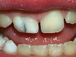
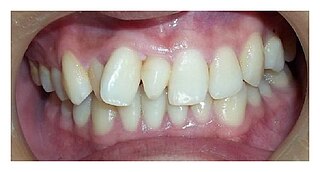
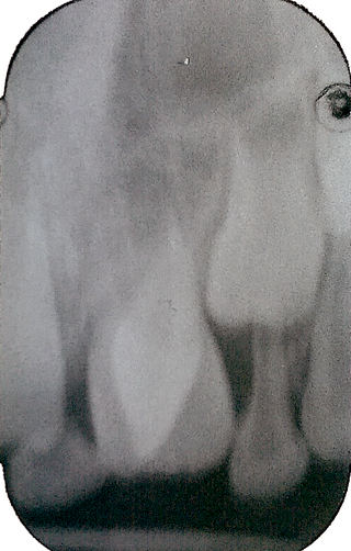
Tooth fusion arises through union of two normally separated tooth germs, and depending upon the stage of development of the teeth at the time of union, it may be either complete or incomplete. On some occasions, two independent pulp chambers and root canals can be seen. However, fusion can also be the union of a normal tooth bud to a supernumerary tooth germ. In these cases, the number of teeth is fewer if the anomalous tooth is counted as one tooth. In geminated teeth, division is usually incomplete and results in a large tooth crown that has a single root and a single canal. Both gemination and fusion are prevalent in primary dentition, with incisors being more affected.
Dens evaginatus is a rare odontogenic developmental anomaly that is found in teeth where the outer surface appears to form an extra bump or cusp.

Dentin dysplasia (DD) is a rare genetic developmental disorder affecting dentine production of the teeth, commonly exhibiting an autosomal dominant inheritance that causes malformation of the root. It affects both primary and permanent dentitions in approximately 1 in every 100,000 patients. It is characterized by the presence of normal enamel but atypical dentin with abnormal pulpal morphology. Witkop in 1972 classified DD into two types which are Type I (DD-1) is the radicular type, and type II (DD-2) is the coronal type. DD-1 has been further divided into 4 different subtypes (DD-1a,1b,1c,1d) based on the radiographic features.
Talon cusp is a rare dental anomaly resulting in an extra cusp or cusp-like projection on an anterior tooth, located on the inside surface of the affected tooth. Sometimes it can also be found on the facial surface of the anterior tooth.
Tooth eruption is a process in tooth development in which the teeth enter the mouth and become visible. It is currently believed that the periodontal ligament plays an important role in tooth eruption. The first human teeth to appear, the deciduous (primary) teeth, erupt into the mouth from around 6 months until 2 years of age, in a process known as "teething". These teeth are the only ones in the mouth until a person is about 6 years old creating the primary dentition stage. At that time, the first permanent tooth erupts and begins a time in which there is a combination of primary and permanent teeth, known as the mixed dentition stage, which lasts until the last primary tooth is lost. Then, the remaining permanent teeth erupt into the mouth during the permanent dentition stage.
Dental anatomy is a field of anatomy dedicated to the study of human tooth structures. The development, appearance, and classification of teeth fall within its purview. Tooth formation begins before birth, and the teeth's eventual morphology is dictated during this time. Dental anatomy is also a taxonomical science: it is concerned with the naming of teeth and the structures of which they are made, this information serving a practical purpose in dental treatment.
Dental pertains to the teeth, including dentistry. Topics related to the dentistry, the human mouth and teeth include:

Enamel hypoplasia is a defect of the teeth in which the enamel is deficient in quantity, caused by defective enamel matrix formation during enamel development, as a result of inherited and acquired systemic condition(s). It can be identified as missing tooth structure and may manifest as pits or grooves in the crown of the affected teeth, and in extreme cases, some portions of the crown of the tooth may have no enamel, exposing the dentin. It may be generalized across the dentition or localized to a few teeth. Defects are categorized by shape or location. Common categories are pit-form, plane-form, linear-form, and localised enamel hypoplasia. Hypoplastic lesions are found in areas of the teeth where the enamel was being actively formed during a systemic or local disturbance. Since the formation of enamel extends over a long period of time, defects may be confined to one well-defined area of the affected teeth. Knowledge of chronological development of deciduous and permanent teeth makes it possible to determine the approximate time at which the developmental disturbance occurred. Enamel hypoplasia varies substantially among populations and can be used to infer health and behavioural impacts from the past. Defects have also been found in a variety of non-human animals.

Dental trauma refers to trauma (injury) to the teeth and/or periodontium, and nearby soft tissues such as the lips, tongue, etc. The study of dental trauma is called dental traumatology.
Tooth ankylosis is the pathological fusion between alveolar bone and the cementum of teeth, which is a rare phenomenon in the deciduous dentition and even more uncommon in permanent teeth. Ankylosis occurs when partial root resorption is followed by repair with either cementum or dentine that unites the tooth root with the alveolar bone, usually after trauma. However, root resorption does not necessarily lead to tooth ankylosis and the causes of tooth ankylosis remain uncertain to a large extent. However, it is evident that the incident rate of ankylosis in deciduous teeth is much higher than that of permanent teeth.
References
- 1 2 3 4 5 6 7 8 Süha Türkaslan, Hasan Suat Gökçe and Mehmet Dalkız (July 2017). "Esthetic Rehabilitation of Bilateral Geminated Teeth: A Case Report". European Journal of Dentistry. 1 (3): 188–191. PMC 2638247 . PMID 19212565.
- 1 2 3 4 5 6 7 Nandini DB, Deepak BS, Selvamani M, Puneeth HK (2014). "Diagnostic dilemma of a double tooth: a rare case report and review". Journal of Clinical and Diagnostic Research. 8 (1): 271–2. doi:10.7860/JCDR/2014/6556.3928. PMC 3939503 . PMID 24596793.
- 1 2 3 4 5 6 "Tooth gemination in dentistry". DentaGama Dental Social Network. DentaGama. Retrieved 1 December 2017.
- ↑ E. Grammatopoulos (11 Aug 2007). "Gemination or fusion?". British Dental Journal. 203 (3): 119–120. doi: 10.1038/bdj.2007.699 . PMID 17694005.
- ↑ Siavash Moushekhian, Masoud Shiehzade, Amir Shammas (June 2014). "Treatment Plan and Clinical Management of a Geminated Maxillary Lateral Incisor: A Case Report". Journal of Dental Materials and Techniques. 3 (2): 87–90.
- ↑ Spuller RL, Harrington M (1986). "Gemination of a maxillary permanent central incisor treated by autogenous transplantation of a supernumerary incisor: case report". The American Academy of Pediatric Dentistry. 8 (4): 299–302. PMID 3472179.