
A fascia is a generic term for macroscopic membranous bodily structures. Fasciae are classified as superficial, visceral or deep, and further designated according to their anatomical location.
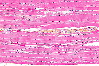
Striated muscle tissue is a muscle tissue that features repeating functional units called sarcomeres. The presence of sarcomeres manifests as a series of bands visible along the muscle fibers, which is responsible for the striated appearance observed in microscopic images of this tissue. There are two types of striated muscle:

Pectoralis minor muscle is a thin, triangular muscle, situated at the upper part of the chest, beneath the pectoralis major in the human body. It arises from ribs III-V; it inserts onto the coracoid process of the scapula. It is innervated by the medial pectoral nerve. Its function is to stabilise the scapula by holding it fast in position against the chest wall.
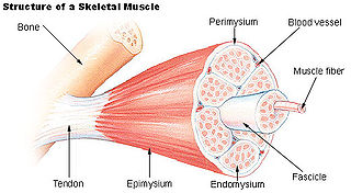
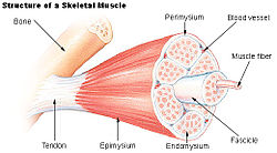
Perimysium is a sheath of dense irregular connective tissue that groups muscle fibers into bundles or fascicles.

The epineurium is the outermost layer of dense irregular connective tissue surrounding a peripheral nerve. It usually surrounds multiple nerve fascicles as well as blood vessels which supply the nerve. Smaller branches of these blood vessels penetrate into the perineurium. In addition to blood vessels which supply the nerve, lymphocytes and fibroblasts are also present and contribute to the production of collagen fibers that form the backbone of the epineurium. In addition to providing structural support, lymphocytes and fibroblasts also play a vital role in maintenance and repair of the surrounding tissues.
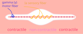
Intrafusal muscle fibers are skeletal muscle fibers that serve as specialized sensory organs (proprioceptors). They detect the amount and rate of change in length of a muscle. They constitute the muscle spindle, and are innervated by both sensory (afferent) and motor (efferent) fibers.

The anterior superior iliac spine (ASIS) is a bony projection of the iliac bone, and an important landmark of surface anatomy. It refers to the anterior extremity of the iliac crest of the pelvis. It provides attachment for the inguinal ligament, and the sartorius muscle. The tensor fasciae latae muscle attaches to the lateral aspect of the superior anterior iliac spine, and also about 5 cm away at the iliac tubercle.
Ground substance is an amorphous gel-like substance in the extracellular space of animals that contains all components of the extracellular matrix (ECM) except for fibrous materials such as collagen and elastin. Ground substance is active in the development, movement, and proliferation of tissues, as well as their metabolism. Additionally, cells use it for support, water storage, binding, and a medium for intercellular exchange. Ground substance provides lubrication for collagen fibers.

The adductor canal is an aponeurotic tunnel in the middle third of the thigh giving passage to parts of the femoral artery, vein, and nerve. It extends from the apex of the femoral triangle to the adductor hiatus.

The intercostal space (ICS) is the anatomic space between two ribs. Since there are 12 ribs on each side, there are 11 intercostal spaces, each numbered for the rib superior to it.
The medial pterygoid nerve (nerve to medial pterygoid, or internal pterygoid nerve) is a nerve of the head. It is a branch of the mandibular nerve (CN V3). It supplies the medial pterygoid muscle, the tensor veli palatini muscle, and the tensor tympani muscle.

The angular artery is an artery of the face. It is the terminal part of the facial artery. It ascends to the medial angle of the eye's orbit. It is accompanied by the angular vein. It ends by anastomosing with the dorsal nasal branch of the ophthalmic artery. It supplies the lacrimal sac, the orbicularis oculi muscle, and the outer side of the nose.
The common extensor tendon is a tendon that attaches to the lateral epicondyle of the humerus.
A muscle fascicle is a bundle of skeletal muscle fibers surrounded by perimysium, a type of connective tissue.
The endoneurium is a layer of delicate connective tissue around the myelin sheath of each myelinated nerve fiber in the peripheral nervous system. Its component cells are called endoneurial cells. The endoneuria with their enclosed nerve fibers are bundled into groups called nerve fascicles, each fascicle within its own protective sheath called a perineurium. In sufficiently large nerves multiple fascicles, each with its blood supply and fatty tissue, may be bundled within yet another sheath, the epineurium.

The buccopharyngeal fascia is a fascia of the pharynx. It represents the posterior portion of the pretracheal fascia. It covers the superior pharyngeal constrictor muscles, and buccinator muscle.
FACIT collagen is a type of collagen and also a proteoglycan that have two or more triple-helical domains that connect to collagen fibrils and share protein domains with non-collagen matrix molecules. FACIT collagens derive their name from their association and interaction with fibrillar collagens. Unlike fibrillar collagens, which form long fibers.

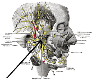
The anterior auricular muscle, the smallest of the three auricular muscles, is thin and fan-shaped, and its fibers are pale and indistinct. It arises from the lateral edge of the epicranial aponeurosis, and its fibers converge to be inserted into a projection on the front of the helix.
A key component in lateral force transmission in skeletal muscle is the extracellular matrix (ECM). Skeletal muscle is a complex biological material that is composed of muscle fibers and an ECM consisting of the epimysium, perimysium, and endomysium. It can be described as a collagen fiber-reinforced composite. The ECM has at least three functions: (1) to provide a framework binding muscle fibers together and ensure their proper alignment, (2) to transmit the forces, either from active muscle contraction or ones passively imposed on it, and (3) providing lubricated surfaces between muscle fibers and bundles enabling the muscle to change shape. The mechanical properties of skeletal muscle depend on both the properties of muscle fibers and the ECM, and the interaction between the two. Contractile forces are transmitted laterally within intramuscular connective tissue to the epimysium and then to the tendon. Due to the nature of skeletal muscle, direct measurements are not possible, but many indirect studies and analyses have shown that the ECM is an important part of force transmission during muscle contraction.