Endovascular coiling

Last updated
Endovascular coiling
Coiled PCA residual aneurysm arteriogram.JPG
Post-embolization arteriogram showing coiled aneurysm (indicated by yellow arrows) of the posteriorcerebral artery with a residual aneurysmal sac.
Other namesEndovascular embolization
Specialty Interventional neuroradiology

Endovascular coiling is an endovascular treatment for intracranial aneurysms and bleeding throughout the body. The procedure reduces blood circulation to the aneurysm through the use of microsurgical detachable platinum wires, with the clinician inserting one or more into the aneurysm until it is determined that blood flow is no longer occurring within the space. It is one of two main treatments for cerebral aneurysms, the other being surgical clipping. Clipping is an alternative to stenting for bleeding.

Contents

Medical uses

Endovascular coiling is used to treat cerebral aneurysms. The main goal is prevention of rupture in unruptured aneurysms, and prevention of rebleeding in ruptured aneurysms by limiting blood circulation to the aneurysm space. Clinically, packing density is recommended to be 20-30% or more of the aneurysm's volume, typically requiring deployment of multiple wires. [1] Higher volumes may be difficult due to the delicate nature of the aneurysm; intraoperative rupture rates are as high as 7.6% for this procedure. [2] In ruptured aneurysms, coiling is performed quickly after rupture because of the high risk of rebleeding within the first few weeks after initial rupture. The patients most suitable for endovascular coiling are those with aneurysms with a small neck size (preferably <4 mm), luminal diameter <25 mm and those that are distinct from the parent vessel. [3] Larger aneurysms are subject to compaction of coils, due to both looser packing densities (more coils are needed) and increased blood flow. Coil compaction renders them unsuitable as they are incapable of stemming blood flow. [4] However, technological advances have made coiling of many other aneurysms possible as well.

Results

A number of studies have questioned the efficacy of endovascular coiling over the more traditional surgical clipping. Most concerns involve the chance of later bleeds or other recanalization. [5] [6] [7] Due to its less invasive nature, endovascular coiling usually presents faster recovery times than surgical clipping, with one study finding a significant decrease in probability of death or dependency compared to a neurosurgical population. [8] Complication rates for coiling as well are generally found to be lower than microsurgery (11.7% and 17.6% for coiling and microsurgery, respectively). Despite this, intraoperative rupture rates for coiling have been documented as being as high as 7.6%. [2] Clinical results are found to be similar at a two-month and one year follow-up between coiling and neurosurgery. [9]

Reported recurrence rates are quite varied, with rates between 20 and 50% of aneurysms recurring within one year of coiling, and with the recurrence rate increasing with time. [2] [10] These results are similar to those previously reported by other endovascular groups. [11] Other studies have questioned whether new matrix coils work better than bare platinum coils. [12]

The International Subarachnoid Aneurysm Trial tested the efficacy of endovascular coiling against the traditional micro-surgical clipping. The study initially found very favorable results for coiling, however its results and methodology were criticized. Since the study's release in 2002, and again in 2005, some studies have found higher recurrence rates with coiling, while others have concluded that there is no clear consensus between which procedure is preferred. [13]

Risks

Risks of endovascular coiling include stroke, aneurysm rupture during the procedure and aneurysm recurrence and rupture after the procedure. [3] Additionally in some patients coiling may not be successful. In general, coiling is only performed when the risk of aneurysm rupture is higher than the risks of the procedure itself.

Similar to patients who experience neurosurgical procedures, coiling results in an increase in resting energy expenditure, albeit at a slightly reduced rate than their neurosurgery counterpart. This can lead to malnutrition if steps are not taken to compensate for the increased metabolic rate. [14]

Mechanism

The treatment works by promoting blood clotting (thrombosis) in the aneurysm, eventually sealing it from the blood flow. This is accomplished by decreasing the amount of blood flow going into the aneurysm, increasing the residence time of the blood (thereby lowering the velocity) in the aneurysm space and reducing the wall shear stress of the aneurysm wall. This change in the blood flow, or hemodynamics, is ultimately dependent on several factors, including:

While these factors are crucial to the success of the procedure, thrombosis ultimately is dependent on biological processes, with the coiling only providing the appropriate conditions for the process to occur, and hopefully closing the aneurysm.

Procedure

Resected middle cerebral artery aneurysm filled with multiple coils. Aneurysma Coil.jpg
Resected middle cerebral artery aneurysm filled with multiple coils.

Endovascular coiling is usually performed by an interventional neuroradiologist or neurosurgeon with the patient under general anaesthesia. The whole procedure is performed under fluoroscopic imaging guidance. A guiding catheter is inserted through the femoral artery and advanced to a site close to the aneurysm after which angiography is performed to localize and assess the aneurysm. After this, a microcatheter is navigated into the aneurysm.

The treatment uses detachable coils made of platinum that are inserted into the aneurysm using the microcatheter. A variety of coils are available, including Guglielmi Detachable Coils (GDC) which are platinum, Matrix coils which are coated with a biopolymer, and hydrogel coated coils. Coils are also available in a variety of diameters, lengths, and cross sections. [16] A coil is first inserted along the aneurysm wall to create a frame, with the core then being filled with more coils. [17] A series of progressively smaller coils may also be used. Success is determined by injecting a contrast dye into parent artery and qualitatively determining if dye is flowing into the aneurysm space during fluoroscopy. If no flow is observed, the procedure is considered completed. [2] In the case of wide-necked aneurysms a stent may be used. [18]

History

Endovascular coiling was a developed through the synthesis of a number of innovations that took place between 1970 and 1990 in the field of electronics, neurosurgery, and interventional radiology. [4] While the procedure itself has been and continues to be compared to surgical clipping, the development of the concept and procedure has resulted in it becoming the gold standard at many centers. [4]

Filling the intravascular compartment

The first documented technique of using metal coils to induce thrombosis was accomplished by Mullan in 1974. Copper coils were inserted into a giant aneurysm through externally puncturing the aneurysm wall via craniotomy. Five patients died, with ten having satisfactory process. [19] It did not gain popularity due to the specialized equipment required, in addition to the technique being unsuitable for many types of aneurysms. [4] Later, in 1980, similar techniques were developed by Alksne and Smith using iron suspended in methyl methacrylate in a limited set of patients. There were no deaths in 22 consecutive cases with low morbidity. [20] This technique also did not gain traction due to advances in clipping. [4]

Endovascular approaches

As a means of avoiding invasive methods, early endovascular interventions involved the usage of detachable and nondetachable balloon catheters to occlude the aneurysm while preserving the parent artery. [21] Despite the innovative approach, the aneurysms were often found to adapt to the shape of the balloon itself resulting in higher incidents of aneurysm rupture. This procedure was deemed "uncontrollable" due to its high morbidity and mortality rate, but it demonstrated that the endovascular approach was feasible for many aneurysms. [4] Endovascular coils would later be used in 1989 by Hilal et al., but these were short, stiff coils that offered no control, preventing dense packing of the aneurysm. [22] Controllable microguidewire systems were later used. [4]

Detachable coil system

In 1983 the use of electrically induced thrombosis for intracranial aneurysms was described for the first time. [23] A stainless steel electrode supplied a positive current to the aneurysm to stimulate electrothrombosis. Minimal occlusion was achieved, but the researchers discovered that the erosion of the electrode due to electrolysis would be useful as a detachment system. [4] Detachable coils were constructed from a platinum coil soldered to a stainless steel delivery wire, first described in 1991 by Guglielmi et al. [3] When combined with a controllable microguide wire system, multiple coils could be inserted to fully pack an aneurysm. [4]

Research

A 3D reconstruction of the Circle of Willis derived from a CT angiogram. Circle of willis from CT angio.gif
A 3D reconstruction of the Circle of Willis derived from a CT angiogram.

Given the complexity of modeling the vasculature, much research has been devoted towards modeling the hemodynamics of an aneurysm before and after an intervention. Techniques such as particle image velocimetry (PIV) and computational fluid dynamics/finite element analysis (CFD/FEA) have yielded results that have influenced the direction of research, but no model to date has been able to account for all factors present. [2] [24] [25] Advantages of the in-silico research method include flexibility of selecting variables, but one comparative study has found that simulations tend to over-emphasis results compared to PIV, and are more beneficial for trends than exact values. [25]

Medical images, particularly CT angiography, can be used to generate 3D reconstructions of patient specific anatomy. When combined with CFD/FEA, hemodynamics can be estimated in patient specific simulations, giving the clinician greater predictive tools for surgical planning and outcome evaluation to best promote thrombus formation. [26] [27] However, most computer models use many assumptions for simplicity, including rigid walls (non-elastic) for vasculature, substituting a porous medium in place of physical coil representations, and navier-stokes for fluid behavior. However, new predictive models are being developed as computational power increases, including algorithms for simulations of coil behavior in-vivo. [16]

Related Research Articles

<span class="mw-page-title-main">Cerebral arteriovenous malformation</span> Medical condition

A cerebral arteriovenous malformation is an abnormal connection between the arteries and veins in the brain—specifically, an arteriovenous malformation in the cerebrum.

<span class="mw-page-title-main">Intracranial aneurysm</span> Cerebrovascular disorder

An intracranial aneurysm, also known as a cerebral aneurysm, is a cerebrovascular disorder in which weakness in the wall of a cerebral artery or vein causes a localized dilation or ballooning of the blood vessel.

<span class="mw-page-title-main">Aneurysm</span> Bulge in the wall of a blood vessel

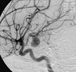
An aneurysm is an outward bulging, likened to a bubble or balloon, caused by a localized, abnormal, weak spot on a blood vessel wall. Aneurysms may be a result of a hereditary condition or an acquired disease. Aneurysms can also be a nidus for clot formation (thrombosis) and embolization. As an aneurysm increases in size, the risk of rupture, which leads to uncontrolled bleeding, increases. Although they may occur in any blood vessel, particularly lethal examples include aneurysms of the circle of Willis in the brain, aortic aneurysms affecting the thoracic aorta, and abdominal aortic aneurysms. Aneurysms can arise in the heart itself following a heart attack, including both ventricular and atrial septal aneurysms. There are congenital atrial septal aneurysms, a rare heart defect.

<span class="mw-page-title-main">Interventional radiology</span> Medical subspecialty

Interventional radiology (IR) is a medical specialty that performs various minimally-invasive procedures using medical imaging guidance, such as x-ray fluoroscopy, computed tomography, magnetic resonance imaging, or ultrasound. IR performs both diagnostic and therapeutic procedures through very small incisions or body orifices. Diagnostic IR procedures are those intended to help make a diagnosis or guide further medical treatment, and include image-guided biopsy of a tumor or injection of an imaging contrast agent into a hollow structure, such as a blood vessel or a duct. By contrast, therapeutic IR procedures provide direct treatment—they include catheter-based medicine delivery, medical device placement, and angioplasty of narrowed structures.

<span class="mw-page-title-main">Subarachnoid hemorrhage</span> Bleeding into the subarachnoid space

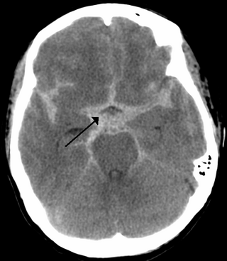
Subarachnoid hemorrhage (SAH) is bleeding into the subarachnoid space—the area between the arachnoid membrane and the pia mater surrounding the brain. Symptoms may include a severe headache of rapid onset, vomiting, decreased level of consciousness, fever, weakness, numbness, and sometimes seizures. Neck stiffness or neck pain are also relatively common. In about a quarter of people a small bleed with resolving symptoms occurs within a month of a larger bleed.

<span class="mw-page-title-main">Subdural hematoma</span> Hematoma usually associated with traumatic brain injury

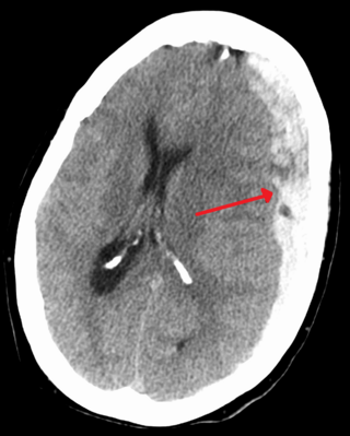
A subdural hematoma (SDH) is a type of bleeding in which a collection of blood—usually but not always associated with a traumatic brain injury—gathers between the inner layer of the dura mater and the arachnoid mater of the meninges surrounding the brain. It usually results from tears in bridging veins that cross the subdural space.

<span class="mw-page-title-main">Intracranial hemorrhage</span> Hemorrhage, or bleeding, within the skull

Intracranial hemorrhage (ICH), also known as intracranial bleed, is bleeding within the skull. Subtypes are intracerebral bleeds, subarachnoid bleeds, epidural bleeds, and subdural bleeds.

<span class="mw-page-title-main">Cerebral angiography</span> Angiography that produces images of blood vessels in and around the brain

Cerebral angiography is a form of angiography which provides images of blood vessels in and around the brain, thereby allowing detection of abnormalities such as arteriovenous malformations and aneurysms. It was pioneered in 1927 by the Portuguese neurologist Egas Moniz at the University of Lisbon, who also helped develop thorotrast for use in the procedure.

The International Subarachnoid Aneurysm Trial (ISAT) was a large multicentre, prospective randomised clinical medical trial, comparing the safety and efficacy of endovascular coil treatment and surgical clipping for the treatment of brain aneurysms. The study began in 1994. The first results were published in The Lancet in 2002, and the 10 year data were published again in The Lancet in early September 2005. 2,143 study participants were mostly drawn from U.K. hospitals with the rest drawn from North American and European hospitals.

<span class="mw-page-title-main">Dural arteriovenous fistula</span> Medical condition

A dural arteriovenous fistula (DAVF) or malformation is an abnormal direct connection (fistula) between a meningeal artery and a meningeal vein or dural venous sinus.

<span class="mw-page-title-main">Anterior clinoid process</span>

The anterior clinoid process is a posterior projection of the sphenoid bone at the junction of the medial end of either lesser wing of sphenoid bone with the body of sphenoid bone. The bilateral processes flank the sella turcica anteriorly.

<span class="mw-page-title-main">External ventricular drain</span> Medical device

An external ventricular drain (EVD), also known as a ventriculostomy or extraventricular drain, is a device used in neurosurgery to treat hydrocephalus and relieve elevated intracranial pressure when the normal flow of cerebrospinal fluid (CSF) inside the brain is obstructed. An EVD is a flexible plastic catheter placed by a neurosurgeon or neurointensivist and managed by intensive care unit (ICU) physicians and nurses. The purpose of external ventricular drainage is to divert fluid from the ventricles of the brain and allow for monitoring of intracranial pressure. An EVD must be placed in a center with full neurosurgical capabilities, because immediate neurosurgical intervention can be needed if a complication of EVD placement, such as bleeding, is encountered.

Joshua B. Bederson is an American neurosurgeon, Leonard I. Malis, MD/Corinne and Joseph Graber Professor of Neurosurgery, and System Chair of Neurosurgery at the Mount Sinai Health System in New York City. He is a Fellow of the American College of Surgeons, and an attending neurosurgeon at The Mount Sinai Hospital.

<span class="mw-page-title-main">Vein of Galen aneurysmal malformations</span> Medical condition

Vein of Galen aneurysmal malformations(VGAMs) and Vein of Galen aneurysmal dilations (VGADs) are the most frequent arteriovenous malformations in infants and fetuses. A VGAM consists of a tangled mass of dilated vessels supplied by an enlarged artery. The malformation increases greatly in size with age, although the mechanism of the increase is unknown. Dilation of the great cerebral vein of Galen is a secondary result of the force of arterial blood either directly from an artery via an arteriovenous fistula or by way of a tributary vein that receives the blood directly from an artery. There is usually a venous anomaly downstream from the draining vein that, together with the high blood flow into the great cerebral vein of Galen causes its dilation. The right sided cardiac chambers and pulmonary arteries also develop mild to severe dilation.

Interventional neuroradiology (INR) also known as neurointerventional surgery (NIS), endovascular therapy (EVT), endovascular neurosurgery, and interventional neurology is a medical subspecialty of neurosurgery, neuroradiology, intervention radiology and neurology specializing in minimally invasive image-based technologies and procedures used in diagnosis and treatment of diseases of the head, neck, and spine.

An infectious intracranial aneurysm is a cerebral aneurysm that is caused by infection of the cerebral arterial wall.

<span class="mw-page-title-main">Flow diverter</span>

A flow diverter is an endovascular prosthesis used to treat intracranial aneurysms. It is placed in the aneurysm's parent artery, covering the neck, in order to divert blood flow and determine a progressive thrombosis of the sac. Flow diverting stents consist of structural Cobalt-chrome or Nitinol alloy wires and often a set of radiopaque wires woven together in a flexible braid.

<span class="mw-page-title-main">William Couldwell</span>

William T. Couldwell is a Canadian neurosurgeon who is professor and Chairman of the Department of Neurosurgery at the University of Utah, a position he assumed in 2001.

Alexander Coon is an American neurosurgeon who is the Director of Endovascular and Cerebrovascular Neurosurgery at the Carondelet Neurological Institute of St. Joseph's and St. Mary's Hospitals in Tucson, Arizona. He was previously the Director of Endovascular Neurosurgery at the Johns Hopkins Hospital and an assistant professor of neurosurgery, Neurology, and Radiology at the Johns Hopkins Hospital. He is known for his work in cerebrovascular and endovascular neurosurgery and his research in neuroendovascular devices and clinical outcomes in the treatment of cerebral aneurysms, subarachnoid hemorrhage, and AVMs.

Luca Paolo Eugenio Regli is a neurosurgeon and full professor and chairman of the Department of Neurosurgery of the University Hospital of Zürich since October 2012. He is the son of Franco Regli, a Swiss professor of neurology and founder of the Foundation Franco Regli for the Research in the Field of Neurodegenerative Diseases.

References

  1. 1 2 Otani, Tomohiro; Nakamura, Masanori; Fujinaka, Toshiyuki; Hirata, Masayuki; Kuroda, Junko; Shibano, Katsuhiko; Wada, Shigeo (2013-03-26). "Computational fluid dynamics of blood flow in coil-embolized aneurysms: effect of packing density on flow stagnation in an idealized geometry". Medical & Biological Engineering & Computing. 51 (8): 901–910. doi:10.1007/s11517-013-1062-5. ISSN   0140-0118. PMID   23529587. S2CID   7811834.
  2. 1 2 3 4 5 6 Babiker, M. H.; Gonzalez, L. F.; Albuquerque, F.; Collins, D.; Elvikis, A.; Zwart, C.; Roszelle, B.; Frakes, D. H. (2013-04-01). "An In Vitro Study of Pulsatile Fluid Dynamics in Intracranial Aneurysm Models Treated with Embolic Coils and Flow Diverters". IEEE Transactions on Biomedical Engineering. 60 (4): 1150–1159. doi:10.1109/TBME.2012.2228002. ISSN   0018-9294. PMID   23192467. S2CID   206612828.
  3. 1 2 3 Currie, S; Mankad, K; Goddard, A (Jan 2011). "Endovascular treatment of intracranial aneurysms: review of current practice". Postgraduate Medical Journal. 87 (1023): 41–50. doi:10.1136/pgmj.2010.105387. PMID   20937736. S2CID   30220296.
  4. 1 2 3 4 5 6 7 8 9 Guglielmi, Guido (2009-03-13). "History of the genesis of detachable coils". Journal of Neurosurgery. 111 (1): 1–8. doi:10.3171/2009.2.JNS081039. ISSN   0022-3085. PMID   19284239.
  5. J Neurointerv Surg. 2011 Apr 27. [Epub ahead of print]
  6. Campi A, Ramzi N, Molyneux AJ, Summers PE, Kerr RS, Sneade M, Yarnold JA, Rischmiller J, Byrne JV (May 2007). "Retreatment of ruptured cerebral aneurysms in patients randomized by coiling or clipping in the International Subarachnoid Aneurysm Trial (ISAT)". Stroke. 38 (5): 1538–44. doi: 10.1161/STROKEAHA.106.466987 . PMID   17395870.
  7. Mitchell P, Kerr R, Mendelow AD, Molyneux A. Could late rebleeding overturn the superiority of cranial aneurysm coil embolization over clip ligation seen in ISAT?" Journal of Neurosurgery 108: 437-442, March 2008. But see, J Mocco, L. Nelson Hopkins, "International Subarachnoid Aneurysm Trial analysis", Journal of Neurosurgery, March 2008 / Vol. 108 / No. 3 / Pages 436-436.
  8. Molyneux, Andrew J; Birks, Jacqueline; Clarke, Alison; Sneade, Mary; Kerr, Richard S C (2015-02-21). "The durability of endovascular coiling versus neurosurgical clipping of ruptured cerebral aneurysms: 18 year follow-up of the UK cohort of the International Subarachnoid Aneurysm Trial (ISAT)". The Lancet. 385 (9969): 691–697. doi:10.1016/s0140-6736(14)60975-2. PMC   4356153 . PMID   25465111.
  9. Li, Jian; Su, Long; Ma, Jian; Kang, Ping; Ma, Liujia; Ma, Lianting (2017). "Endovascular coiling versus microsurgical clipping for patients with ruptured very small intracranial aneurysms: Management strategies and clinical outcomes- of 162 cases". World Neurosurgery. 99: 763–769. doi:10.1016/j.wneu.2015.11.079. PMID   26732968.
  10. Piotin, M; Spelle, L; Mounayer, C; Salles-Rezende, MT; Giansante-Abud, D; Vanzin-Santos, R; Moret, J (May 2007). "Intracranial aneurysms: treatment with bare platinum coils—aneurysm packing, complex coils, and angiographic recurrence". Radiology. 243 (2): 500–8. doi:10.1148/radiol.2431060006. PMID   17293572.
  11. Raymond, J; Guilbert, F; Weill, A; Georganos, SA; Juravsky, L; Lambert, A; Lamoureux, J; Chagnon, M; Roy, D (June 2003). "Long-term angiographic recurrences after selective endovascular treatment of aneurysms with detachable coils". Stroke. 34 (6): 1398–1403. doi: 10.1161/01.STR.0000073841.88563.E9 . PMID   12775880.
  12. Piotin M, Spelle L, Mounayer C, Loureiros C, Ghorbani A, Moret J. Intracranial aneurysms coiling with matrix. Immediate results in 152 patients and midterm anatomic follow-up from 115 patients. Stroke November 2008 (e-pub ahead of print)
  13. Raja PV, Huang J, Germanwala AV, Gailloud P, Murphy KP, Tamargo RJ (2008). "Microsurgical clipping and endovascular coiling of intracranial aneurysms: A critical review of the literature". Neurosurgery. 62 (6): 1187–1202. doi:10.1227/01.neu.0000333291.67362.0b. PMID   18824986. S2CID   21058347.
  14. Nagano, Ayano; Yamada, Yoshitaka; Miyake, Hiroji; Domen, Kazuhisa; Koyama, Tetsuo (April 2016). "Increased Resting Energy Expenditure after Endovascular Coiling for Subarachnoid Hemorrhage". Journal of Stroke and Cerebrovascular Diseases. 25 (4): 813–818. doi:10.1016/j.jstrokecerebrovasdis.2015.12.008. PMID   26796057.
  15. Hoi, Yiemeng; Meng, Hui; Woodward, Scott H.; Bendok, Bernard R.; Hanel, Ricardo A.; Guterman, Lee R.; Hopkins, L. Nelson (2004-10-01). "Effects of arterial geometry on aneurysm growth: three-dimensional computational fluid dynamics study". Journal of Neurosurgery. 101 (4): 676–681. doi:10.3171/jns.2004.101.4.0676. ISSN   0022-3085. PMID   15481725. S2CID   6565151.
  16. 1 2 Morales, H. G.; Kim, M.; Vivas, E. E.; Villa-Uriol, M.-C.; Larrabide, I.; Sola, T.; Guimaraens, L.; Frangi, A. F. (2011-11-01). "How Do Coil Configuration and Packing Density Influence Intra-Aneurysmal Hemodynamics?". American Journal of Neuroradiology. 32 (10): 1935–1941. doi: 10.3174/ajnr.A2635 . ISSN   0195-6108. PMC   7965998 . PMID   21885712.
  17. Morales, Hernan (2012). Endovascular coiling and its influence on intra-aneurysmal hemodynamics by image-based modeling. Barcelona, Spain. p. 19. ISBN   978-84-615-9824-3.{{cite book}}: CS1 maint: location missing publisher (link)
  18. Oushy, Soliman; Rinaldo, Lorenzo; Brinjikji, Waleed; Cloft, Harry; Lanzino, Giuseppe (June 2020). "Recent advances in stent-assisted coiling of cerebral aneurysms". Expert Review of Medical Devices. 17 (6): 519–532. doi:10.1080/17434440.2020.1778463. ISSN   1745-2422. PMID   32500761. S2CID   219328499.
  19. Mullan, S. (1974-12-01). "Experiences with surgical thrombosis of intracranial berry aneurysms and carotid cavernous fistulas". Journal of Neurosurgery. 41 (6): 657–670. doi:10.3171/jns.1974.41.6.0657. ISSN   0022-3085. PMID   4609023.
  20. Alksne, J. F.; Smith, R. W. (1980-06-01). "Stereotaxic occlusion of 22 consecutive anterior communicating artery aneurysms". Journal of Neurosurgery. 52 (6): 790–793. doi:10.3171/jns.1980.52.6.0790. ISSN   0022-3085. PMID   6991648.
  21. Serbinenko, F. A. (1974-08-01). "Balloon catheterization and occlusion of major cerebral vessels". Journal of Neurosurgery. 41 (2): 125–145. doi:10.3171/jns.1974.41.2.0125. ISSN   0022-3085. PMID   4841872.
  22. Group, British Medical Journal Publishing (1963-03-16). "Obliteration of Intracranial Aneurysms by Pilojection". Br Med J. 1 (5332): 700. doi:10.1136/bmj.1.5332.700-a. ISSN   0007-1447. S2CID   30623125.
  23. Guglielmi, G; Guerrisi, G; Guidetti, B (1983). "L'elettrotrombosi intravasale nelle malformazioni vascolari sperimentalmente provocate". Proceedings of III Congress of the Italian Society of Neuroradiology. Bari: Associazione Italiana di Neuroradiologia: 139–146.
  24. Wong, George K. C.; Poon, W. S. (2011-10-01). "Current status of computational fluid dynamics for cerebral aneurysms: the clinician's perspective". Journal of Clinical Neuroscience. 18 (10): 1285–1288. doi:10.1016/j.jocn.2011.02.014. ISSN   1532-2653. PMID   21795051. S2CID   22669620.
  25. 1 2 Ford, Matthew D.; Nikolov, Hristo N.; Milner, Jaques S.; Lownie, Stephen P.; DeMont, Edwin M.; Kalata, Wojciech; Loth, Francis; Holdsworth, David W.; Steinman, David A. (2008-04-03). "PIV-Measured Versus CFD-Predicted Flow Dynamics in Anatomically Realistic Cerebral Aneurysm Models". Journal of Biomechanical Engineering. 130 (2): 021015–021015–9. doi:10.1115/1.2900724. ISSN   0148-0731. PMID   18412502. S2CID   31375424.
  26. Kakalis, N. M. P.; Mitsos, A. P.; Byrne, J. V.; Ventikos, Y. (2008-06-01). "The Haemodynamics of Endovascular Aneurysm Treatment: A Computational Modelling Approach for Estimating the Influence of Multiple Coil Deployment". IEEE Transactions on Medical Imaging. 27 (6): 814–824. doi:10.1109/TMI.2008.915549. ISSN   0278-0062. PMID   18541488. S2CID   27015964.
  27. Cheng, Xiao Qing; Chen, Qian; Zhou, Chang Sheng; Li, Jian Rui; Zhang, Zong Jun; Zhang, Long Jiang; Huang, Wei; Lu, Guang Ming (April 2016). "Whole-brain CT perfusion combined with CT angiography for ischemic complications following microsurgical clipping and endovascular coiling of ruptured intracranial aneurysms". Journal of Clinical Neuroscience. 26: 50–56. doi:10.1016/j.jocn.2015.05.067. PMID   26775148. S2CID   24464063.