Intracranial aneurysm

Last updated
Intracranial aneurysm
Other namesCerebral aneurysm, brain aneurysm
Cerebellar aneurysm.png
Aneurysm of the basilar artery and the vertebral arteries
Specialty Interventional neuroradiology, neurosurgery, neurology   OOjs UI icon edit-ltr-progressive.svg
Symptoms None, severe headache, visual problems, nausea and vomiting, confusion [1]
Usual onset30–60 years old
Causes Hypertension, infection, head trauma [2]
Risk factors old age, family history, smoking, alcoholism, cocaine use [1]
Diagnostic method Angiography, CT scan
Treatment Endovascular coiling, surgical clipping, cerebral bypass surgery, pipeline embolization

An intracranial aneurysm, also known as a cerebral aneurysm, is a cerebrovascular disorder in which weakness in the wall of a cerebral artery or vein causes a localized dilation or ballooning of the blood vessel.

Contents

Aneurysms in the posterior circulation (basilar artery, vertebral arteries and posterior communicating artery) have a higher risk of rupture. Basilar artery aneurysms represent only 3–5% of all intracranial aneurysms but are the most common aneurysms in the posterior circulation.

Classification

Diagram of cerebral aneurysm. Cerebral aneurysm NIH.jpg
Diagram of cerebral aneurysm.

Cerebral aneurysms are classified both by size and shape. Small aneurysms have a diameter of less than 15 mm. Larger aneurysms include those classified as large (15 to 25 mm), giant (25 to 50 mm) (0.98 inches to 1.97 inches), and super-giant (over 50 mm). [3]

Berry (saccular) aneurysms

Saccular aneurysms, also known as berry aneurysms, appear as a round outpouching and are the most common form of cerebral aneurysm. [3] [4] Causes include connective tissue disorders, polycystic kidney disease, arteriovenous malformations, untreated hypertension, tobacco smoking, cocaine and amphetamines, intravenous drug abuse (can cause infectious mycotic aneurysms), alcoholism, heavy caffeine intake, head trauma, and infection in the arterial wall from bacteremia (mycotic aneurysms). [5]

Fusiform aneurysms

Fusiform dolichoectatic aneurysms represent a widening of a segment of an artery around the entire blood vessel, rather than just arising from a side of an artery's wall. They have an estimated annual risk of rupture between 1.6 and 1.9 percent. [6] [7]

Microaneurysms

Microaneurysms, also known as Charcot–Bouchard aneurysms, typically occur in small blood vessels (less than 300 micrometre diameter), most often the lenticulostriate vessels of the basal ganglia, and are associated with chronic hypertension. [8] Charcot–Bouchard aneurysms are a common cause of intracranial hemorrhage. [9]

Signs and symptoms

A small, unchanging aneurysm will produce few, if any, symptoms. Before a larger aneurysm ruptures, the individual may experience such symptoms as a sudden and unusually severe headache, nausea, vision impairment, vomiting, and loss of consciousness, or no symptoms at all. [10]

Subarachnoid bleed

If an aneurysm ruptures, blood leaks into the space around the brain. This is called a subarachnoid hemorrhage. Onset is usually sudden without prodrome, classically presenting as a "thunderclap headache" worse than previous headaches. [11] [12] Symptoms of a subarachnoid hemorrhage differ depending on the site and size of the aneurysm. [12] Symptoms of a ruptured aneurysm can include: [13]

Almost all aneurysms rupture at their apex. This leads to hemorrhage in the subarachnoid space and sometimes in brain parenchyma. Minor leakage from aneurysm may precede rupture, causing warning headaches. About 60% of patients die immediately after rupture. [14] Larger aneurysms have a greater tendency to rupture, though most ruptured aneurysms are less than 10 mm in diameter. [12]

Microaneurysms

A ruptured microaneurysm may cause an intracerebral hemorrhage, presenting as a focal neurological deficit. [12]

Rebleeding, hydrocephalus (the excessive accumulation of cerebrospinal fluid), vasospasm (spasm, or narrowing, of the blood vessels), or multiple aneurysms may also occur. The risk of rupture from a cerebral aneurysm varies according to the size of an aneurysm, with the risk rising as the aneurysm size increases. [15]

Vasospasm

Vasospasm, referring to blood vessel constriction, can occur secondary to subarachnoid hemorrhage following a ruptured aneurysm. This is most likely to occur within 21 days and is seen radiologically within 60% of such patients. The vasospasm is thought to be secondary to the apoptosis of inflammatory cells such as macrophages and neutrophils that become trapped in the subarachnoid space. These cells initially invade the subarachnoid space from the circulation in order to phagocytose the hemorrhaged red blood cells. Following apoptosis, it is thought there is a massive degranulation of vasoconstrictors, including endothelins and free radicals, that cause the vasospasm. [16]

Risk factors

Intracranial aneurysms may result from diseases acquired during life, or from genetic conditions. Hypertension, smoking, alcoholism, and obesity are associated with the development of brain aneurysms. [11] [12] [17] Cocaine use has also been associated with the development of intracranial aneurysms. [12]

Other acquired associations with intracranial aneurysms include head trauma and infections. [11]

Genetic associations

Coarctation of the aorta is also a known risk factor, [11] as is arteriovenous malformation. [14] Genetic conditions associated with connective tissue disease may also be associated with the development of aneurysms. [11] This includes: [18]

Specific genes have also had reported association with the development of intracranial aneurysms, including perlecan, elastin, collagen type 1 A2, endothelial nitric oxide synthase, endothelin receptor A and cyclin dependent kinase inhibitor. Recently, several genetic loci have been identified as relevant to the development of intracranial aneurysms. These include 1p34–36, 2p14–15, 7q11, 11q25, and 19q13.1–13.3. [19]

Pathophysiology

Aneurysm means an outpouching of a blood vessel wall that is filled with blood. Aneurysms occur at a point of weakness in the vessel wall. This can be because of acquired disease or hereditary factors. The repeated trauma of blood flow against the vessel wall presses against the point of weakness and causes the aneurysm to enlarge. [20] As described by the law of Young-Laplace, the increasing area increases tension against the aneurysmal walls, leading to enlargement. [21] [22] [23] In addition, a combination of computational fluid dynamics and morphological indices have been proposed as reliable predictors of cerebral aneurysm rupture. [24]

Both high and low wall shear stress of flowing blood can cause aneurysm and rupture. However, the mechanism of action is still unknown. It is speculated that low shear stress causes growth and rupture of large aneurysms through inflammatory response while high shear stress causes growth and rupture of small aneurysm through mural response (response from the blood vessel wall). Other risk factors that contributes to the formation of aneurysm are: cigarette smoking, hypertension, female gender, family history of cerebral aneurysm, infection, and trauma. Damage to structural integrity of the arterial wall by shear stress causes an inflammatory response with the recruitment of T cells, macrophages, and mast cells. The inflammatory mediators are: interleukin 1 beta, interleukin 6, tumor necrosis factor alpha (TNF alpha), MMP1, MMP2, MMP9, prostaglandin E2, complement system, reactive oxygen species (ROS), and angiotensin II. However, smooth muscle cells from the tunica media layer of the artery moved into the tunica intima, where the function of the smooth muscle cells changed from contractile function into pro-inflammatory function. This causes the fibrosis of the arterial wall, with reduction of number of smooth muscle cells, abnormal collagen synthesis, resulting in a thinning of the arterial wall and the formation of aneurysm and rupture. No specific gene loci has been identified to be associated with cerebral aneurysms. [25]

Generally, aneurysms larger than 7 mm in diameter should be treated because they are prone for rupture. Meanwhile, aneurysms less than 7 mm arise from the anterior and posterior communicating artery and are more easily ruptured when compared to aneurysms arising from other locations. [25]

Saccular aneurysms

The most common sites of intracranial saccular aneurysms Wikipedia intracranial aneurysms - inferior view - heat map.jpg
The most common sites of intracranial saccular aneurysms

Saccular aneurysms are almost always the result of hereditary weaknesses in blood vessels and typically occur within the arteries of the circle of Willis, [20] [26] in order of frequency affecting the following arteries: [27]

Saccular aneurysms tend to have a lack of tunica media and elastic lamina around their dilated locations (congenital), with a wall of sac made up of thickened hyalinized intima and adventitia. [14] In addition, some parts of the brain vasculature are inherently weak—particularly areas along the circle of Willis, where small communicating vessels link the main cerebral vessels. These areas are particularly susceptible to saccular aneurysms. [11] Approximately 25% of patients have multiple aneurysms, predominantly when there is a familial pattern. [12]

Diagnosis

CT angiography showing aneurysm measuring 2.6 mm in diameter at the ACOM (anterior communicating artery). CT angiography showing aneurysm at the ACOM.jpg
CT angiography showing aneurysm measuring 2.6 mm in diameter at the ACOM (anterior communicating artery).

Once suspected, intracranial aneurysms can be diagnosed radiologically using magnetic resonance or CT angiography. [28] But these methods have limited sensitivity for diagnosis of small aneurysms, and often cannot be used to specifically distinguish them from infundibular dilations without performing a formal angiogram. [28] [29] The determination of whether an aneurysm is ruptured is critical to diagnosis. Lumbar puncture (LP) is the gold standard technique for determining aneurysm rupture (subarachnoid hemorrhage). Once an LP is performed, the CSF is evaluated for RBC count, and presence or absence of xanthochromia. [30]

Treatment

A selection of Mayfield and Drake aneurysm clips ready for implantation. AneurysmClips.jpg
A selection of Mayfield and Drake aneurysm clips ready for implantation.

Emergency treatment for individuals with a ruptured cerebral aneurysm generally includes restoring deteriorating respiration and reducing intracranial pressure. Currently there are two treatment options for securing intracranial aneurysms: surgical clipping or endovascular coiling. If possible, either surgical clipping or endovascular coiling is typically performed within the first 24 hours after bleeding to occlude the ruptured aneurysm and reduce the risk of recurrent hemorrhage. [31]

While a large meta-analysis found the outcomes and risks of surgical clipping and endovascular coiling to be statistically similar, no consensus has been reached. [32] In particular, the large randomised control trial International Subarachnoid Aneurysm Trial appears to indicate a higher rate of recurrence when intracerebral aneurysms are treated using endovascular coiling. Analysis of data from this trial has indicated a 7% lower eight-year mortality rate with coiling, [33] a high rate of aneurysm recurrence in aneurysms treated with coiling—from 28.6 to 33.6% within a year, [34] [35] a 6.9 times greater rate of late retreatment for coiled aneurysms, [36] and a rate of rebleeding 8 times higher than surgically clipped aneurysms. [37]

Surgical clipping

Aneurysms can be treated by clipping the base of the aneurysm with a specially-designed clip. Whilst this is typically carried out by craniotomy, a new endoscopic endonasal approach is being trialled. [38] Surgical clipping was introduced by Walter Dandy of the Johns Hopkins Hospital in 1937. [39] After clipping, a catheter angiogram or CTA can be performed to confirm complete clipping. [40]

Endovascular coiling

Endovascular coiling refers to the insertion of platinum coils into the aneurysm. A catheter is inserted into a blood vessel, typically the femoral artery, and passed through blood vessels into the cerebral circulation and the aneurysm. Coils are pushed into the aneurysm, or released into the blood stream ahead of the aneurysm. Upon depositing within the aneurysm, the coils expand and initiate a thrombotic reaction within the aneurysm. If successful, this prevents further bleeding from the aneurysm. [41] In the case of broad-based aneurysms, a stent may be passed first into the parent artery to serve as a scaffold for the coils. [42]

Cerebral bypass surgery

Cerebral bypass surgery was developed in the 1960s in Switzerland by Gazi Yaşargil. When a patient has an aneurysm involving a blood vessel or a tumor at the base of the skull wrapping around a blood vessel, surgeons eliminate the problem vessel by replacing it with an artery from another part of the body. [43]

Prognosis

Outcomes depend on the size of the aneurysm. [44] Small aneurysms (less than 7 mm) have a low risk of rupture and increase in size slowly. [44] The risk of rupture is less than one percent for aneurysms of this size. [44]

The prognosis for a ruptured cerebral aneurysm depends on the extent and location of the aneurysm, the person's age, general health, and neurological condition. Some individuals with a ruptured cerebral aneurysm die from the initial bleeding. Other individuals with cerebral aneurysm recover with little or no neurological deficit. The most significant factors in determining outcome are the Hunt and Hess grade, and age. Generally patients with Hunt and Hess grade I and II hemorrhage on admission to the emergency room and patients who are younger within the typical age range of vulnerability can anticipate a good outcome, without death or permanent disability. Older patients and those with poorer Hunt and Hess grades on admission have a poor prognosis. Generally, about two-thirds of patients have a poor outcome, death, or permanent disability. [20] [45] [46]

Increased availability and greater access to medical imaging has caused a rising number of asymptomatic, unruptured cerebral aneurysms to be discovered incidentally during medical imaging investigations. [47] Unruptured aneurysms may be managed by endovascular clipping or stenting. For those subjects that underwent follow-up for the unruptured aneurysm, computed tomography angiography (CTA) or magnetic resonance angiography (MRA) of the brain can be done yearly. [48] Recently, an increasing number of aneurysm features have been evaluated in their ability to predict aneurysm rupture status, including aneurysm height, aspect ratio, height-to-width ratio, inflow angle, deviations from ideal spherical or elliptical forms, and radiomics morphological features. [49]

Epidemiology

The prevalence of intracranial aneurysm is about 1–5% (10 million to 12 million persons in the United States) and the incidence is 1 per 10,000 persons per year in the United States (approximately 27,000), with 30- to 60-year-olds being the age group most affected. [10] [20] Intracranial aneurysms occur more in women, by a ratio of 3 to 2, and are rarely seen in pediatric populations. [10] [17]

See also

Related Research Articles

<span class="mw-page-title-main">Arteriovenous malformation</span> Vascular anomaly

An arteriovenous malformation (AVM) is an abnormal connection between arteries and veins, bypassing the capillary system. Usually congenital, this vascular anomaly is widely known because of its occurrence in the central nervous system, but can appear anywhere in the body. The symptoms of AVMs can range from none at all to intense pain or bleeding, and they can lead to other serious medical problems.

<span class="mw-page-title-main">Cerebral arteriovenous malformation</span> Medical condition

A cerebral arteriovenous malformation is an abnormal connection between the arteries and veins in the brain—specifically, an arteriovenous malformation in the cerebrum.

<span class="mw-page-title-main">Cerebrovascular disease</span> Condition that affects the arteries that supply the brain

Cerebrovascular disease includes a variety of medical conditions that affect the blood vessels of the brain and the cerebral circulation. Arteries supplying oxygen and nutrients to the brain are often damaged or deformed in these disorders. The most common presentation of cerebrovascular disease is an ischemic stroke or mini-stroke and sometimes a hemorrhagic stroke. Hypertension is the most important contributing risk factor for stroke and cerebrovascular diseases as it can change the structure of blood vessels and result in atherosclerosis. Atherosclerosis narrows blood vessels in the brain, resulting in decreased cerebral perfusion. Other risk factors that contribute to stroke include smoking and diabetes. Narrowed cerebral arteries can lead to ischemic stroke, but continually elevated blood pressure can also cause tearing of vessels, leading to a hemorrhagic stroke.

<span class="mw-page-title-main">Aneurysm</span> Bulge in the wall of a blood vessel

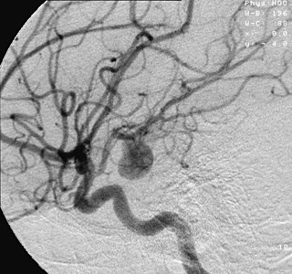
An aneurysm is an outward bulging, likened to a bubble or balloon, caused by a localized, abnormal, weak spot on a blood vessel wall. Aneurysms may be a result of a hereditary condition or an acquired disease. Aneurysms can also be a nidus for clot formation (thrombosis) and embolization. As an aneurysm increases in size, the risk of rupture, which leads to uncontrolled bleeding, increases. Although they may occur in any blood vessel, particularly lethal examples include aneurysms of the circle of Willis in the brain, aortic aneurysms affecting the thoracic aorta, and abdominal aortic aneurysms. Aneurysms can arise in the heart itself following a heart attack, including both ventricular and atrial septal aneurysms. There are congenital atrial septal aneurysms, a rare heart defect.

<span class="mw-page-title-main">Interventional radiology</span> Medical subspecialty

Interventional radiology (IR) is a medical specialty that performs various minimally-invasive procedures using medical imaging guidance, such as x-ray fluoroscopy, computed tomography, magnetic resonance imaging, or ultrasound. IR performs both diagnostic and therapeutic procedures through very small incisions or body orifices. Diagnostic IR procedures are those intended to help make a diagnosis or guide further medical treatment, and include image-guided biopsy of a tumor or injection of an imaging contrast agent into a hollow structure, such as a blood vessel or a duct. By contrast, therapeutic IR procedures provide direct treatment—they include catheter-based medicine delivery, medical device placement, and angioplasty of narrowed structures.

<span class="mw-page-title-main">Subarachnoid hemorrhage</span> Bleeding into the subarachnoid space

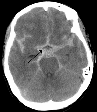
Subarachnoid hemorrhage (SAH) is bleeding into the subarachnoid space—the area between the arachnoid membrane and the pia mater surrounding the brain. Symptoms may include a severe headache of rapid onset, vomiting, decreased level of consciousness, fever, weakness, numbness, and sometimes seizures. Neck stiffness or neck pain are also relatively common. In about a quarter of people a small bleed with resolving symptoms occurs within a month of a larger bleed.

<span class="mw-page-title-main">Intracranial hemorrhage</span> Hemorrhage, or bleeding, within the skull

Intracranial hemorrhage (ICH), also known as intracranial bleed, is bleeding within the skull. Subtypes are intracerebral bleeds, subarachnoid bleeds, epidural bleeds, and subdural bleeds.

<span class="mw-page-title-main">Cerebral angiography</span> Angiography that produces images of blood vessels in and around the brain

Cerebral angiography is a form of angiography which provides images of blood vessels in and around the brain, thereby allowing detection of abnormalities such as arteriovenous malformations and aneurysms. It was pioneered in 1927 by the Portuguese neurologist Egas Moniz at the University of Lisbon, who also helped develop thorotrast for use in the procedure.

A thunderclap headache is a headache that is severe and has a sudden onset. It is defined as a severe headache that takes seconds to minutes to reach maximum intensity. Although approximately 75% are attributed to "primary" headaches—headache disorder, non-specific headache, idiopathic thunderclap headache, or uncertain headache disorder—the remainder are secondary to other causes, which can include some extremely dangerous acute conditions, as well as infections and other conditions. Usually, further investigations are performed to identify the underlying cause.

The International Subarachnoid Aneurysm Trial (ISAT) was a large multicentre, prospective randomised clinical medical trial, comparing the safety and efficacy of endovascular coil treatment and surgical clipping for the treatment of brain aneurysms. The study began in 1994. The first results were published in The Lancet in 2002, and the 10 year data were published again in The Lancet in early September 2005. 2,143 study participants were mostly drawn from U.K. hospitals with the rest drawn from North American and European hospitals.

<span class="mw-page-title-main">Intracerebral hemorrhage</span> Type of intracranial bleeding that occurs within the brain tissue itself

Intracerebral hemorrhage (ICH), also known as hemorrhagic stroke, is a sudden bleeding into the tissues of the brain, into its ventricles, or into both. An ICH is a type of bleeding within the skull and one kind of stroke. Symptoms can vary dramatically depending on the severity, acuity, and location (anatomically) but can include headache, one-sided weakness, numbness, tingling, or paralysis, speech problems, vision or hearing problems, memory loss, attention problems, coordination problems, balance problems, dizziness or lightheadedness or vertigo, nausea/vomiting, seizures, decreased level of consciousness or total loss of consciousness, neck stiffness, and fever.

<span class="mw-page-title-main">Dural arteriovenous fistula</span> Medical condition

A dural arteriovenous fistula (DAVF) or malformation is an abnormal direct connection (fistula) between a meningeal artery and a meningeal vein or dural venous sinus.

<span class="mw-page-title-main">Vertebral artery dissection</span> Tear of the inner lining of the vertebral artery

Vertebral artery dissection (VAD) is a flap-like tear of the inner lining of the vertebral artery, which is located in the neck and supplies blood to the brain. After the tear, blood enters the arterial wall and forms a blood clot, thickening the artery wall and often impeding blood flow. The symptoms of vertebral artery dissection include head and neck pain and intermittent or permanent stroke symptoms such as difficulty speaking, impaired coordination, and visual loss. It is usually diagnosed with a contrast-enhanced CT or MRI scan.

The Center for Cerebrovascular Research at the University of California, San Francisco is a collective of faculty and staff investigating matters related to cerebral circulation, particularly cerebrovascular disease resulting from narrowing of major blood vessels in the brain and vascular malformation of the brain. While research offices are located on Parnassus campus, San Francisco General Hospital hosts the center's laboratories and facilities. The center coordinates with additional faculty in various fields of neuroscience and vascular biology. Sponsors include the National Institute of Neurological Disorders and Stroke and the UCSF departments of Anesthesia, Neurological Surgery and Neurology.

Joshua B. Bederson is an American neurosurgeon, Leonard I. Malis, MD/Corinne and Joseph Graber Professor of Neurosurgery, and System Chair of Neurosurgery at the Mount Sinai Health System in New York City. He is a Fellow of the American College of Surgeons, and an attending neurosurgeon at The Mount Sinai Hospital.

<span class="mw-page-title-main">Endovascular coiling</span>

Endovascular coiling is an endovascular treatment for intracranial aneurysms and bleeding throughout the body. The procedure reduces blood circulation to the aneurysm through the use of microsurgical detachable platinum wires, with the clinician inserting one or more into the aneurysm until it is determined that blood flow is no longer occurring within the space. It is one of two main treatments for cerebral aneurysms, the other being surgical clipping. Clipping is an alternative to stenting for bleeding.

Interventional neuroradiology (INR) also known as neurointerventional surgery (NIS), endovascular therapy (EVT), endovascular neurosurgery, and interventional neurology is a medical subspecialty of neurosurgery, neuroradiology, intervention radiology and neurology specializing in minimally invasive image-based technologies and procedures used in diagnosis and treatment of diseases of the head, neck, and spine.

An infectious intracranial aneurysm is a cerebral aneurysm that is caused by infection of the cerebral arterial wall.

Alexander Coon is an American neurosurgeon who is the Director of Endovascular and Cerebrovascular Neurosurgery at the Carondelet Neurological Institute of St. Joseph's and St. Mary's Hospitals in Tucson, Arizona. He was previously the Director of Endovascular Neurosurgery at the Johns Hopkins Hospital and an assistant professor of neurosurgery, Neurology, and Radiology at the Johns Hopkins Hospital. He is known for his work in cerebrovascular and endovascular neurosurgery and his research in neuroendovascular devices and clinical outcomes in the treatment of cerebral aneurysms, subarachnoid hemorrhage, and AVMs.

<span class="mw-page-title-main">Clazosentan</span> Chemical compound

Clazosentan is a drug belonging to the class of endothelin receptor antagonists.

References

  1. 1 2 "Brain aneurysm - Symptoms and causes". Mayo Clinic .
  2. "What is an Aneurysm?".
  3. 1 2 "What You Should Know About Cerebral Aneurysms". www.hopkinsmedicine.org. 8 August 2021.
  4. Bhidayasiri, Roongroj; Waters, Michael F.; Giza, Christopher C. (2005). Neurological differential diagnosis : a prioritized approach (3. Dr. ed.). Oxford: Blackwell Publishing. p.  133. ISBN   978-1-4051-2039-5.
  5. Koutsothanasis G.A.; Sampath R., Berry Aneurysm (update: October 2, 2020). Link https://www.ncbi.nlm.nih.gov/books/NBK557480/
  6. Xu, David S.; Levitt, Michael R.; Kalani, M. Yashar S.; Rangel-Castilla, Leonardo; Mulholland, Celene B.; Abecassis, Isaac J.; Morton, Ryan P.; Nerva, John D.; Siddiqui, Adnan H.; Levy, Elad I.; Spetzler, Robert F. (2017-04-07). "Dolichoectatic aneurysms of the vertebrobasilar system: clinical and radiographic factors that predict poor outcomes". Journal of Neurosurgery. 128 (2): 560–66. doi: 10.3171/2016.10.JNS161041 . ISSN   1933-0693. PMID   28387624.
  7. Fusiform and dolichoectatic aneurysms By James R Brorson MD (Dr. Brorson of the University of Chicago received consultation fees from CVS-Caremark, National Peer Review Corporation, and Medico-legal Consulting.) Originally released October 28, 1997; last updated January 29, 2017; expires January 29, 2020 https://tcapp.org/wp-content/uploads/2017/09/Fusiform-and-Dolichoectatic-Aneurysms.pdf
  8. Kumar; Abbas; Fausto, eds. (2005). Robbins and Cotran Pathologic Basis of Disease (7th ed.). China: Elsevier. ISBN   978-0-7216-0187-8.[ page needed ]
  9. Gupta, Kashvi; M Das, Joe (2021), Charcot Bouchard Aneurysm, Treasure Island (FL): StatPearls Publishing, PMID   31971704 , retrieved 2021-05-06
  10. 1 2 3 Brisman, JL; Song, JK; Newell, DW (Aug 31, 2006). "Cerebral aneurysms". The New England Journal of Medicine. 355 (9): 928–39. doi:10.1056/nejmra052760. PMID   16943405.
  11. 1 2 3 4 5 6 Goljan, Edward F. (2006). Rapid Review Pathology (2nd ed.). St. Louis: Mosby. p. 158. ISBN   978-0-323-04414-1.
  12. 1 2 3 4 5 6 7 Alway, David; Cole, John Walden, eds. (2009). Stroke Essentials for Primary Care: A Practical Guide . New York: Humana Press. pp.  86–88, 153. ISBN   978-1-934115-01-5.
  13. Publishing, Harvard Health (18 December 2018). "Subarachnoid Hemorrhage". Harvard Health. Retrieved 2021-05-06.
  14. 1 2 3 DiMaio, Vincent J.; DiMaio, Dominick (2001). Forensic pathology (2nd ed.). Boca Raton, FL: CRC Press. p.  61. ISBN   978-0-8493-0072-1.
  15. Korja Miikka; Lehto Hanna; Juvela Seppo (2014-07-01). "Lifelong Rupture Risk of Intracranial Aneurysms Depends on Risk Factors". Stroke. 45 (7): 1958–63. doi: 10.1161/STROKEAHA.114.005318 . PMID   24851875.
  16. Gallia, Gary L.; Tamargo, Rafael J. (1 October 2006). "Leukocyte-endothelial cell interactions in chronic vasospasm after subarachnoid hemorrhage". Neurological Research. 28 (7): 750–58. doi:10.1179/016164106X152025. PMID   17164038. S2CID   27713975.
  17. 1 2 Brown, Walter L. Kemp, Dennis K. Burns, Travis G. (2008). Pathology the big picture. New York: McGraw-Hill Medical. p. 148. ISBN   978-0-07-159379-3.{{cite book}}: CS1 maint: multiple names: authors list (link)
  18. Caranci, F.; Briganti, F.; Cirillo, L.; Leonardi, M.; Muto, M. (2012). "Epidemiology and genetics of intracranial aneurysms". European Journal of Radiology. 82 (10): 1598–605. doi:10.1016/j.ejrad.2012.12.026. PMID   23399038.
  19. van der Voet, M; Olson, J; Kuivaniemi, H; Dudek, D; Skunca, M; Ronkainen, A; Niemelä, M; Jääskeläinen, J; Hernesniemi, J; Helin, K (1 March 2004). "Intracranial Aneurysms in Finnish Families: Confirmation of Linkage and Refinement of the Interval to Chromosome 19q13.3". The American Journal of Human Genetics. 74 (3): 564–71. doi:10.1086/382285. PMC   1182270 . PMID   14872410.
  20. 1 2 3 4 Haberland, Catherine (2007). Clinical neuropathology : text and color atlas (Online-Ausg. ed.). New York: Demos. p.  70. ISBN   978-1-888799-97-2.
  21. Humphrey, J. D.; Kyriacou, S. K. (June 1996). "The use of Laplace's equation in aneurysm mechanics". Neurological Research. 18 (3): 204–08. doi:10.1080/01616412.1996.11740404. ISSN   0161-6412. PMID   8837052.
  22. "Laplace's Law and Aneurysms". sciencedemonstrations.fas.harvard.edu. Retrieved 2021-05-06.
  23. Hademenos, George J. (2008-01-11). "The Physics of Cerebral Aneurysms". Physics Today. 48 (2): 24. doi:10.1063/1.881442. ISSN   0031-9228.
  24. Amigo, Nicolás; Valencia, Alvaro; Wu, Wei; Patnaik, Sourav; Finol, Ender (2021-03-08). "Cerebral aneurysm rupture status classification using statistical and machine learning methods". Proceedings of the Institution of Mechanical Engineers, Part H. 235 (6): 655–662. doi:10.1177/09544119211000477. ISSN   2041-3033. PMID   33685288. S2CID   232161029.
  25. 1 2 Chalouhi, Nohra; Loh, Brian L; Hasan, David (25 November 2013). "Review of Cerebral Aneurysm Formation, Growth, and Rupture". Stroke. 44 (12): 3613–22. doi: 10.1161/STROKEAHA.113.002390 . PMID   24130141.
  26. "berry aneurysm" "at Dorland's Medical Dictionary
  27. Howlett, David; Ayers, Brian (2004). The hands-on guide to imaging . Oxford: Blackwell. p.  204. ISBN   978-1-4051-1551-3.
  28. 1 2 White, Philip M.; Teasdale, Evelyn M.; Wardlaw, Joanna M.; Easton, Valerie (2001-06-01). "Intracranial Aneurysms: CT Angiography and MR Angiography for Detection – Prospective Blinded Comparison in a Large Patient Cohort". Radiology. 219 (3): 739–49. doi:10.1148/radiology.219.3.r01ma16739. ISSN   0033-8419. PMID   11376263.
  29. Greenberg, Mark (2010). Handbook of neurosurgery. Greenberg Graphics. ISBN   978-1604063264. OCLC   892183792.[ page needed ]
  30. Mark, Dustin G.; Kene, Mamata V.; Offerman, Steven R.; Vinson, David R.; Ballard, Dustin W. (2015). "Validation of cerebrospinal fluid findings in aneurysmal subarachnoid hemorrhage". The American Journal of Emergency Medicine. 33 (9): 1249–52. doi:10.1016/j.ajem.2015.05.012. PMID   26022754.
  31. Park, Jaechan; Woo, Hyunjin; Kang, Dong-Hun; Kim, Yong-Sun; Kim, Min Young; Shin, Im Hee; Kwak, Sang Gyu (2015-02-01). "Formal protocol for emergency treatment of ruptured intracranial aneurysms to reduce in-hospital rebleeding and improve clinical outcomes". Journal of Neurosurgery. 122 (2): 383–91. doi: 10.3171/2014.9.JNS131784 . ISSN   1933-0693. PMID   25403841.
  32. Raja, PV; Huang, J; Germanwala, AV; Gailloud, P; Murphy, KP; Tamargo, RJ (June 2008). "Microsurgical clipping and endovascular coiling of intracranial aneurysms: a critical review of the literature". Neurosurgery. 62 (6): 1187–202, discussion 1202–03. doi:10.1227/01.neu.0000333291.67362.0b. PMID   18824986.
  33. Mitchell P, Kerr R, Mendelow AD, Molyneux A. "Could late rebleeding overturn the superiority of cranial aneurysm coil embolization over clip ligation seen in ISAT?" Journal of Neurosurgery 108: 437–42, March 2008
  34. Piotin, M; Spelle, L; Mounayer, C; Salles-Rezende, MT; Giansante-Abud, D; Vanzin-Santos, R; Moret, J (May 2007). "Intracranial aneurysms: treatment with bare platinum coils – aneurysm packing, complex coils, and angiographic recurrence". Radiology. 243 (2): 500–08. doi:10.1148/radiol.2431060006. PMID   17293572.
  35. Raymond, J; Guilbert, F; Weill, A; Georganos, SA; Juravsky, L; Lambert, A; Lamoureux, J; Chagnon, M; Roy, D (June 2003). "Long-term angiographic recurrences after selective endovascular treatment of aneurysms with detachable coils". Stroke. 34 (6): 1398–1403. doi: 10.1161/01.STR.0000073841.88563.E9 . PMID   12775880.
  36. Campi A, Ramzi N, Molyneux AJ, Summers PE, Kerr RS, Sneade M, Yarnold JA, Rischmiller J, Byrne JV (May 2007). "Retreatment of ruptured cerebral aneurysms in patients randomized by coiling or clipping in the International Subarachnoid Aneurysm Trial (ISAT)". Stroke. 38 (5): 1538–44. doi: 10.1161/STROKEAHA.106.466987 . PMID   17395870.
  37. Mitchell P, Kerr R, Mendelow AD, Molyneux A (March 2008). "Could late rebleeding overturn the superiority of cranial aneurysm coil embolization over clip ligation seen in the International Subarachnoid Aneurysm Trial?". J. Neurosurg. 108 (3): 437–42. doi:10.3171/JNS/2008/108/3/0437. PMID   18312088. S2CID   24304414.
  38. "Germanwala Presents First Aneurysm Patient Treated Through Nose". UNC. June 25, 2009.
  39. Sasidharan, Gopalakrishnan Madhavan; Sastri, Savitr B V; Pandey, Paritosh (1 January 2015). "Aneurysm clips: What every resident should know". Neurology India. 63 (1) via www.neurologyindia.com.
  40. Bharatha A, Yeung R, Durant D, Fox AJ, Aviv RI, Howard P, Thompson AL, Bartlett ES, Symons SP. "Comparison of computed tomography angiography with digital subtraction angiography in the assessment of clipped intracranial aneurysms". Journal of Computer Assisted Tomography. 2010 May–June; 34(3): 440–45.
  41. Brilstra EH, Rinkel GJ, van der Graaf Y, van Rooij WJ, Algra A (February 1999). "Treatment of intracranial aneurysms by embolization with coils: a systematic review". Stroke. 30 (2): 470–76. doi: 10.1161/01.STR.30.2.470 . PMID   9933290.
  42. Oushy, Soliman; Rinaldo, Lorenzo; Brinjikji, Waleed; Cloft, Harry; Lanzino, Giuseppe (June 2020). "Recent advances in stent-assisted coiling of cerebral aneurysms". Expert Review of Medical Devices. 17 (6): 519–32. doi:10.1080/17434440.2020.1778463. ISSN   1745-2422. PMID   32500761. S2CID   219328499.
  43. Spetzler, Robert; Chater, Norman (1 November 1976). "Microvascular bypass surgery". Journal of Neurosurgery. 45 (5): 508–13. doi:10.3171/jns.1976.45.5.0508. PMID   972334.
  44. 1 2 3 Malhotra A, Wu X, Forman HP, Grossetta Nardini HK, Matouk CC, Gandhi D, Moore C, Sanelli P (July 2017). "Growth and Rupture Risk of Small Unruptured Intracranial Aneurysms: A Systematic Review". Ann. Intern. Med. 167 (1): 26–33. doi:10.7326/M17-0246. PMID   28586893.
  45. Hop, J. W.; Rinkel, G. J.E.; Algra, A.; van Gijn, J. (1 March 1997). "Case-Fatality Rates and Functional Outcome After Subarachnoid Hemorrhage : A Systematic Review". Stroke. 28 (3): 660–64. doi:10.1161/01.STR.28.3.660. PMID   9056628.
  46. Ljunggren, B; Sonesson, B; Säveland, H; Brandt, L (May 1985). "Cognitive impairment and adjustment in patients without neurological deficits after aneurysmal SAH and early operation". Journal of Neurosurgery. 62 (5): 673–79. doi:10.3171/jns.1985.62.5.0673. PMID   3989590. S2CID   26649695.
  47. Vernooij, MW; Ikram, MA; Tanghe, HL (2007). "Incidental findings on brain MRI in the general population.Incidental findings on brain MRI in the general population". N Engl J Med. 357 (5): 1821–28. doi: 10.1056/NEJMoa070972 . PMID   17978290.
  48. Thompson, B. Gregory; Brown, Robert D.; Amin-Hanjani, Sepideh; Broderick, Joseph P.; Cockroft, Kevin M.; Connolly, E. Sander; Duckwiler, Gary R.; Harris, Catherine C.; Howard, Virginia J.; Johnston, S. Claiborne (Clay); Meyers, Philip M.; Molyneux, Andrew; Ogilvy, Christopher S.; Ringer, Andrew J.; Torner, James (August 2015). "Guidelines for the Management of Patients With Unruptured Intracranial Aneurysms: A Guideline for Healthcare Professionals From the American Heart Association/American Stroke Association". Stroke. 46 (8): 2368–2400. doi: 10.1161/STR.0000000000000070 . ISSN   0039-2499.
  49. Ludwig, CG; Lauric, A; Malek, JA; Mulligan, R; Malek, AM (2020). "Performance of Radiomics derived morphological features for prediction of aneurysm rupture status". Journal of NeuroInterventional Surgery. 13 (8). doi:10.1136/neurintsurg-2020-016808. PMID   33158993. S2CID   226274492.