Radiomics

Last updated

In the field of medicine, radiomics is a method that extracts a large number of features from medical images using data-characterisation algorithms. [1] [2] [3] [4] [5] These features, termed radiomic features, have the potential to uncover tumoral patterns and characteristics that fail to be appreciated by the naked eye. [6] The hypothesis of radiomics is that the distinctive imaging features between disease forms may be useful for predicting prognosis and therapeutic response for various cancer types, thus providing valuable information for personalized therapy. [1] [7] [8] Radiomics emerged from the medical fields of radiology and oncology [3] [9] [10] and is the most advanced in applications within these fields. However, the technique can be applied to any medical study where a pathological process can be imaged.

Contents

Process

Image acquisition

The image data is provided by radiological modalities as CT, [11] MRI, [12] PET/CT or even PET/MR. [13] The produced raw data volumes are used to find different pixel/voxel characteristics through extraction tools. [2]

The extracted features are saved in large databases where clinics have access so as to enable broadly collaborative and cumulative work in which all can benefit from growing amounts of data, ideally enabling a more precise workflow.

Image segmentation

After the images have been saved in the database, they have to be reduced to the essential parts, in this case the tumors, which are called “volumes of interest”. [2]

Because of the large image data that needs to be processed, it would be too much work to perform the segmentation manually for every single image if a radiomics database with lots of data is created. Instead of manual segmentation, an automated process has to be used. A possible solution are automatic and semiautomatic segmentation algorithms. Before it can be applied on a big scale an algorithm must score as high as possible in the following four tasks:

Feature extraction and qualification

After the segmentation, many features can be extracted and the relative net change from longitudinal images (delta-radiomics) can be computed. Radiomic features can be divided into five groups: size and shape based–features, descriptors of the image intensity histogram, descriptors of the relationships between image voxels (e.g. gray-level co-occurrence matrix (GLCM), run length matrix (RLM), size zone matrix (SZM), and neighborhood gray tone difference matrix (NGTDM) derived textures), textures extracted from filtered images, and fractal features. The mathematical definitions of these features are independent of imaging modality and can be found in the literature. [14] [15] [16] [17] A detailed description of texture features for radiomics can be found in Parekh et al. (2016) [4] and Depeursinge et al. (2017). [18]

Due to its massive variety, feature reductions need to be implemented to eliminate redundant information. Hundreds of different features need to be evaluated with a selection algorithms to accelerate this process. Additionally, features that are unstable and non-reproducible should be eliminated since features with low-fidelity will likely lead to spurious findings and unrepeatable models. [19] [20]

Analysis

After the selection of features that are important for our task it is crucial to analyze the chosen data. Before the actual analysis, the clinical and molecular (sometimes even the genetic) data needs to be integrated because it has a big impact on what can be deducted from the analysis. There are different methods to finally analyze the data. First, the different features are compared to one another to find out whether they have any information in common and to reveal what it means when they all occur at the same time.

Another way is Supervised or Unsupervised Analysis. Supervised Analysis uses an outcome variable to be able to create prediction models. Unsupervised Analysis summarizes the information we have and can be represented graphically. So that the conclusion of our results is clearly visible.

Databases

Creation

Several steps are necessary to create an integrated radiomics database. The imaging data needs to be exported from the clinics. This is already a very challenging step because the patient information is very sensitive and governed by Privacy laws, such as HIPAA. At the same time the exported data must not lose any of its integrity when compressed so that the database only incorporates data of the same quality. The integration of clinical and molecular data is important as well and a large image storage location is needed.

Use

The goal of radiomics is to be able to use this database for new patients. This means that we need algorithms that run new input data through the database which return a result with information about what the course of the patients’ disease might look like. For example, how fast the tumor will grow or how good the chances are that the patient survives for a certain time, whether distant metastases are possible and where. This determines how the further treatment (like surgery, chemotherapy, radiotherapy or targeted drugs etc.) and the best solution which maximizes survival or improvement is selected. The algorithm has to recognize correlations between the images and the features, so that it is possible to extrapolate from the data base material to the input data.

Applications

Prediction of clinical outcomes

Aerts et al. (2014) [21] performed the first large-scale radiomic study that included three lung and two head-and-neck cancer cohorts, consisting of over 1000 patients. They assessed the prognostic values of over 400 textural and shape- and intensity-based features extracted from the computed tomography (CT) images acquired before any treatment. Tumor volumes were defined either by expert radiation oncologists or using semiautomatic segmentation methods. [22] [23] Their results identified a subset of radiomic features that may be useful for predicting patient survival and describing intratumoural heterogeneity. They also confirmed that the prognostic ability of these radiomics features may be transferred from lung to head-and-neck cancer. However, Parmar et al. (2015) [24] demonstrated that prognostic value of some radiomic features may be cancer type dependent. Particularly, they observed that not every radiomic feature that significantly predicted the survival of lung cancer patients could also predict the survival of head-and-neck cancer patients and vice versa.

Nasief et al. (2019) [20] showed that changes of radiomic features over time in longitudinal images (delta-radiomic features, DRFs) can potentially be used as a biomarker to predict treatment response for pancreatic cancer. Their results showed that a Bayesian regularization neural network can be used to identify a subset of DRFs that demonstrated significant changes between good- and bad- responders following 2–4 weeks of treatment with an AUC = 0.94. They also showed (Nasief et al., 2020) that DRFs are independent predictor of survival and if combined with the clinical biomarker CA19-9 can improve treatment response prediction and increase the possibility for response-based treatment adaptation . [25]

Several studies have also showed that radiomic features are better at predicting treatment response than conventional measures, such as tumor volume and diameter, and the maximum radiotracer uptake on positron emission tomography (PET) imaging. [26] [27] [28] [29] [30] [31] [32] Using this technique an algorithm has been developed, after initial training based on intra tumor lymphocyte density, to predict the probability of tumor response to immunotherapy, providing a demonstration of the clinical potential of radiomics as a powerful tool for personalized therapy in the emerging field of immunooncology. [33] Other studies have also demonstrated the utility of radiomics for predicting immunotherapy response of NSCLC patients using pre-treatment CT [34] and PET/CT images. [35]

Radiomics remains inferior to conventional techniques in some applications, suggesting the necessity of continued improvement and manipulation of Radiomics features to different clinical scenarios. For instance, Ludwig et al. (2020) [36] demonstrated that morphological Radiomics features were inferior to previously established features in the discrimination of intracranial aneurysm rupture status from 3-dimensional rotational angiography.

Prognostication

Radiomic studies have shown that image-based markers have the potential to provide information orthogonal to staging and biomarkers and improve prognostication. [21] [37] [38]

Prediction risk of distant metastasis

Metastatic potential of tumors may also be predicted by radiomic features. [39] [40] For example, thirty-five CT-based radiomic features were identified to be predictive of distant metastasis of lung cancer in a study by Coroller et al. in 2015. [39] They thus concluded that radiomic features can be useful to identify patients with high risk of developing distant metastasis, guiding physicians to select the effective treatment for individual patients.

Assessment of cancer genetics

Lung tumor biological mechanisms may demonstrate distinct and complex imaging patterns. [41] [42] [1] In particular, Aerts et al. (2014) [1] showed that radiomic features were associated with biological gene sets, such as cell cycle phase, DNA recombination, regulation of immune system process, etc. Moreover, various mutations of glioblastoma (GBM), such as 1p/19q deletion, MGMT methylation, TP53, EGFR, and NF1, have been shown to be significantly predicted by magnetic resonance imaging (MRI) volumetric measures, including tumor volume, necrosis volume, and contrast enhancing volume. [43] [44] [45] In addition, the tumor mutation burden in recurrent gliomas was also associated with a unique radiomic signature [46]

Image guided radiotherapy

Radiomics offers the advantage to be non invasive and can therefore be repeated prospectively for a given patient more easily than invasive tumor biopsies. It has been suggested that radiomics could be a mean to monitor tumor dynamic changes along the course of radiotherapy and to define sub volumes at risk for which dose escalation could be beneficial. [47] [48]

Distinguishing true progression from radionecrosis

Treatment effect or radiation necrosis after stereotactic radiosurgery (SRS) for brain metastases is a common phenomenon often indistinguishable from true progression. Radiomics demonstrated significant differences in a set of 82 treated lesions in 66 patients with pathological outcomes. Top-ranked Radiomic features feed into an optimized IsoSVM classifier resulted in a sensitivity and specificity of 65.38% and 86.67%, respectively, with an area under the curve of 0.81 on leave-one-out cross-validation. Only 73% of cases were classifiable by the neuroradiologist, with a sensitivity of 97% and specificity of 19%. These results show that radiomics holds promise for differentiating between treatment effect and true progression in brain metastases treated with SRS. [49]

Prediction of physiological events

Radiomics can also be used to identify challenging physiological events such as brain activity, which is usually studied with imaging techniques such as functional MRI "fMRI". FMRI raw images can undergo radiomic analysis to generate imaging features that can be later correlated with meaningful brain activity. [50]

Imaging genomics

In imaging genomics, radiogenomics can be used to create imaging biomarkers that can identify the genomics of a disease, especially cancer without the use of a biopsy. Various techniques for dealing with high-dimensional data are used to find statistically significant correlations between MRI, CT, and PET imaging features and the genomics of disease, including SAM, VAMPIRE, and GSEA.

The imaging radiogenomic approach has proven successful [51] in determining the MRI phenotype associated genetics of glioblastoma, a highly aggressive type of brain tumor with low prognosis. The first large-scale MR-imaging microRNA-mRNA correlative study in GBM was published by Zinn et al. in 2011 [52] Similar studies in liver cancer have successfully determined much of the liver cancer genome from non-invasive imaging features. [53] Gevaert et al. at Stanford University have shown the potential to link image features of non-small cell lung nodules in CT scans to predict survival by leveraging publicly available gene expression data. [54] This publication was accompanied by an editorial discussing the synergy between imaging and genomics. [55] More recently, Mu Zhou et al. at Stanford University have showed that multiple associations between semantic image features and metagenes that represented canonical molecular pathways, and it can result in noninvasive identification of molecular properties of non-small cell lung cancer. [56]

Several radiogenomic studies have now been carried out in prostate cancer, [57] [58] [59] Some have noted that genetic features correlated with MRI signal are often also associated with more aggressive prostate cancer. [60] A systematic review of the genetic features found in more visible lesions on MRI identified multiple studies which had found loss of the tumour suppressor PTEN, increased gene expression linked to cell proliferation as well as cell-ECM interactions. [61] This may indicate that certain genetic features drives cellular changes which ultimately effect fluid movement which can be seen on MRI and these features are predominantly associated with poor prognosis. [61] The combination of more dangerous genetic alterations, histology and clinical outcomes for patients with prostate tumours which are visible on mpMRI, has led to suggestions that the definition of 'clinically significant cancer' should be at least in part based on mpMRI findings. [62]

The radiogenomic approach has been also successfully applied in breast cancer. In 2014, Mazurowski et al. [63] showed that enhancement dynamics in MRI, computed using computer vision algorithms, are associated with gene expression-based tumor molecular subtype in breast cancer patients.

Programs that study the connections between radiology and genomics are active at the University of Pennsylvania, UCLA, MD Anderson Cancer Center, Stanford University and at Baylor College of Medicine in Houston, Texas.

Multiparametric radiomics

Multiparametric radiological imaging is vital for detection, characterization and diagnosis of many different diseases. However, current methods in radiomics are limited to using single images for the extraction of these textural features and may limit the applicable scope of radiomics in different clinical settings. Thus, in the current form, they are not capable of capturing the true underlying tissue characteristics in high dimensional multiparametric imaging space.

Recently, a Multiparametric imaging radiomic framework termed MPRAD for extraction of radiomic features from high dimensional datasets was developed. [64] The Multiparametric Radiomics was tested on two different organs and diseases; breast cancer and cerebrovascular accidents in brain, commonly referred to as stroke.

Breast cancer

In breast cancer, The MPRAD framework classified malignant from benign breast lesions with excellent sensitivity and specificity of 87% and 80.5% respectively with an AUC of 0.88. MPRAD provided a 9%-28% increase in AUC over single radiomic parameters. More importantly, in breast, normal glandular tissue MPRAD were similar between each group with no significance differences. [64]

Stroke

Similarly, the MPRAD features in brain stroke demonstrated increased performance in distinguishing the perfusion-diffusion mismatch compared to single parameter radiomics and there were no differences within the white and gray matter tissue. [64] The majority of the single radiomic second order features (GLCM) did not show any significant textural difference between infarcted tissue and tissue at risk on the ADC map. Whereas the same second order multiparametric radiomic features (TSPM) were significantly different for the DWI dataset. Similarly, multiparametric radiomic values for the TTP and PWI dataset demonstrated excellent results for the MPRAD. The MPRAD TSPM Entropy exhibited significant difference between infarcted tissue and potential tissue-at-risk: (6.6±0.5 vs 8.4±0.3, p=0.01).

See also

Related Research Articles

<span class="mw-page-title-main">Positron emission tomography</span> Medical imaging technique

Positron emission tomography (PET) is a functional imaging technique that uses radioactive substances known as radiotracers to visualize and measure changes in metabolic processes, and in other physiological activities including blood flow, regional chemical composition, and absorption. Different tracers are used for various imaging purposes, depending on the target process within the body.

<span class="mw-page-title-main">Radiology</span> Branch of medicine

Radiology is the medical specialty that uses medical imaging to diagnose diseases and guide their treatment, within the bodies of humans and other animals. It began with radiography, but today it includes all imaging modalities, including those that use no ionizing electromagnetic radiation, as well as others that do, such as computed tomography (CT), fluoroscopy, and nuclear medicine including positron emission tomography (PET). Interventional radiology is the performance of usually minimally invasive medical procedures with the guidance of imaging technologies such as those mentioned above.

<span class="mw-page-title-main">Pheochromocytoma</span> Type of neuroendocrine tumor

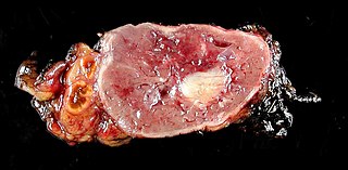
Pheochromocytoma is a rare tumor of the adrenal medulla composed of chromaffin cells, also known as pheochromocytes. When a tumor composed of the same cells as a pheochromocytoma develops outside the adrenal gland, it is referred to as a paraganglioma. These neuroendocrine tumors typically release massive amounts of catecholamines which result in the most common symptoms, including hypertension, tachycardia, and sweating. Rarely, some tumors may secrete little to no catecholamines, making diagnosis difficult. While tumors of the head and neck are parasympathetic, their sympathetic counterparts are predominantly located in the abdomen and pelvis, particularly concentrated at the organ of Zuckerkandl.

<span class="mw-page-title-main">Hemangiosarcoma</span> Medical condition

Hemangiosarcoma is a rapidly growing, highly invasive variety of cancer that occurs almost exclusively in dogs, and only rarely in cats, horses, mice, or humans. It is a sarcoma arising from the lining of blood vessels; that is, blood-filled channels and spaces are commonly observed microscopically. A frequent cause of death is the rupturing of this tumor, causing the patient to rapidly bleed to death.

<span class="mw-page-title-main">Gastrointestinal stromal tumor</span> Human disease (cancer)

Gastrointestinal stromal tumors (GISTs) are the most common mesenchymal neoplasms of the gastrointestinal tract. GISTs arise in the smooth muscle pacemaker interstitial cell of Cajal, or similar cells. They are defined as tumors whose behavior is driven by mutations in the KIT gene (85%), PDGFRA gene (10%), or BRAF kinase (rare). 95% of GISTs stain positively for KIT (CD117). Most (66%) occur in the stomach and gastric GISTs have a lower malignant potential than tumors found elsewhere in the GI tract.

In medical or research imaging, an incidental imaging finding is an unanticipated finding which is not related to the original diagnostic inquiry. As with other types of incidental medical findings, they may represent a diagnostic, ethical, and philosophical dilemma because their significance is unclear. While some coincidental findings may lead to beneficial diagnoses, others may lead to overdiagnosis that results in unnecessary testing and treatment, sometimes called the "cascade effect".

<span class="mw-page-title-main">Neuroendocrine tumor</span> Medical condition

Neuroendocrine tumors (NETs) are neoplasms that arise from cells of the endocrine (hormonal) and nervous systems. They most commonly occur in the intestine, where they are often called carcinoid tumors, but they are also found in the pancreas, lung, and the rest of the body.

<span class="mw-page-title-main">18F-EF5</span> Chemical compound

EF5 is a nitroimidazole derivative used in oncology research. Due to its similarity in chemical structure to etanidazole, EF5 binds in cells displaying hypoxia.

<span class="mw-page-title-main">Lung nodule</span> Medical condition

A lung nodule or pulmonary nodule is a relatively small focal density in the lung. A solitary pulmonary nodule (SPN) or coin lesion, is a mass in the lung smaller than three centimeters in diameter. A pulmonary micronodule has a diameter of less than three millimetres. There may also be multiple nodules.

<span class="mw-page-title-main">Bone metastasis</span> Medical condition

Bone metastasis, or osseous metastatic disease, is a category of cancer metastases that result from primary tumor invasions into bones. Bone-originating primary tumors such as osteosarcoma, chondrosarcoma, and Ewing sarcoma are rare; the most common bone tumor is a metastasis. Bone metastases can be classified as osteolytic, osteoblastic, or both. Unlike hematologic malignancies which originate in the blood and form non-solid tumors, bone metastases generally arise from epithelial tumors and form a solid mass inside the bone. Bone metastases, especially in a state of advanced disease, can cause severe pain, characterized by a dull, constant ache with periodic spikes of incident pain.

<span class="mw-page-title-main">PET-MRI</span>

Positron emission tomography–magnetic resonance imaging (PET–MRI) is a hybrid imaging technology that incorporates magnetic resonance imaging (MRI) soft tissue morphological imaging and positron emission tomography (PET) functional imaging.

<span class="mw-page-title-main">Positron emission mammography</span> Imaging procedure used to detect breast cancer

Positron emission mammography (PEM) is a nuclear medicine imaging modality used to detect or characterise breast cancer. Mammography typically refers to x-ray imaging of the breast, while PEM uses an injected positron emitting isotope and a dedicated scanner to locate breast tumors. Scintimammography is another nuclear medicine breast imaging technique, however it is performed using a gamma camera. Breasts can be imaged on standard whole-body PET scanners, however dedicated PEM scanners offer advantages including improved resolution.

<span class="mw-page-title-main">FMISO</span> Chemical compound

18F-FMISO or fluoromisonidazole is a radiopharmaceutical used for PET imaging of hypoxia. It consists of a 2-nitroimidazole molecule labelled with the positron-emitter fluorine-18.

PI-RADS is an acronym for Prostate Imaging Reporting and Data System, defining standards of high-quality clinical service for multi-parametric magnetic resonance imaging (mpMRI), including image creation and reporting.

CAPP-Seq is a next-generation sequencing based method used to quantify circulating DNA in cancer (ctDNA). The method was introduced in 2014 by Ash Alizadeh and Maximilian Diehn’s laboratories at Stanford, as a tool for measuring Cell-free tumor DNA which is released from dead tumor cells into the blood and thus may reflect the entire tumor genome. This method can be generalized for any cancer type that is known to have recurrent mutations. CAPP-Seq can detect one molecule of mutant DNA in 10,000 molecules of healthy DNA. The original method was further refined in 2016 for ultra sensitive detection through integration of multiple error suppression strategies, termed integrated Digital Error Suppression (iDES). The use of ctDNA in this technique should not be confused with circulating tumor cells (CTCs); these are two different entities.

Fluciclovine (<sup>18</sup>F) Chemical compound

Fluciclovine (18F), also known as anti-1-amino-3-18F-fluorocyclobutane-1-carboxylic acid, or as Axumin, is a diagnostic agent indicated for positron emission tomography (PET) imaging in men with suspected prostate cancer recurrence based on elevated prostate specific antigen (PSA) levels.

<span class="mw-page-title-main">Andreas Kjær (scientist)</span> Danish physician-scientist

Andreas Kjær is a Danish physician-scientist and European Research Council (ERC) advanced grantee. He is professor at the University of Copenhagen and chief physician at Rigshospitalet, the National University Hospital of Denmark. He is board certified in Nuclear Medicine and his research is focused on molecular imaging with PET and PET/MRI and targeted radionuclide therapies (theranostics) in cancer. His achievements include development of several new PET tracers that have reached first-in-human clinical use. He has published more than 400 peer-review articles, filed 10 patents, supervised more than 40 PhD students and received numerous prestigious scientific awards over the years. He is a member of the Danish Academy of Technical Sciences

Jason S. Lewis is a British radiochemist whose work relates to oncologic therapy and diagnosis. His research focus is a molecular imaging-based program focused on radiopharmaceutical development as well as the study of multimodality small- and biomolecule-based agents and their clinical translation. He has worked on the development of small molecules as well as radiolabeled peptides and antibodies probing the overexpression of receptors and antigens on tumors.

<span class="mw-page-title-main">Papillary renal cell carcinoma</span> Medical condition

Papillary renal cell carcinoma (PRCC) is a malignant, heterogeneous tumor originating from renal tubular epithelial cells of the kidney, which comprises approximately 10-15% of all kidney neoplasms. Based on its morphological features, PRCC can be classified into two main subtypes, which are type 1 (basophilic) and type 2 (eosinophilic).

Vicky Goh is a professor, chair of clinical cancer imaging, and head of cancer imaging department at the King's College London, England, United Kingdom. She joined King's College London in 2011. She is also a consultant radiologist at Guy's and St Thomas' Hospital in London.

References

  1. 1 2 3 4 Lambin P, Rios-Velazquez E, Leijenaar R, Carvalho S, van Stiphout RG, Granton P, et al. (March 2012). "Radiomics: extracting more information from medical images using advanced feature analysis". European Journal of Cancer. 48 (4): 441–6. doi:10.1016/j.ejca.2011.11.036. PMC   4533986 . PMID   22257792.
  2. 1 2 3 Kumar V, Gu Y, Basu S, Berglund A, Eschrich SA, Schabath MB, et al. (November 2012). "Radiomics: the process and the challenges". Magnetic Resonance Imaging. 30 (9): 1234–48. doi:10.1016/j.mri.2012.06.010. PMC   3563280 . PMID   22898692.
  3. 1 2 Gillies RJ, Kinahan PE, Hricak H (February 2016). "Radiomics: Images Are More than Pictures, They Are Data". Radiology. 278 (2): 563–77. doi:10.1148/radiol.2015151169. PMC   4734157 . PMID   26579733.
  4. 1 2 Parekh V, Jacobs MA (2016). "Radiomics: a new application from established techniques". Expert Review of Precision Medicine and Drug Development. 1 (2): 207–226. doi:10.1080/23808993.2016.1164013. PMC   5193485 . PMID   28042608.
  5. Yip SS, Aerts HJ (July 2016). "Applications and limitations of radiomics". Physics in Medicine and Biology. 61 (13): R150-66. Bibcode:2016PMB....61R.150Y. doi:10.1088/0031-9155/61/13/R150. PMC   4927328 . PMID   27269645.
  6. Yip SS, Liu Y, Parmar C, Li Q, Liu S, Qu F, et al. (June 2017). "Associations between radiologist-defined semantic and automatically computed radiomic features in non-small cell lung cancer". Scientific Reports. 7 (1): 3519. Bibcode:2017NatSR...7.3519Y. doi:10.1038/s41598-017-02425-5. PMC   5471260 . PMID   28615677.
  7. Chicklore S, Goh V, Siddique M, Roy A, Marsden PK, Cook GJ (January 2013). "Quantifying tumour heterogeneity in 18F-FDG PET/CT imaging by texture analysis". European Journal of Nuclear Medicine and Molecular Imaging. 40 (1): 133–40. doi:10.1007/s00259-012-2247-0. PMID   23064544. S2CID   24695383.
  8. Cook GJ, Siddique M, Taylor BP, Yip C, Chicklore S, Goh V (2014). "Radiomics in PET: Principles and applications". Clinical and Translational Imaging. 2 (3): 269–276. doi: 10.1007/s40336-014-0064-0 .
  9. Parekh VS, Jacobs MA (2017-11-14). "Integrated radiomic framework for breast cancer and tumor biology using advanced machine learning and multiparametric MRI". npj Breast Cancer. 3 (1): 43. doi:10.1038/s41523-017-0045-3. PMC   5686135 . PMID   29152563.
  10. Parekh VS, Jacobs MA (2019-03-04). "Deep learning and radiomics in precision medicine". Expert Review of Precision Medicine and Drug Development. 4 (2): 59–72. doi:10.1080/23808993.2019.1585805. PMC   6508888 . PMID   31080889.
  11. Kumar, Virendra; Gu, Yuhua; Basu, Satrajit; Berglund, Anders; Eschrich, Steven A.; Schabath, Matthew B.; Forster, Kenneth; Aerts, Hugo J.W.L.; Dekker, Andre; Fenstermacher, David; Goldgof, Dmitry B. (November 2012). "Radiomics: the process and the challenges". Magnetic Resonance Imaging. 30 (9): 1234–1248. doi:10.1016/j.mri.2012.06.010. PMC   3563280 . PMID   22898692.
  12. Schick, Ulrike; Lucia, François; Dissaux, Gurvan; Visvikis, Dimitris; Badic, Bogdan; Masson, Ingrid; Pradier, Olivier; Bourbonne, Vincent; Hatt, Mathieu (December 2019). "MRI-derived radiomics: methodology and clinical applications in the field of pelvic oncology". The British Journal of Radiology. 92 (1104): 20190105. doi:10.1259/bjr.20190105. ISSN   0007-1285. PMC   6913356 . PMID   31538516.
  13. Amorim, Barbara Juarez; Torrado-Carvajal, Angel; Esfahani, Shadi A; Marcos, Sara S; Vangel, Mark; Stein, Dan; Groshar, David; Catalano, Onofrio A (October 2020). "PET/MRI Radiomics in Rectal Cancer: a Pilot Study on the Correlation Between PET- and MRI-Derived Image Features with a Clinical Interpretation". Molecular Imaging and Biology. 22 (5): 1438–1445. doi:10.1007/s11307-020-01484-x. ISSN   1536-1632. PMID   32270337. S2CID   215411812.
  14. Galloway, Mary M (1975). "Texture analysis using gray level run lengths". Computer Graphics and Image Processing. 4 (2): 172–179. doi:10.1016/S0146-664X(75)80008-6.
  15. Pentland AP (June 1984). "Fractal-based description of natural scenes". IEEE Transactions on Pattern Analysis and Machine Intelligence. 6 (6): 661–74. doi:10.1109/TPAMI.1984.4767591. PMID   22499648. S2CID   17415943.
  16. Amadasun M, King R (1989). "Textural features corresponding to textural properties". IEEE Transactions on Systems, Man, and Cybernetics. 19 (5): 1264–1274. doi:10.1109/21.44046.
  17. Thibault G, Angulo J, Meyer F (March 2014). "Advanced statistical matrices for texture characterization: application to cell classification". IEEE Transactions on Bio-Medical Engineering. 61 (3): 630–7. doi:10.1109/TBME.2013.2284600. PMID   24108747. S2CID   11319154.
  18. Ranjbar S, Mitchell JR (2017). "An Introduction to Radiomics: An Evolving Cornerstone of Precision Medicine". Biomedical Texture Analysis. pp. 223–245. doi:10.1016/B978-0-12-812133-7.00008-9. ISBN   9780128121337.
  19. Tunali, Ilke; Hall, Lawrence O.; Napel, Sandy; Cherezov, Dmitry; Guvenis, Albert; Gillies, Robert J.; Schabath, Matthew B. (23 September 2019). "Stability and reproducibility of computed tomography radiomic features extracted from peritumoral regions of lung cancer lesions". Medical Physics. 46 (11): 5075–5085. Bibcode:2019MedPh..46.5075T. doi:10.1002/mp.13808. PMC   6842054 . PMID   31494946.
  20. 1 2 Nasief, Haidy; Zheng, Cheng; Schott, Diane; Hall, William; Tsai, Susan; Erickson, Beth; Allen Li, X. (4 October 2019). "A machine learning based delta-radiomics process for early prediction of treatment response of pancreatic cancer". npj Precision Oncology. 3 (1): 25. doi:10.1038/s41698-019-0096-z. PMC   6778189 . PMID   31602401.
  21. 1 2 Aerts HJ, Velazquez ER, Leijenaar RT, Parmar C, Grossmann P, Carvalho S, Bussink J, Monshouwer R, Haibe-Kains B, Rietveld D, et al. (Jun 2014). "Decoding tumour phenotype by noninvasive imaging using a quantitative radiomics approach". Nat Commun. 5: 4006. Bibcode:2014NatCo...5.4006A. doi:10.1038/ncomms5006. PMC   4059926 . PMID   24892406.
  22. Gu Y, Kumar V, Hall LO, Goldgof DB, Li CY, Korn R, et al. (March 2013). "Automated Delineation of Lung Tumors from CT Images Using a Single Click Ensemble Segmentation Approach". Pattern Recognition. 46 (3): 692–702. Bibcode:2013PatRe..46..692G. doi:10.1016/j.patcog.2012.10.005. PMC   3580869 . PMID   23459617.
  23. Velazquez ER, Parmar C, Jermoumi M, Mak RH, van Baardwijk A, Fennessy FM, et al. (December 2013). "Volumetric CT-based segmentation of NSCLC using 3D-Slicer". Scientific Reports. 3: 3529. Bibcode:2013NatSR...3E3529V. doi:10.1038/srep03529. PMC   3866632 . PMID   24346241.
  24. Parmar C, Leijenaar RT, Grossmann P, Rios Velazquez E, Bussink J, Rietveld D, et al. (June 2015). "Radiomic feature clusters and prognostic signatures specific for Lung and Head & Neck cancer". Scientific Reports. 5: 11044. Bibcode:2015NatSR...511044P. doi:10.1038/srep11044. PMC   4937496 . PMID   26251068.
  25. Nasief, Haidy; Hall, William; Zheng, Cheng; Tsai, Susan; Wang, Liang; Erickson, Beth; Li, X. Allen (8 January 2020). "Improving Treatment Response Prediction for Chemoradiation Therapy of Pancreatic Cancer Using a Combination of Delta-Radiomics and the Clinical Biomarker CA19-9". Frontiers in Oncology. 9: 1464. doi: 10.3389/fonc.2019.01464 . PMC   6960122 . PMID   31970088.
  26. Tixier F, Le Rest CC, Hatt M, Albarghach N, Pradier O, Metges JP, et al. (March 2011). "Intratumor heterogeneity characterized by textural features on baseline 18F-FDG PET images predicts response to concomitant radiochemotherapy in esophageal cancer". Journal of Nuclear Medicine. 52 (3): 369–78. doi:10.2967/jnumed.110.082404. PMC   3789272 . PMID   21321270.
  27. Hatt M, Majdoub M, Vallières M, Tixier F, Le Rest CC, Groheux D, et al. (January 2015). "18F-FDG PET uptake characterization through texture analysis: investigating the complementary nature of heterogeneity and functional tumor volume in a multi-cancer site patient cohort". Journal of Nuclear Medicine. 56 (1): 38–44. doi: 10.2967/jnumed.114.144055 . PMID   25500829.
  28. van Rossum PS, Fried DV, Zhang L, Hofstetter WL, van Vulpen M, Meijer GJ, et al. (May 2016). "The Incremental Value of Subjective and Quantitative Assessment of 18F-FDG PET for the Prediction of Pathologic Complete Response to Preoperative Chemoradiotherapy in Esophageal Cancer". Journal of Nuclear Medicine. 57 (5): 691–700. doi: 10.2967/jnumed.115.163766 . PMID   26795288.
  29. Yip SS, Coroller TP, Sanford NN, Mamon H, Aerts HJ, Berbeco RI (2016). "Relationship between the Temporal Changes in Positron-Emission-Tomography-Imaging-Based Textural Features and Pathologic Response and Survival in Esophageal Cancer Patients". Frontiers in Oncology. 6: 72. doi: 10.3389/fonc.2016.00072 . PMC   4810033 . PMID   27066454.
  30. Zhang H, Tan S, Chen W, Kligerman S, Kim G, D'Souza WD, et al. (January 2014). "Modeling pathologic response of esophageal cancer to chemoradiation therapy using spatial-temporal 18F-FDG PET features, clinical parameters, and demographics". International Journal of Radiation Oncology, Biology, Physics. 88 (1): 195–203. doi:10.1016/j.ijrobp.2013.09.037. PMC   3875172 . PMID   24189128.
  31. Cheng NM, Fang YH, Lee LY, Chang JT, Tsan DL, Ng SH, et al. (March 2015). "Zone-size nonuniformity of 18F-FDG PET regional textural features predicts survival in patients with oropharyngeal cancer". European Journal of Nuclear Medicine and Molecular Imaging. 42 (3): 419–28. doi:10.1007/s00259-014-2933-1. PMID   25339524. S2CID   6167165.
  32. Cook GJ, Yip C, Siddique M, Goh V, Chicklore S, Roy A, et al. (January 2013). "Are pretreatment 18F-FDG PET tumor textural features in non-small cell lung cancer associated with response and survival after chemoradiotherapy?". Journal of Nuclear Medicine. 54 (1): 19–26. doi: 10.2967/jnumed.112.107375 . PMID   23204495.
  33. Sun R, Limkin EJ, Vakalopoulou M, Dercle L, Champiat S, Han SR, et al. (September 2018). "A radiomics approach to assess tumour-infiltrating CD8 cells and response to anti-PD-1 or anti-PD-L1 immunotherapy: an imaging biomarker, retrospective multicohort study". The Lancet. Oncology. 19 (9): 1180–1191. doi:10.1016/S1470-2045(18)30413-3. PMID   30120041. S2CID   52032552.
  34. Tunali I, Gray JE, Qi J, Abdallah M, Jeong DK, Guvenis A, Gillies RJ, Gillies RJ (Jan 2019). "Novel Clinical and Radiomic Predictors of Rapid Disease Progression Phenotypes among Lung Cancer Patients Treated with Immunotherapy: An Early Report". Lung Cancer. 129: 75–79. doi:10.1016/j.lungcan.2019.01.010. PMC   6450086 . PMID   30797495.
  35. Mu W, Tunali I, Gray JE, Qi J, Gillies RJ, Gillies RJ (Dec 2019). "Radiomics of 18F-FDG PET/CT images predicts clinical benefit of advanced NSCLC patients to checkpoint blockade immunotherapy". Eur J Nucl Med Mol Imaging. 47 (5): 1168–1182. doi:10.1007/s00259-019-04625-9. PMC   8663718 . PMID   31807885. S2CID   208650067.
  36. Ludwig, CG; Lauric, A; Malek, JA; Mulligan, R; Malek, AM (2020). "Performance of Radiomics derived morphological features for prediction of aneurysm rupture status". Journal of NeuroInterventional Surgery. 13 (8). doi:10.1136/neurintsurg-2020-016808. PMID   33158993. S2CID   226274492.
  37. Tunali I, Stringfield O, Guvenis A, Wang H, Liu Y, et al. (Aug 2017). "Radial gradient and radial deviation radiomic features from pre-surgical CT scans are associated with survival among lung adenocarcinoma patients". Oncotarget. 8 (56): 96013–96026. doi:10.18632/oncotarget.21629. PMC   5707077 . PMID   29221183.
  38. Huang P, Park S, Yan R, Lee J, Chu LC, Lin CT, Hussien A, Rathmell J, Thomas B, Chen C, et al. (Sep 2018). "Added Value of Computer-aided CT Image Features for Early Lung Cancer Diagnosis with Small Pulmonary Nodules: A Matched Case-Control Study". Radiology. 286 (1): 286–295. doi:10.1148/radiol.2017162725. PMC   5779085 . PMID   28872442.
  39. 1 2 Coroller TP, Grossmann P, Hou Y, Rios Velazquez E, Leijenaar RT, Hermann G, et al. (March 2015). "CT-based radiomic signature predicts distant metastasis in lung adenocarcinoma". Radiotherapy and Oncology. 114 (3): 345–50. doi:10.1016/j.radonc.2015.02.015. PMC   4400248 . PMID   25746350.
  40. Vallières M, Freeman CR, Skamene SR, El Naqa I (July 2015). "A radiomics model from joint FDG-PET and MRI texture features for the prediction of lung metastases in soft-tissue sarcomas of the extremities". Physics in Medicine and Biology. 60 (14): 5471–96. Bibcode:2015PMB....60.5471V. doi:10.1088/0031-9155/60/14/5471. PMID   26119045. S2CID   4709680.
  41. Rios Velazquez E, Parmar C, Liu Y, Coroller TP, Cruz G, Stringfield O, et al. (July 2017). "Somatic Mutations Drive Distinct Imaging Phenotypes in Lung Cancer". Cancer Research. 77 (14): 3922–3930. doi:10.1158/0008-5472.CAN-17-0122. PMC   5528160 . PMID   28566328.
  42. Yip SS, Kim J, Coroller TP, Parmar C, Velazquez ER, Huynh E, et al. (April 2017). "Associations Between Somatic Mutations and Metabolic Imaging Phenotypes in Non-Small Cell Lung Cancer". Journal of Nuclear Medicine. 58 (4): 569–576. doi:10.2967/jnumed.116.181826. PMC   5373502 . PMID   27688480.
  43. Brown R, Zlatescu M, Sijben A, Roldan G, Easaw J, Forsyth P, et al. (April 2008). "The use of magnetic resonance imaging to noninvasively detect genetic signatures in oligodendroglioma". Clinical Cancer Research. 14 (8): 2357–62. doi: 10.1158/1078-0432.CCR-07-1964 . PMID   18413825.
  44. Drabycz S, Roldán G, de Robles P, Adler D, McIntyre JB, Magliocco AM, et al. (January 2010). "An analysis of image texture, tumor location, and MGMT promoter methylation in glioblastoma using magnetic resonance imaging". NeuroImage. 49 (2): 1398–405. doi:10.1016/j.neuroimage.2009.09.049. PMID   19796694. S2CID   18857965.
  45. Gutman DA, Dunn WD, Grossmann P, Cooper LA, Holder CA, Ligon KL, et al. (December 2015). "Somatic mutations associated with MRI-derived volumetric features in glioblastoma". Neuroradiology. 57 (12): 1227–37. doi:10.1007/s00234-015-1576-7. PMC   4648958 . PMID   26337765.
  46. Hassan, Islam, et al. "NIMG-28. Increased Mutation Burden (Hypermutation) in Gliomas is Associated With a Unique Radiomic Texture Signature in Magnetic Resonance Imaging." Neuro-oncology 19.Suppl 6 (2017): vi147.
  47. Sun R, Orlhac F, Robert C, Reuzé S, Schernberg A, Buvat I, et al. (August 2016). "In Regard to Mattonen et al". International Journal of Radiation Oncology, Biology, Physics. 95 (5): 1544–1545. doi:10.1016/j.ijrobp.2016.03.038. PMID   27479727.
  48. Yip SS, Coroller TP, Sanford NN, Huynh E, Mamon H, Aerts HJ, Berbeco RI (January 2016). "Use of registration-based contour propagation in texture analysis for esophageal cancer pathologic response prediction". Physics in Medicine and Biology. 61 (2): 906–22. Bibcode:2016PMB....61..906Y. doi:10.1088/0031-9155/61/2/906. PMID   26738433. S2CID   21799373.
  49. Peng L, Parekh V, Huang P, Lin DD, Sheikh K, Baker B, et al. (November 2018). "Distinguishing True Progression From Radionecrosis After Stereotactic Radiation Therapy for Brain Metastases With Machine Learning and Radiomics". International Journal of Radiation Oncology, Biology, Physics. 102 (4): 1236–1243. doi:10.1016/j.ijrobp.2018.05.041. PMC   6746307 . PMID   30353872.
  50. Hassan I, Kotrotsou A, Bakhtiari AS, Thomas GA, Weinberg JS, Kumar AJ, et al. (May 2016). "Radiomic Texture Analysis Mapping Predicts Areas of True Functional MRI Activity". Scientific Reports. 6: 25295. Bibcode:2016NatSR...625295H. doi:10.1038/srep25295. PMC   4858648 . PMID   27151623.
  51. Diehn, Maximilian; Nardini, Christine; Wang, David S.; McGovern, Susan; Jayaraman, Mahesh; Liang, Yu; Aldape, Kenneth; Cha, Soonmee; Kuo, Michael D. (2008). "Identification of noninvasive imaging surrogates for brain tumor gene-expression modules". Proceedings of the National Academy of Sciences. 105 (13): 5213–8. doi: 10.1073/pnas.0801279105 . PMC   2278224 . PMID   18362333.
  52. Zinn, Pascal O.; Mahajan, Bhanu; Sathyan, Pratheesh; Singh, Sanjay K.; Majumder, Sadhan; Jolesz, Ferenc A.; Colen, Rivka R. (2011). Deutsch, Eric (ed.). "Radiogenomic Mapping of Edema/Cellular Invasion MRI-Phenotypes in Glioblastoma Multiforme". PLOS ONE. 6 (10): e25451. Bibcode:2011PLoSO...625451Z. doi: 10.1371/journal.pone.0025451 . PMC   3187774 . PMID   21998659.
  53. Rutman, Aaron M.; Kuo, Michael D. (2009). "Radiogenomics: Creating a link between molecular diagnostics and diagnostic imaging". European Journal of Radiology. 70 (2): 232–41. doi:10.1016/j.ejrad.2009.01.050. PMID   19303233.
  54. Gevaert, O.; Xu, J.; Hoang, C. D.; Leung, A.N.; Xu, Y.; Quon, A.; Rubin, D.L.; Napel, S.; Plevritis, S.K. (2012). "Non-small cell lung cancer: identifying prognostic imaging biomarkers by leveraging public gene expression microarray data--methods and preliminary results". Radiology. 264 (2): 387–96. doi:10.1148/radiol.12111607. PMC   3401348 . PMID   22723499.
  55. Jaffe, C. (2012). "Imaging and genomics: is there a synergy?". Radiology. 264 (2): 329–31. doi: 10.1148/radiol.12120871 . PMID   22821693.
  56. Zhou, M.; Leung, A.; Echegaray, S.; Gentles, A.; Shrager, J.; Plevritis, S.; Rubin, D.L.; Napel, S.; Gevaert, O. (2017). "Non-Small Cell Lung Cancer Radiogenomics Map Identifies Relationships between Molecular and Imaging Phenotypes with Prognostic Implications". Radiology. 286 (1): 307–15. doi:10.1148/radiol.2017161845. PMC   5749594 . PMID   28727543.
  57. Houlahan, Kathleen E.; Salmasi, Amirali; Sadun, Taylor Y.; Pooli, Aydin; Felker, Ely R.; Livingstone, Julie; Huang, Vincent; Raman, Steven S.; Ahuja, Preeti; Sisk, Anthony E.; Boutros, Paul C. (July 2019). "Molecular Hallmarks of Multiparametric Magnetic Resonance Imaging Visibility in Prostate Cancer". European Urology. 76 (1): 18–23. doi:10.1016/j.eururo.2018.12.036. ISSN   1873-7560. PMC   10228592 . PMID   30685078. S2CID   59304426.
  58. Li, Ping; You, Sungyong; Nguyen, Christopher; Wang, Yanping; Kim, Jayoung; Sirohi, Deepika; Ziembiec, Asha; Luthringer, Daniel; Lin, Shih-Chieh; Daskivich, Timothy; Wu, Jonathan (2018). "Genes involved in prostate cancer progression determine MRI visibility". Theranostics. 8 (7): 1752–1765. doi:10.7150/thno.23180. ISSN   1838-7640. PMC   5858498 . PMID   29556354.
  59. Purysko, Andrei S.; Magi-Galluzzi, Cristina; Mian, Omar Y.; Sittenfeld, Sarah; Davicioni, Elai; du Plessis, Marguerite; Buerki, Christine; Bullen, Jennifer; Li, Lin; Madabhushi, Anant; Stephenson, Andrew (September 2019). "Correlation between MRI phenotypes and a genomic classifier of prostate cancer: preliminary findings". European Radiology. 29 (9): 4861–4870. doi:10.1007/s00330-019-06114-x. ISSN   1432-1084. PMC   6684343 . PMID   30847589.
  60. Norris, Joseph M.; Simpson, Benjamin S.; Parry, Marina A.; Kasivisvanathan, Veeru; Allen, Clare; Ball, Rhys; Freeman, Alex; Kelly, Daniel; Kirkham, Alex; Whitaker, Hayley C.; Emberton, Mark (March 2020). "Genetic correlates of prostate cancer visibility (and invisibility) on multiparametric magnetic resonance imaging: it's time to take stock". BJU International. 125 (3): 340–342. doi: 10.1111/bju.14919 . ISSN   1464-410X. PMID   31600865.
  61. 1 2 Norris, Joseph M.; Simpson, Benjamin S.; Parry, Marina A.; Allen, Clare; Ball, Rhys; Freeman, Alex; Kelly, Daniel; Kim, Hyung L.; Kirkham, Alex; You, Sungyong; Kasivisvanathan, Veeru (2020-07-01). "Genetic Landscape of Prostate Cancer Conspicuity on Multiparametric Magnetic Resonance Imaging: A Systematic Review and Bioinformatic Analysis". European Urology Open Science. 20: 37–47. doi: 10.1016/j.euros.2020.06.006 . ISSN   2666-1683. PMC   7497895 . PMID   33000006.
  62. Norris, Joseph M.; Simpson, Benjamin S.; Freeman, Alex; Kirkham, Alex; Whitaker, Hayley C.; Emberton, Mark (2020). "Conspicuity of prostate cancer on multiparametric magnetic resonance imaging: A cross-disciplinary translational hypothesis". The FASEB Journal. 34 (11): 14150–14159. doi: 10.1096/fj.202001466R . ISSN   1530-6860. PMC   8436756 . PMID   32920937.
  63. Mazurowski, M. A.; Zhang, J.; Grimm, L. J.; Yoon, S. C.; Silber, J. I. (2014). "Radiogenomic Analysis of Breast Cancer: Luminal B Molecular Subtype Is Associated with Enhancement Dynamics at MR Imaging". Radiology. 273 (2): 365–72. doi: 10.1148/radiol.14132641 . PMID   25028781.
  64. 1 2 3 Parekh VS, Jacobs MA (2018-09-25). "MPRAD: A Multiparametric Radiomics Framework". Breast Cancer Research and Treatment. 180 (2): 407–421. arXiv: 1809.09973 . Bibcode:2018arXiv180909973P. doi:10.1007/s10549-020-05533-5. PMC   7066290 . PMID   32020435.