Arteritis

Last updated
Arteritis
Artery.svg
Artery (normal)
Specialty Rheumatology   OOjs UI icon edit-ltr-progressive.svg

Arteritis is a vascular disorder characterized by inflammation of the walls of arteries, [1] usually as a result of infection or autoimmune responses. Arteritis, a complex disorder, is still not entirely understood. [2] Arteritis may be distinguished by its different types, based on the organ systems affected by the disease. [2] A complication of arteritis is thrombosis, which can be fatal. Arteritis and phlebitis are forms of vasculitis.

Contents

Signs and Symptoms

Symptoms of general arteritis may include: [3]

Diagnosis

Diagnosis of arteritis is based on unusual medical symptoms. [4] Similar symptoms may be caused by a number of other conditions, such as Ehlers-Danlos syndrome and Marfan syndrome (both heritable disorders of connective tissue), tuberculosis, syphilis, spondyloarthropathies, Cogans' syndrome, Buerger's, Behcet's, and Kawasaki disease. [4] Various imaging techniques may be used to diagnose and monitor disease progression. Imaging modalities may include direct angiography, magnetic resonance angiography, and ultrasonography. [4]

Angiography is commonly used in the diagnosis of Takayasu arteritis, [4] especially in the advanced stages of the disease, when arterial stenosis, occlusion, and aneurysms may be observed. [4] However, angiography is a relatively invasive investigation, exposing patients to large doses of radiation, [4] so is not recommended for routine, long-term monitoring of disease progression in patients with Takayasu arteritis. [4]

Computed tomography angiography can determine the size of the aorta and its surrounding branches, and can identify vessel wall lesions in middle to late stages of arteritis. [4] CTA can also show the blood flow within the blood vessels. [4] Like angiography, CTA exposes patients to high dosages of radiation. [4]

Magnetic resonance angiography is used to diagnose Takayasu arteritis in the early stages, showing changes such as the thickening of the vessel wall. [4] Even small changes may be measured, making MRA a useful tool for monitoring disease progression without exposing patients to the radiation of direct angiography or CTA. [4] MRA is an expensive investigation, and shows calcification of the aorta and distal branches less clearly than other imaging methods. [4]

Ultrasonography is an ideal method of diagnosing patients in early stages of arteritis when inflammation in the vessel walls occurs. [4] It can also show the blood flow within the blood vessels. [4] Ultrasonography is a popular first-line investigation for diagnosis because it is relatively quick, cheap, noninvasive, and does not expose patients to radiation. [4] It is also used for long-term monitoring of disease progression in Takayasu arteritis. Not all vascular lesions are visible on ultrasound, and the accuracy of the scan depends, to some extent, on the person reading the scan, as the results are observed in real time. [4]

Types

Arteritis may be primary or secondary to some other disease process. The primary types are:

Comparison of major types of arteritis
ArteritisAffected organsHistopathology
Takayasu arteritis Large vessels, [3] including aorta and arch branches [5] Histiocytes, giant cells [5]
Giant cell arteritis, also often called temporal arteritis (although they differ slightly) Superficial temporal artery, other medium- and large-sized vessels, [6] e.g. those supplying the head, eyes and optic nerves Lymphocytes, macrophages, and multinucleated giant cells [6]
Polyarteritis nodosa Medium-sized vessels, CNS, PNS damage, kidneys, gastrointestinal tract, skeletal muscle, heart [5] Neutrophils, fibrinoid necrosis [5]

An example of a secondary arteritis is arteritis caused by infection with the fungal pathogen Candida albicans . [7]

Giant cell arteritis

Giant cell arteritis contains two different types of arteritides that are almost indistinguishable from one another. [2] It includes two types, temporal arteritis and Takayasu arteritis. Both types contain an occupancy of medium- and larger-sized arteries which are categorized based on the infiltration of the giant cells. [2]

Takayasu arteritis

This type of arteritis is most common in females, with a median age of 25 years. [3] Takayasu arteritis is more common in women of Asian descent who are in their reproductive years. [3] However, over the past decades, its incidence in Africa, Europe, and North America has been increasing. [3] Takayasu arteritis is an inflammatory disease that mainly affects the larger vessels such as the aorta and its surrounding branches. [3] Research focused on Takayasu arteritis in the western parts of the world remains limited. An estimation suggests that, each year, the number of cases per million people is 2.6. [3]

Temporal arteritis

Temporal arteritis, the second type of giant cell arteritis, is also a chronic, inflammatory disease involving mid- to large-sized arteries. [8] Temporal arteritis has a higher incidence in people of Scandinavian descent. [8] However, the incidence rate differs based on population, region and races. [8] Temporal arteritis is not uncommon in North America. [8] The incidence rate is around 0.017% for individuals over 50 years of age. [8]

Symptoms of temporal arteritis are classified as specific and nonspecific. [8]

Nonspecific symptoms: [8]

  • Headache
  • Low grade fever
  • Sweating
  • Anorexia (loss of appetite)
  • Weight loss
  • General malaise

Specific symptoms: [8]

  • Claudication of the jaw
  • Engorged, tender vessels

Specific symptoms usually develop in the advanced stages of temporal arteritis. [8] These symptoms can include damage to eyesight and sudden blindness in one or both eyes. [9]

Polyarteritis nodosa of unknown mechanism can cause testicular pain. It is often associated with aneurysms and Hepatitis B.

Treatment

Medications

The first-line treatment for arteritis is oral glucocorticoid (steroid) medication, such as prednisone, taken daily for a period of three months. [3] After this initial phase, the medication may be reduced in dose or frequency, e.g. every other day, if possible. [3] If the disease worsens with the new treatment schedule, a cytotoxic medication may be given, in addition to the glucocorticoid. [3] Commonly used cytotoxic agents include azathioprine, methotrexate, or cyclophosphamide. [3] The dose of glucocorticoid medication may be decreased if response to treatment is good. [3] This medication may be reduced gradually once the disease becomes inactive, slowly tapering the dose (to allow the body time to adjust) until the medication may be stopped completely. [3] Conversely, if the disease remains active, the medication will need to be increased. [3] After six months, if the medication cannot be reduced in frequency to alternate days, or if in 12 months the medications cannot be stopped completely, then treatment is deemed to have failed. [3]

Pulsed therapy is an alternative method of administering the medications above, using much higher doses over a short period of time (a pulse), to reduce the inflammation within the arteries. Methylprednisolone, a glucocorticoid, is often used for pulse therapy; cyclophosphamide is an alternative. This method has been shown to be successful for some patients. [10] Immunosuppressive pulse therapy, such as with cyclophosphamide, has also demonstrated relief of symptoms associated with arteritis. [11]

Related Research Articles

<span class="mw-page-title-main">Giant cell arteritis</span> Medical condition

Giant cell arteritis (GCA), also called temporal arteritis, is an inflammatory autoimmune disease of large blood vessels. Symptoms may include headache, pain over the temples, flu-like symptoms, double vision, and difficulty opening the mouth. Complication can include blockage of the artery to the eye with resulting blindness, as well as aortic dissection, and aortic aneurysm. GCA is frequently associated with polymyalgia rheumatica.

<span class="mw-page-title-main">Aortic dissection</span> Injury to the innermost layer of the aorta

Aortic dissection (AD) occurs when an injury to the innermost layer of the aorta allows blood to flow between the layers of the aortic wall, forcing the layers apart. In most cases, this is associated with a sudden onset of severe chest or back pain, often described as "tearing" in character. Vomiting, sweating, and lightheadedness may also occur. Damage to other organs may result from the decreased blood supply, such as stroke, lower extremity ischemia, or mesenteric ischemia. Aortic dissection can quickly lead to death from insufficient blood flow to the heart or complete rupture of the aorta.

<span class="mw-page-title-main">Interventional radiology</span> Medical subspecialty

Interventional radiology (IR) is a medical specialty that performs various minimally-invasive procedures using medical imaging guidance, such as x-ray fluoroscopy, computed tomography, magnetic resonance imaging, or ultrasound. IR performs both diagnostic and therapeutic procedures through very small incisions or body orifices. Diagnostic IR procedures are those intended to help make a diagnosis or guide further medical treatment, and include image-guided biopsy of a tumor or injection of an imaging contrast agent into a hollow structure, such as a blood vessel or a duct. By contrast, therapeutic IR procedures provide direct treatment—they include catheter-based medicine delivery, medical device placement, and angioplasty of narrowed structures.

<span class="mw-page-title-main">Vasculitis</span> Medical disorders that destroy blood vessels by inflammation

Vasculitis is a group of disorders that destroy blood vessels by inflammation. Both arteries and veins are affected. Lymphangitis is sometimes considered a type of vasculitis. Vasculitis is primarily caused by leukocyte migration and resultant damage. Although both occur in vasculitis, inflammation of veins (phlebitis) or arteries (arteritis) on their own are separate entities.

<span class="mw-page-title-main">Granulomatosis with polyangiitis</span> Medical condition

Granulomatosis with polyangiitis (GPA), also known as Wegener's granulomatosis (WG), after the German physician Friedrich Wegener, is a rare long-term systemic disorder that involves the formation of granulomas and inflammation of blood vessels (vasculitis). It is an autoimmune disease and a form of vasculitis that affects small- and medium-size vessels in many organs but most commonly affects the upper respiratory tract, lungs and kidneys. The signs and symptoms of GPA are highly varied and reflect which organs are supplied by the affected blood vessels. Typical signs and symptoms include nosebleeds, stuffy nose and crustiness of nasal secretions, and inflammation of the uveal layer of the eye. Damage to the heart, lungs and kidneys can be fatal.

<span class="mw-page-title-main">Eosinophilic granulomatosis with polyangiitis</span> Medical condition

Eosinophilic granulomatosis with polyangiitis (EGPA), formerly known as allergic granulomatosis, is an extremely rare autoimmune condition that causes inflammation of small and medium-sized blood vessels (vasculitis) in persons with a history of airway allergic hypersensitivity (atopy).

<span class="mw-page-title-main">Polyarteritis nodosa</span> Medical condition

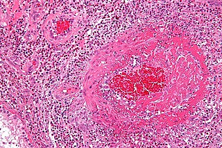
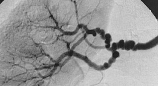
Polyarteritis nodosa (PAN) is a systemic necrotizing inflammation of blood vessels (vasculitis) affecting medium-sized muscular arteries, typically involving the arteries of the kidneys and other internal organs but generally sparing the lungs' circulation. Small aneurysms are strung like the beads of a rosary, therefore making this "rosary sign" an important diagnostic feature of the vasculitis. PAN is sometimes associated with infection by the hepatitis B or hepatitis C virus. The condition may be present in infants.

<span class="mw-page-title-main">Aortoiliac occlusive disease</span> Medical condition

In medicine, aortoiliac occlusive disease is a form of central artery disease involving the blockage of the abdominal aorta as it transitions into the common iliac arteries.

<span class="mw-page-title-main">Takayasu's arteritis</span> Medical condition

Takayasu's arteritis (TA), also known as aortic arch syndrome, nonspecific aortoarteritis, and pulseless disease, is a form of large vessel granulomatous vasculitis with massive intimal fibrosis and vascular narrowing, most commonly affecting young or middle-aged women of Asian descent, though anyone can be affected. It mainly affects the aorta and its branches, as well as the pulmonary arteries. Females are about 8–9 times more likely to be affected than males.

<span class="mw-page-title-main">Polymyalgia rheumatica</span> Medical condition

Polymyalgia rheumatica (PMR) is a syndrome experienced as pain or stiffness, usually in the neck, shoulders, upper arms, and hips, but which may occur all over the body. The pain can be sudden or can occur gradually over a period. Most people with PMR wake up in the morning with pain in their muscles; however, cases have occurred in which the person has developed the pain during the evenings or has pain and stiffness all day long.

Posterior ischemic optic neuropathy (PION) is a medical condition characterized by damage to the retrobulbar portion of the optic nerve due to inadequate blood flow (ischemia) to the optic nerve. Despite the term posterior, this form of damage to the eye's optic nerve due to poor blood flow also includes cases where the cause of inadequate blood flow to the nerve is anterior, as the condition describes a particular mechanism of visual loss as much as the location of damage in the optic nerve. In contrast, anterior ischemic optic neuropathy (AION) is distinguished from PION by the fact that AION occurs spontaneously and on one side in affected individuals with predisposing anatomic or cardiovascular risk factors.

<span class="mw-page-title-main">Fibromuscular dysplasia</span> Human arterial disease

Fibromuscular dysplasia (FMD) is a non-atherosclerotic, non-inflammatory disease of the blood vessels that causes abnormal growth within the wall of an artery. FMD has been found in nearly every arterial bed in the body, although the most commonly affected are the renal and carotid arteries.

<span class="mw-page-title-main">Subclavian steal syndrome</span> Medical condition

Subclavian steal syndrome (SSS), also called subclavian steal steno-occlusive disease, is a constellation of signs and symptoms that arise from retrograde (reversed) blood flow in the vertebral artery or the internal thoracic artery, due to a proximal stenosis (narrowing) and/or occlusion of the subclavian artery. This flow reversal is called the subclavian steal or subclavian steal phenomenon, regardless of signs/symptoms being present. The arm may be supplied by blood flowing in a retrograde direction down the vertebral artery at the expense of the vertebrobasilar circulation. It is more severe than typical vertebrobasilar insufficiency.

<span class="mw-page-title-main">Popliteal artery entrapment syndrome</span> Abnormal popliteal artery compression by surrounding structures

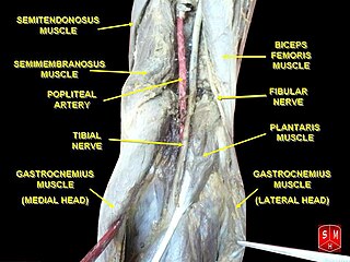
The popliteal artery entrapment syndrome (PAES) is an uncommon pathology that occurs when the popliteal artery is compressed by the surrounding popliteal fossa myofascial structures. This results in claudication and chronic leg ischemia. This condition mainly occurs more in young athletes than in the elderlies. Elderlies, who present with similar symptoms, are more likely to be diagnosed with peripheral artery disease with associated atherosclerosis. Patients with PAES mainly present with intermittent feet and calf pain associated with exercises and relieved with rest. PAES can be diagnosed with a combination of medical history, physical examination, and advanced imaging modalities such as duplex ultrasound, computer tomography, or magnetic resonance angiography. Management can range from non-intervention to open surgical decompression with a generally good prognosis. Complications of untreated PAES can include stenotic artery degeneration, complete popliteal artery occlusion, distal arterial thromboembolism, or even formation of an aneurysm.

<span class="mw-page-title-main">Computed tomography angiography</span> Medical investigation technique

Computed tomography angiography is a computed tomography technique used for angiography—the visualization of arteries and veins—throughout the human body. Using contrast injected into the blood vessels, images are created to look for blockages, aneurysms, dissections, and stenosis. CTA can be used to visualize the vessels of the heart, the aorta and other large blood vessels, the lungs, the kidneys, the head and neck, and the arms and legs. CTA can also be used to localise arterial or venous bleed of the gastrointestinal system.

Aortitis is the inflammation of the aortic wall. The disorder is potentially life-threatening and rare. It is reported that there are only 1–3 new cases of aortitis per year per million people in the United States and Europe. Aortitis is most common in people 10 to 40 years of age.

The following outline is provided as an overview of and topical guide to cardiology, the branch of medicine dealing with disorders of the human heart. The field includes medical diagnosis and treatment of congenital heart defects, coronary artery disease, heart failure, valvular heart disease and electrophysiology. Physicians who specialize in cardiology are called cardiologists.

Cerebral vasculitis is vasculitis involving the brain and occasionally the spinal cord. It affects all of the vessels: very small blood vessels (capillaries), medium-size blood vessels, or large blood vessels. If blood flow in a vessel with vasculitis is reduced or stopped, the parts of the body that receive blood from that vessel begins to die. It may produce a wide range of neurological symptoms, such as headache, skin rashes, feeling very tired, joint pains, difficulty moving or coordinating part of the body, changes in sensation, and alterations in perception, thought or behavior, as well as the phenomena of a mass lesion in the brain leading to coma and herniation. Some of its signs and symptoms may resemble multiple sclerosis. 10% have associated bleeding in the brain.

<span class="mw-page-title-main">Systemic vasculitis</span> Medical condition

Necrotizing vasculitis, also called systemic necrotizing vasculitis, is a general term for the inflammation of veins and arteries that develops into necrosis and narrows the vessels.

<span class="mw-page-title-main">Coronary CT angiography</span> Use of computed tomography angiography to assess the coronary arteries of the heart

Coronary CT angiography is the use of computed tomography (CT) angiography to assess the coronary arteries of the heart. The patient receives an intravenous injection of radiocontrast and then the heart is scanned using a high speed CT scanner, allowing physicians to assess the extent of occlusion in the coronary arteries, usually in order to diagnose coronary artery disease.

References

  1. "Arteritis" "at Dorland's Medical Dictionary
  2. 1 2 3 4 Hollier, L. H. (1 January 1989). "Arteritis". Perspectives in Vascular Surgery and Endovascular Therapy. 2 (1): 1–8. doi:10.1177/153100358900200101.
  3. 1 2 3 4 5 6 7 8 9 10 11 12 13 14 15 Kerr GS, Hallahan CW, Giordano J, Leavitt RY, Fauci AS, Rottem M, Hoffman GS (June 1994). "Takayasu arteritis". Ann. Intern. Med. 120 (11): 919–29. doi:10.7326/0003-4819-120-11-199406010-00004. PMID   7909656. S2CID   21784938.
  4. 1 2 3 4 5 6 7 8 9 10 11 12 13 14 15 16 17 Wen, Dan; Du, Xin; Ma, Chang-Sheng (1 December 2012). "Takayasu Arteritis: Diagnosis, Treatment and Prognosis". International Reviews of Immunology. 31 (6): 462–473. doi:10.3109/08830185.2012.740105. PMID   23215768. S2CID   5434700.
  5. 1 2 3 4 Stevens & Lowe: Pathology. At Fleshandbones.com
  6. 1 2 eMedicine Specialties > Temporal Arteritis Author: Christopher H Lee, MD. Coauthor(s): Jean Marie Hammel, MD. Updated: Sep 8, 2009
  7. Nagi-Miura, N; Harada, T; Shinohara, H; et al. (June 2006). "Lethal and severe coronary arteritis in DBA/2 mice induced by fungal pathogen, CAWS, Candida albicans water-soluble fraction". Atherosclerosis. 186 (2): 310–320. doi:10.1016/j.atherosclerosis.2005.08.014. PMID   16157343.
  8. 1 2 3 4 5 6 7 8 9 Chen, Chun-Hsiung; Kung, Shih-Ya; Tsai, Ying-Yang; Liao, Hsien-Tzung; Chou, Chung-Tei; Huang, De-Feng (2005). "Temporal Arteritis". Journal of the Chinese Medical Association. 68 (7): 333–335. doi:10.1016/S1726-4901(09)70170-4. PMID   16038374.
  9. Feilchenfeld, Zac (Nov 2011). "Answer: Can you identify this condition?". Canadian Family Physician . 57 (11): 1296–1297. PMC   3215611 .
  10. Chevalet, P; Barrier, J. H.; Pottier, P; Magadur-Joly, G; Pottier, M. A.; Hamidou, M; Planchon, B; El Kouri, D; Connan, L; Dupond, J. L.; De Wazieres, B; Dien, G; Duhamel, E; Grosbois, B; Jego, P; Le Strat, A; Capdeville, J; Letellier, P; Agron, L (2000). "A randomized, multicenter, controlled trial using intravenous pulses of methylprednisolone in the initial treatment of simple forms of giant cell arteritis: A one year follow-up study of 164 patients". The Journal of Rheumatology. 27 (6): 1484–91. PMID   10852275.
  11. Bose, P. (29 November 2012). "Takayasu's Arteritis". Journal of Neurology, Neurosurgery & Psychiatry. 83 (Suppl 2): A1.2–A1. doi:10.1136/jnnp-2012-304200a.2. S2CID   219209165.