Pseudoxanthoma elasticum

Last updated
Pseudoxanthoma elasticum
Other namesGrönblad–Strandberg syndrome; [1] Groenblad-Strandberg syndrome
Pseudoxanthoma elasticum 3.JPG
Pseudoxanthoma elasticum of the posterior lateral neck. Note the yellowish slightly raised bumps characteristic of this condition.
Specialty Medical genetics   OOjs UI icon edit-ltr-progressive.svg

Pseudoxanthoma elasticum (PXE) is a genetic disease that causes mineralization of elastic fibers in some tissues. The most common problems arise in the skin and eyes, and later in blood vessels in the form of premature atherosclerosis. [2] [3] [4] PXE is caused by autosomal recessive mutations in the ABCC6 gene on the short arm of chromosome 16 (16p13.1). [3] [5] [6]

Contents

Signs and symptoms

Dermatological manifestations

Usually, pseudoxanthoma elasticum affects the skin first, often in childhood or early adolescence. [7] Small, yellowish papular lesions form and cutaneous laxity mainly affect the neck, axillae (armpits), groin, and flexural creases (the inside parts of the elbows and knees). [3] [8] Skin may become lax and redundant. Many individuals have "oblique mental creases" (horizontal grooves of the chin). [9] The lesions are asymptomatic and have a stochastic pattern of growth and spread. [10]

Ocular manifestations

PXE first affects the retina through a dimpling of the Bruch membrane (a thin membrane separating the blood vessel-rich layer from the pigmented layer of the retina), that is only visible during ophthalmologic examinations. [11] This is called peau d'orange (a French term meaning "skin of the orange"). Eventually the mineralization of the elastic fibers in the Bruch membrane create cracks called angioid streaks that radiate out from the optic nerve. Angioid streaks themselves do not cause distortion of vision, even if they cross into the foveal area. This symptom is present in almost all PXE patients and is usually noticed a few years after the onset of cutaneous lesions. These cracks may allow small blood vessels that were originally held back by Bruch's membrane to penetrate the retina. These blood vessels sometimes leak, and these retinal hemorrhages may lead to the loss of central vision. Vision loss is a major issue in many PXE patients. [12] [3] [4]

Additionally, patients with PXE are at a greater risk of developing optic nerve head (ONH) drusen, which are calcified lesions within the optic nerve. Careful monitoring should be considered to prevent further complication, such as retinal artery occlusion and optic neuropathy. [13]

Cardiovascular manifestations

PXE is caused by mineralization in connective tissues in mainly the skin, eyes, and blood vessels. [14] As a result of mineralized buildup in the vascular wall, patients may. be at a greater risk for intermittent claudication, a condition in which cramping pain in the leg is induced by exercise, [15] and peripheral artery disease [14] Occlusions in cerebral arteries can lead to reduced or blocked blood flow, resulting in serious complications including transient ischemic attack (TIA) and stroke. Similarly, blockages in the coronary circulatory system, also called coronary artery disease may develop, leading to angina and myocardial infarction (heart attack). [16] Cerebral ischemia in PXE is caused by small vessel occlusive disease.

Other manifestations

Although not much is known about potential gastroenterological manifestations, gastrointestinal bleeding is a rare symptom and usually involved bleeding from the stomach.

Other rare neurological complications may include intracranial aneurysms, subarachnoid and intracerebral hemorrhages. [17]

Genetics

Pseudoxanthoma elasticum has an autosomal recessive pattern of inheritance. Autorecessive.svg
Pseudoxanthoma elasticum has an autosomal recessive pattern of inheritance.

80% of clinical cases of pseudoxanthoma elasticum have detectable mutations in the ABCC6 gene. [18] [19] [20] [21] Mutations in almost all parts of the gene have been described, of all types (missense, nonsense, splice alteration, insertion, small deletion or large deletion). Although there have been reports of autosomal dominant inheritance, the inheritance is typically autosomal recessive (both parents need to be carriers, and there is a 25% chance that a child will inherit both abnormal copies of the gene and therefore develop the condition). [3]

Strong genetic linkage was found with mutations in the ABCC6 gene, which codes for the ABCC6 protein, which is a membrane transporter from the large ATP-binding cassette transporter family. The protein is expressed in most organs, but mainly in the liver and kidney. ABCC6 mediates ATP release in the liver. This is the main source of circulating pyrophosphate (PPi), and individuals affected by PXE have strongly reduced plasma PPi levels, explaining their mineralization disorder. [22] One study suggested that mutations causing total absence of an ABCC6 protein caused a more severe disease, [23] but this could not be confirmed in a subsequent case series. [24] Given the variations in age of onset and severity it is likely that other unknown risk factors (genetic, environmental, and lifestyle) may be involved. [3]

Premature atherosclerosis is also associated with mutations in the ABCC6 gene, even in those without PXE. [25] A syndrome almost indistinguishable from hereditary PXE has been described in patients with hemoglobinopathies (sickle-cell disease and thalassemia) through a poorly understood mechanism. [3] In addition, there appears to be another PXE-like syndrome with a similar phenotype but as a result of problems with another gene, gamma-glutamyl carboxylase. [26] Mutations in ABCC6 can also cause generalized arterial calcification of infancy. [27] In some cases of PXE, mutations in ABCC6 cannot be found, and other genes such as ENPP1 may be implicated. [28]

Pathophysiology

In PXE, there is mineralization (accumulation of calcium and other minerals) and fragmentation of the elastin-containing fibers in connective tissue, but primarily in the midlaminar layer of the dermis, Bruch's membrane and the midsized arteries. [29] Recent studies have confirmed that PXE is a metabolic disease, and that its features arise because metabolites of vitamin K cannot reach peripheral tissues. [30] Low levels of PPi cause mineralization in peripheral tissues. [22]

Diagnosis

Histopathology of pseudoxanthoma elasticum: Fragmentation and calcification of middermal elastic fibers on Alizarin Red staining. Histopathology of pseudoxanthoma elasticum.jpg
Histopathology of pseudoxanthoma elasticum: Fragmentation and calcification of middermal elastic fibers on Alizarin Red staining.

The diagnostic criteria for PXE are the typical skin biopsy appearance and the presence of angioid streaks in the retina. Criteria were established by consensus of clinicians and researchers at the 2010 biennial research meeting of the PXE Research Consortium. [32] and confirmed at the 2014 meeting [33] These consensus criteria state that definitive PXE is characterized by two pathogenic mutations in the ABCC6 or ocular findings – angioid streaks > 1 DD or peau d’orange in an individual <20 years of age together with skin findings:

Differential diagnosis

The differential diagnosis PXE-like papillary dermal elastolysis: Small flesh-colored papules in the neck region. PXE-like papillary dermal elastolysis.jpg
The differential diagnosis PXE-like papillary dermal elastolysis: Small flesh-colored papules in the neck region.
Histopathology of PXE-like papillary dermal elastolysis: Loss of elastic fibers in the papillary dermis and abnormal pattern in the reticular dermis (arrow) Histopathology of PXE-like papillary dermal elastolysis.jpg
Histopathology of PXE-like papillary dermal elastolysis: Loss of elastic fibers in the papillary dermis and abnormal pattern in the reticular dermis (arrow)
Pseudoxanthoma elasticum versus histopathological differential diagnoses
on light microscopy (LM) and electron microscopy (EM) [31]
Pseudoxanthoma elasticumLM: Mid-dermal calcification and fragmentation of elastic fibers
EM: Mineralization in elastic fiber core
PXE-like disease with coagulation deficiency LM: Middermal calcification and fragmentation of elastic fibers
EM: Mineralization in elastic fiber periphery
Hemoglobinopathies LM: Middermal calcification and fragmentation of elastic fibers
EM: Mineralization in elastic fiber core
PXE-like papillary dermal elastolysisLM: Selective elastic tissue elimination in the papillary dermis and presence of melanophages
White fibrous papulosis of the neckLM: Dermal fibrosis in papillary and mid-reticular dermis
Late-onset focal dermal elastosis EM: Decrease of elastic fibers; fragmentation of remaining fibers
LM: Accumulation of elastic fibers in mid- and reticular dermis without fragmentation or calcification
Perforating calcific elastosis LM: Middermal calcification and degeneration of elastic fibers with transepidermal elimination
Buschke–Ollendorff syndrome LM: Increased amount of hypertrophic elastic fibers in dermis
EM: Altered translucent elastic fibers
Elastosis perforans serpiginosa LM: Transepidermal or perifollicular perforating canals
Papular elastorrhexis LM: Thickening of collagen bundles next to loss and fragmentation of elastic fibers
Upper dermal elastolysis LM: Complete loss of elastic fibers in the upper dermis
Middermal elastolysis LM: Complete absence of elastic fibers in the middermis
Linear focal elastosis LM: Massive basophilic fibers; clumping of elastic fibers in papillary dermis
Elastoderma LM: Increased, intertwining thin elastic fibers in papillary and upper reticular dermis
Calcinosis cutis LM: Deposits of calcium in the dermis

Treatment

There is no confirmed treatment that directly interferes with the disease process. [5] [6]

Cosmetic surgery to remove excessive skin has been used to improve aesthetic appearance in PXE patients [5] but because of the non-life-threatening nature of these symptoms, should be used with caution. [6]

One of the most critical symptom of PXE is choroidal neovascularization which can lead to deterioration of central vision. Photodynamic therapy has been used as a treatment, but this has been replaced with endothelial growth factor (VEGF) inhibitors (such as bevacizumab, ranibizumab, and aflibercept) [6] with efficacy similar to their use in treatment of age-related macular degeneration. [4]

To limit cardiovascular symptoms, reduction of cardiovascular risk factors through lifestyle changes is recommended. [6] Generally clinicians recommend avoidance of non-steroidal anti-inflammatory drugs (NSAIDS) that increase bleeding risk, such as aspirin, and ibuprofen to prevent eye and gastrointestinal bleeding. [6]

Formerly, dietary restriction of calcium was tried with no benefit, and in fact accelerated mineralization in mice. [34] There are a number of potential treatments that are currently being tested or have just undergone testing including magnesium, [35] etidronate, [36] PPi, [37] and tissue-nonspecific alkaline phosphatase inhibitors. [38]

Given that ABCC6 heterozygous mutations result in few symptoms of PXE, this disease is a candidate for gene therapy. Some initial proof-of-principle experiments have been done in mice that have relieved some of symptoms of PXE, but as with all gene therapy treatments, there are many hurdles that must be overcome including insuring that the treatment will be long-lasting and reducing the risk of insertional mutagenesis and severe immune reactions. [6]

Epidemiology

The reported prevalence of pseudoxanthoma elasticum is about 1:25,000. Females are twice as likely to be affected as males. The disease occurs in all ethnicities, but Afrikaners are more likely to have PXE as a result of a founder effect (i.e., higher prevalence in the small group of people from whom Afrikaners descend). [39]

History

The first description of PXE that distinguished it from other xanthoma conditions was by Dr Ferdinand-Jean Darrier in 1896. [40] The eponym "Grönblad-Strandberg syndrome" is used in older literature, after two physicians who made further discoveries in the disease manifestations. [41]

PXE has the distinction of being the only disease for which a layperson is the discover of the mutated gene. The ABCC6 gene mutation was discovered simultaneously by four research teams, all of which published at the same time. The principal investigators were (in order of the date of publication): Jouni Uitto, [18] Arthur Bergen, [19] Charles Boyd, [20] and Klaus Lindpainter. [21] The gene was patented by Charles D. Boyd, Katalin Csiszar, Olivier LeSaux, Zsolt Urban, Sharon Terry, and assigned to PXE International by these co-inventors. Between the filing and 2013, when the Supreme Court of the United States declared that genes may not be patented. [42] PXE International freely licensed the gene to any lab for clinical testing and research. PXE International continues to hold and maintain other patents (diagnosis and treatment patents). [43] [44]

PXE International, a support organization, was founded in 1995, by Patrick and Sharon Terry, [45] [46] [47] [48] following the diagnosis of their two children. It has a registry of 4,600 affected individuals.

Images

See also

Related Research Articles

<span class="mw-page-title-main">Ehlers–Danlos syndrome</span> Group of genetic connective tissues disorders

Ehlers–Danlos syndromes (EDS) are a group of 13 genetic connective-tissue disorders. Symptoms often include loose joints, joint pain, stretchy velvety skin, and abnormal scar formation. These may be noticed at birth or in early childhood. Complications may include aortic dissection, joint dislocations, scoliosis, chronic pain, or early osteoarthritis. The current classification was last updated in 2017, when a number of rarer forms of EDS were added.

<span class="mw-page-title-main">Epidermolysis bullosa</span> Rare medical conditions that result in easy blistering of the skin and mucous membranes

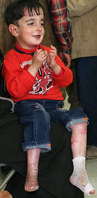
Epidermolysis bullosa (EB) is a group of rare medical conditions that result in easy blistering of the skin and mucous membranes. Blisters occur with minor trauma or friction and are painful. Its severity can range from mild to fatal. Inherited EB is a rare disease with a prevalence in the United States of 8.2 per million live births. Those with mild cases may not develop symptoms until they start to crawl or walk. Complications may include esophageal narrowing, squamous cell skin cancer, and the need for amputations.

<span class="mw-page-title-main">Elastin</span> Protein allowing tissue in the body to resume shape after stretching

Elastin is a protein that in humans is encoded by the ELN gene. Elastin is a key component of the extracellular matrix in gnathostomes. It is highly elastic and present in connective tissue allowing many tissues in the body to resume their shape after stretching or contracting. Elastin helps skin to return to its original position when it is poked or pinched. Elastin is also an important load-bearing tissue in the bodies of vertebrates and used in places where mechanical energy is required to be stored.

<span class="mw-page-title-main">Elastic fiber</span> Type of connective tissue in animals

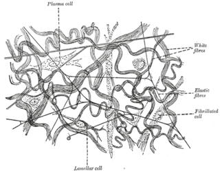
Elastic fibers are an essential component of the extracellular matrix composed of bundles of proteins (elastin) which are produced by a number of different cell types including fibroblasts, endothelial, smooth muscle, and airway epithelial cells. These fibers are able to stretch many times their length, and snap back to their original length when relaxed without loss of energy. Elastic fibers include elastin, elaunin and oxytalan.

<span class="mw-page-title-main">ABCC6</span> Protein-coding gene in the species Homo sapiens

Multidrug resistance-associated protein 6 (MRP6) also known as ATP-binding cassette sub-family C member 6 (ABCC6) and multi-specific organic anion transporter E (MOAT-E) is a protein that in humans is encoded by the ABCC6 gene. The protein encoded by the ABCC6 gene is a member of the superfamily of ATP-binding cassette (ABC) transporters.

<span class="mw-page-title-main">Kindler syndrome</span> Medical condition

Kindler syndrome is a rare congenital disease of the skin caused by a mutation in the KIND1 gene.

<span class="mw-page-title-main">Genetic Alliance</span>

Genetic Alliance is a nonprofit organization, founded in 1986 by Joan O. Weiss, working with Victor A. McKusick, to advocate for health benefits in the accelerating field of genomic research. This organization is a network of over 1,000 disease advocacy organizations, universities, government organizations, private companies, and public policy organizations. They aim to advance genetic research agendas toward health benefit by engaging a broad range of stakeholders, including healthcare providers, researchers, industry professionals, public policy leaders, as well as individuals, families and communities. They create programs using a collaborative approach, and aim to increase efficiency and reduce obstacles in genetic research, while ensuring that voices from the involved disease communities are heard. They also promote public policies to advance healthcare. Genetic Alliance provides technical support and informational resources to guide disease-specific advocacy organizations in being their own research advocates. They also maintain a biobank as a central storage facility for several organizations who otherwise would not have the infrastructure to maintain their own repository.

<span class="mw-page-title-main">Keratin 5</span>

Keratin 5, also known as KRT5, K5, or CK5, is a protein that is encoded in humans by the KRT5 gene. It dimerizes with keratin 14 and forms the intermediate filaments (IF) that make up the cytoskeleton of basal epithelial cells. This protein is involved in several diseases including epidermolysis bullosa simplex and breast and lung cancers.

<span class="mw-page-title-main">Ablepharon macrostomia syndrome</span> Medical condition

Ablepharon macrostomia syndrome (AMS) is an extremely rare, autosomal dominant genetic disorder characterized by abnormal phenotypic appearances that primarily affect the head and face as well as the skull, skin, fingers and genitals. AMS generally results in abnormal ectoderm-derived structures. The most prominent abnormality is the underdevelopment (microblepharon) or absence of eyelids – signifying the ablepharon aspect of the disease – and a wide, fish-like mouth – macrostomia. Recent scholars and surgeons have called into question the naming of the condition as "Ablepharon" on account of recent investigation and histology showing consistent evidence of at least some eyelid tissue. Infants presenting with AMS may also have malformations of the abdominal wall and nipples. Children with AMS might also experience issues with learning development, language difficulties and intellectual disabilities.

<span class="mw-page-title-main">Epidermolysis bullosa dystrophica</span> Medical condition

Epidermolysis bullosa dystrophica or dystrophic EB (DEB) is an inherited disease affecting the skin and other organs.

<span class="mw-page-title-main">Buschke–Ollendorff syndrome</span> Medical condition

Buschke–Ollendorff syndrome (BOS) is a rare genetic skin disorder associated with LEMD3 that typically presents with widespread painless papules.

<span class="mw-page-title-main">Collagen, type VII, alpha 1</span> Protein found in humans

Collagen alpha-1(VII) chain is a protein that in humans is encoded by the COL7A1 gene. It is composed of a triple helical, collagenous domain flanked by two non-collagenous domains, and functions as an anchoring fibril between the dermal-epidermal junction in the basement membrane. Mutations in COL7A1 cause all types of dystrophic epidermolysis bullosa, and the exact mutations vary based on the specific type or subtype. It has been shown that interactions between the NC-1 domain of collagen VII and several other proteins, including laminin-5 and collagen IV, contribute greatly to the overall stability of the basement membrane.

<span class="mw-page-title-main">Dystonin</span> Neurologically significant human protein

Dystonin(DST), also known as bullous pemphigoid antigen 1 (BPAG1), isoforms 1/2/3/4/5/8, is a protein that in humans is encoded by the DST gene.

<span class="mw-page-title-main">Myosin-11</span> Protein-coding gene in the species Homo sapiens

Myosin-11 is a protein that in humans is encoded by the MYH11 gene.

<span class="mw-page-title-main">NOMO1</span> Protein-coding gene in the species Homo sapiens

Nodal modulator 1 is a protein that in humans is encoded by the NOMO1 gene.

<span class="mw-page-title-main">Urbach–Wiethe disease</span> Rare recessive genetic disorder

Urbach–Wiethe disease is a very rare recessive genetic disorder, with approximately 400 reported cases since its discovery. It was first officially reported in 1929 by Erich Urbach and Camillo Wiethe, although cases may be recognized dating back as early as 1908.

Congenital generalized lipodystrophy is an extremely rare autosomal recessive condition, characterized by an extreme scarcity of fat in the subcutaneous tissues. It is a type of lipodystrophy disorder where the magnitude of fat loss determines the severity of metabolic complications. Only 250 cases of the condition have been reported, and it is estimated that it occurs in 1 in 10 million people worldwide.

Generalized arterial calcification of infancy (GACI) is an extremely rare genetic disorder. It is caused by mutations in the ENPP1 gene in 75% of the subjects or in mutations in the ABCC6 genes in 10% of patients. However, sometimes individuals affected with GACI do not have mutations in the ENPP1 or ABCC6 gene and in those cases the cause of the disorder is unknown.

<span class="mw-page-title-main">ACDC (medicine)</span> Medical condition

Arterial calcification due to deficiency of CD73 (ACDC) is a rare genetic disorder that causes calcium buildup in the arteries and joints of the hands and feet, and other areas below the waist. Although patients exhibiting these symptoms have been identified as early as 1914, this disorder had not been studied extensively until recently. The identification of the specific ACDC gene and mutations occurred in 2011. ACDC is caused by a mutation in the NT5E gene, which prevents calcium-removing agents from functioning,. Patients with this mutation experience chronic pain, difficulty moving, and increased risk of cardiovascular problems. In experiments at the molecular level, treatment with adenosine or a phosphatase inhibitor reversed and prevented calcification, suggesting they could be used as possible treatment methods. There is currently no cure for ACDC, and patients have limited treatment options which focus primarily on removal of blood calcium and improving mobility.

INZ-701 is a recombinant ENPP1 enzyme developed to treat some genetic disorders that prevent normal production of ENPP1. It is developed by Inozyme Pharma.

References

  1. Rapini RP, Bolognia JL, Jorizzo JL (2007). Dermatology: 2-Volume Set. St. Louis: Mosby. ISBN   978-1-4160-2999-1.
  2. Li Q, van de Wetering K, Uitto J (February 2019). "Pseudoxanthoma Elasticum as a Paradigm of Heritable Ectopic Mineralization Disorders: Pathomechanisms and Treatment Development". review. The American Journal of Pathology. 189 (2): 216–225. doi:10.1016/j.ajpath.2018.09.014. PMC   6412714 . PMID   30414410.
  3. 1 2 3 4 5 6 7 Chassaing N, Martin L, Calvas P, Le Bert M, Hovnanian A (December 2005). "Pseudoxanthoma elasticum: a clinical, pathophysiological and genetic update including 11 novel ABCC6 mutations". review. Journal of Medical Genetics. 42 (12): 881–92. doi:10.1136/jmg.2004.030171. PMC   1735972 . PMID   15894595.
  4. 1 2 3 Finger RP, Charbel Issa P, Ladewig MS, Götting C, Szliska C, Scholl HP, Holz FG (2009). "Pseudoxanthoma elasticum: genetics, clinical manifestations and therapeutic approaches". review. Survey of Ophthalmology. 54 (2): 272–85. doi:10.1016/j.survophthal.2008.12.006. PMID   19298904.
  5. 1 2 3 Marconi B, Bobyr I, Campanati A, Molinelli E, Consales V, Brisigotti V, et al. (August 2015). "Pseudoxanthoma elasticum and skin: Clinical manifestations, histopathology, pathomechanism, perspectives of treatment". review. Intractable & Rare Diseases Research. 4 (3): 113–22. doi:10.5582/irdr.2015.01014. PMC   4561240 . PMID   26361562.
  6. 1 2 3 4 5 6 7 Germain DP (May 2017). "Pseudoxanthoma elasticum". review. Orphanet Journal of Rare Diseases. 12 (1): 85. doi: 10.1186/s13023-017-0639-8 . PMC   5424392 . PMID   28486967.
  7. Naouri M, Boisseau C, Bonicel P, Daudon P, Bonneau D, Chassaing N, Martin L (September 2009). "Manifestations of pseudoxanthoma elasticum in childhood". The British Journal of Dermatology. 161 (3): 635–9. doi:10.1111/j.1365-2133.2009.09298.x. PMID   19519828. S2CID   24795259.
  8. Neldner KH, Struk B (2003). "Pseudoxanthoma Elasticum". Connective Tissue and Its Heritable Disorders: Molecular, Genetic, and Medical Aspects. John Wiley & Sons. pp. 561–583. doi:10.1002/0471221929.ch11. ISBN   9780471221920. S2CID   32736212.
  9. Lebwohl M, Lebwohl E, Bercovitch L (April 2003). "Prominent mental (chin) crease: a new sign of pseudoxanthoma elasticum". primary. Journal of the American Academy of Dermatology. 48 (4): 620–2. doi:10.1067/mjd.2003.195. PMID   12664032. S2CID   4831796.
  10. Marconi, Barbara; Bobyr, Ivan; Campanati, Anna; Molinelli, Elisa; Consales, Veronica; Brisigotti, Valerio; Scarpelli, Marina; Racchini, Stefano; Offidani, Annamaria (2015). "Pseudoxanthoma elasticum and skin: Clinical manifestations, histopathology, pathomechanism, perspectives of treatment". Intractable & Rare Diseases Research. 4 (3): 113–122. doi:10.5582/irdr.2015.01014. ISSN   2186-361X. PMC   4561240 . PMID   26361562.
  11. Gliem M, Müller PL, Birtel J, Hendig D, Holz FG, Charbel Issa P (June 2016). "Frequency, Phenotypic Characteristics and Progression of Atrophy Associated With a Diseased Bruch's Membrane in Pseudoxanthoma Elasticum". primary. Investigative Ophthalmology & Visual Science. 57 (7): 3323–30. doi: 10.1167/iovs.16-19388 . PMID   27367499.
  12. Risseeuw S, Ossewaarde-van Norel J, Klaver CC, Colijn JM, Imhof SM, van Leeuwen R (August 2019). "Visual Acuity in Pseudoxanthoma Elasticum". primary. Retina. 39 (8): 1580–1587. doi:10.1097/IAE.0000000000002173. PMID   29652691. S2CID   4893600.
  13. Ahmed, Harris; Khazaeni, Leila (2024), "Optic Disc Drusen", StatPearls, Treasure Island (FL): StatPearls Publishing, PMID   35593854 , retrieved 2024-03-07
  14. 1 2 Germain, Dominique P. (2017-05-10). "Pseudoxanthoma elasticum". Orphanet Journal of Rare Diseases. 12 (1): 85. doi: 10.1186/s13023-017-0639-8 . ISSN   1750-1172. PMC   5424392 . PMID   28486967.
  15. Lefthériotis G, Abraham P, Le Corre Y, Le Saux O, Henrion D, Ducluzeau PH, et al. (November 2011). "Relationship between ankle brachial index and arterial remodeling in pseudoxanthoma elasticum". primary. Journal of Vascular Surgery. 54 (5): 1390–4. doi:10.1016/j.jvs.2011.04.041. PMC   5529101 . PMID   21723076.
  16. Kranenburg G, de Jong PA, Mali WP, Attrach M, Visseren FL, Spiering W (January 2017). "Prevalence and severity of arterial calcifications in pseudoxanthoma elasticum (PXE) compared to hospital controls. Novel insights into the vascular phenotype of PXE". primary. Atherosclerosis. 256: 7–14. doi:10.1016/j.atherosclerosis.2016.11.012. PMID   27940376.
  17. Kauw F, Kranenburg G, Kappelle LJ, Hendrikse J, Koek HL, Visseren FL, et al. (February 2017). "Cerebral disease in a nationwide Dutch pseudoxanthoma elasticum cohort with a systematic review of the literature". review. Journal of the Neurological Sciences. 373: 167–172. doi:10.1016/j.jns.2016.12.053. PMID   28131180. S2CID   206292331.
  18. 1 2 Ringpfeil F, Lebwohl MG, Christiano AM, Uitto J (May 2000). "Pseudoxanthoma elasticum: mutations in the MRP6 gene encoding a transmembrane ATP-binding cassette (ABC) transporter". primary. Proceedings of the National Academy of Sciences of the United States of America. 97 (11): 6001–6. Bibcode:2000PNAS...97.6001R. doi: 10.1073/pnas.100041297 . PMC   18548 . PMID   10811882.
  19. 1 2 Bergen AA, Plomp AS, Schuurman EJ, Terry S, Breuning M, Dauwerse H, et al. (June 2000). "Mutations in ABCC6 cause pseudoxanthoma elasticum". primary. Nature Genetics. 25 (2): 228–31. doi:10.1038/76109. PMID   10835643. S2CID   510024.
  20. 1 2 Le Saux O, Urban Z, Tschuch C, Csiszar K, Bacchelli B, Quaglino D, et al. (June 2000). "Mutations in a gene encoding an ABC transporter cause pseudoxanthoma elasticum". primary. Nature Genetics. 25 (2): 223–7. doi:10.1038/76102. PMID   10835642. S2CID   8883528.
  21. 1 2 Struk B, Cai L, Zäch S, Ji W, Chung J, Lumsden A, et al. (2000). "Mutations of the gene encoding the transmembrane transporter protein ABC-C6 cause pseudoxanthoma elasticum". primary. Journal of Molecular Medicine. 78 (5): 282–6. doi:10.1007/s001090000114. PMID   10954200. S2CID   8062022.
  22. 1 2 Jansen RS, Duijst S, Mahakena S, Sommer D, Szeri F, Váradi A, et al. (September 2014). "ABCC6-mediated ATP secretion by the liver is the main source of the mineralization inhibitor inorganic pyrophosphate in the systemic circulation-brief report". primary. Arteriosclerosis, Thrombosis, and Vascular Biology. 34 (9): 1985–9. doi:10.1161/ATVBAHA.114.304017. PMC   6743317 . PMID   24969777.
  23. Schulz V, Hendig D, Szliska C, Götting C, Kleesiek K (June 2005). "Novel mutations in the ABCC6 gene of German patients with pseudoxanthoma elasticum". primary. Human Biology. 77 (3): 367–84. doi:10.1353/hub.2005.0054. PMID   16392638. S2CID   44925940.
  24. Pfendner EG, Vanakker OM, Terry SF, Vourthis S, McAndrew PE, McClain MR, et al. (October 2007). "Mutation detection in the ABCC6 gene and genotype-phenotype analysis in a large international case series affected by pseudoxanthoma elasticum". primary. Journal of Medical Genetics. 44 (10): 621–8. doi:10.1136/jmg.2007.051094. PMC   2597973 . PMID   17617515.
  25. Trip MD, Smulders YM, Wegman JJ, Hu X, Boer JM, ten Brink JB, et al. (August 2002). "Frequent mutation in the ABCC6 gene (R1141X) is associated with a strong increase in the prevalence of coronary artery disease". primary. Circulation. 106 (7): 773–5. doi:10.1161/01.CIR.0000028420.27813.C0. PMID   12176944. S2CID   2107131.
  26. Vanakker OM, Martin L, Gheduzzi D, Leroy BP, Loeys BL, Guerci VI, et al. (March 2007). "Pseudoxanthoma elasticum-like phenotype with cutis laxa and multiple coagulation factor deficiency represents a separate genetic entity". primary. The Journal of Investigative Dermatology. 127 (3): 581–7. doi: 10.1038/sj.jid.5700610 . PMID   17110937.
  27. Li Q, Brodsky JL, Conlin LK, Pawel B, Glatz AC, Gafni RI, et al. (March 2014). "Mutations in the ABCC6 gene as a cause of generalized arterial calcification of infancy: genotypic overlap with pseudoxanthoma elasticum". primary. The Journal of Investigative Dermatology. 134 (3): 658–665. doi:10.1038/jid.2013.370. PMC   3945730 . PMID   24008425.
  28. Jin L, Jiang Q, Wu Z, Shao C, Zhou Y, Yang L, et al. (May 2015). "Genetic heterogeneity of pseudoxanthoma elasticum: the Chinese signature profile of ABCC6 and ENPP1 mutations". primary. The Journal of Investigative Dermatology. 135 (5): 1294–1302. doi:10.1038/jid.2015.10. PMC   4402129 . PMID   25615550.
  29. Gheduzzi D, Sammarco R, Quaglino D, Bercovitch L, Terry S, Taylor W, Ronchetti IP (2003). "Extracutaneous ultrastructural alterations in pseudoxanthoma elasticum". primary. Ultrastructural Pathology. 27 (6): 375–84. doi:10.1080/716100800. PMID   14660276.
  30. Li Q, Jiang Q, Pfendner E, Váradi A, Uitto J (January 2009). "Pseudoxanthoma elasticum: clinical phenotypes, molecular genetics and putative pathomechanisms". review. Experimental Dermatology. 18 (1): 1–11. doi:10.1111/j.1600-0625.2008.00795.x. PMC   3349969 . PMID   19054062.
  31. 1 2 3 4 Hosen, Mohammad J.; Lamoen, Anouck; De Paepe, Anne; Vanakker, Olivier M. (2012). "Histopathology of Pseudoxanthoma Elasticum and Related Disorders: Histological Hallmarks and Diagnostic Clues". Scientifica. 2012: 1–15. doi: 10.6064/2012/598262 . ISSN   2090-908X. PMC   3820553 . PMID   24278718.
    -Creative Commons Attribution 3.0 Unported license
  32. Uitto J, Bercovitch L, Terry SF, Terry PF (July 2011). "Pseudoxanthoma elasticum: progress in diagnostics and research towards treatment : Summary of the 2010 PXE International Research Meeting". review. American Journal of Medical Genetics. Part A. 155A (7): 1517–26. doi:10.1002/ajmg.a.34067. PMC   3121926 . PMID   21671388.
  33. Uitto J, Jiang Q, Váradi A, Bercovitch LG, Terry SF (June 2014). "Visual Acuity in Pseudoxanthoma Elasticum". review. Expert Opinion on Orphan Drugs. 2 (6): 567–577. doi:10.1517/21678707.2014.908702. PMC   4219573 . PMID   25383264.
  34. Jiang Q, Uitto J (September 2012). "Restricting dietary magnesium accelerates ectopic connective tissue mineralization in a mouse model of pseudoxanthoma elasticum (Abcc6(-/-) )". primary. Experimental Dermatology. 21 (9): 694–9. doi:10.1111/j.1600-0625.2012.01553.x. PMC   3422765 . PMID   22897576.
  35. Rose S, On SJ, Fuchs W, Chen C, Phelps R, Kornreich D, et al. (July 2019). "Magnesium supplementation in the treatment of pseudoxanthoma elasticum: A randomized trial". primary. Journal of the American Academy of Dermatology. 81 (1): 263–265. doi: 10.1016/j.jaad.2019.02.055 . PMID   30826287.
  36. Kranenburg G, de Jong PA, Bartstra JW, Lagerweij SJ, Lam MG, Ossewaarde-van Norel J, et al. (March 2018). "Etidronate for Prevention of Ectopic Mineralization in Patients With Pseudoxanthoma Elasticum". primary. Journal of the American College of Cardiology. 71 (10): 1117–1126. doi: 10.1016/j.jacc.2017.12.062 . PMID   29519353.
  37. Dedinszki D, Szeri F, Kozák E, Pomozi V, Tőkési N, Mezei TR, et al. (November 2017). "Oral administration of pyrophosphate inhibits connective tissue calcification". primary. EMBO Molecular Medicine. 9 (11): 1463–1470. doi:10.15252/emmm.201707532. PMC   5666306 . PMID   28701330.
  38. Li Q, Huang J, Pinkerton AB, Millan JL, van Zelst BD, Levine MA, et al. (February 2019). "-/- Mouse Model of PXE but Not in the Enpp1 Mutant Mouse Models of GACI". primary. The Journal of Investigative Dermatology. 139 (2): 360–368. doi:10.1016/j.jid.2018.07.030. PMC   6342656 . PMID   30130617.
  39. Le Saux O, Beck K, Sachsinger C, Treiber C, Göring HH, Curry K, et al. (October 2002). "Evidence for a founder effect for pseudoxanthoma elasticum in the Afrikaner population of South Africa". primary. Human Genetics. 111 (4–5): 331–8. doi:10.1007/s00439-002-0808-1. PMID   12384774. S2CID   11117332.
  40. Darrier FJ (1896). "Pseudoxanthoma elasticum". Monatschr Prakt Dermatol (in German). 23: 609–17.
  41. synd/1059 at Who Named It?
  42. Liptak A (2013-06-13). "Justices, 9-0, Bar Patenting Human Genes". The New York Times. ISSN   0362-4331 . Retrieved 2019-08-24.
  43. Katalin Csiszar; Olivier LeSaux; Zsolt Urban; Sharon Terry; Charles D. Boyd. "Methods and Composition for Diagnosing and Treating Pseudoxanthoma Elasticum and Related Conditions". assignment.uspto.gov. United States Patent and Trademark Office. Retrieved 2019-08-24.
  44. Charles D. Boyd; Katalin Csiszar; Olivier LeSaux; Zsolt Urban; Sharon Terry. "Methods for Diagnosing Pseudoxanthoma Elasticum". assignment.uspto.gov. United States Patent and Trademark Office. Retrieved 2019-08-24.
  45. Terry SF (2003). "Learning genetics". Health Affairs. 22 (5): 166–71. doi:10.1377/hlthaff.22.5.166. PMID   14515892.
  46. Terry SF (March 2016). "Life as a numerator: Putting the person in personal genomics". Applied & Translational Genomics. 8: 40–1. doi:10.1016/j.atg.2016.01.007. PMC   4796705 . PMID   27047760.
  47. Terry S (15 June 2017), Science didn't understand my kids' rare disease until I decided to study it , retrieved 2019-08-24
  48. "Sharon Terry: When Siblings Get A Rare Diagnosis, Can Their Parents Find the Cure? TED Radio Hour". NPR.org. Retrieved 2019-08-24.