
A gallstone is a stone formed within the gallbladder from precipitated bile components. The term cholelithiasis may refer to the presence of gallstones or to any disease caused by gallstones, and choledocholithiasis refers to the presence of migrated gallstones within bile ducts.

Bowel obstruction, also known as intestinal obstruction, is a mechanical or functional obstruction of the intestines which prevents the normal movement of the products of digestion. Either the small bowel or large bowel may be affected. Signs and symptoms include abdominal pain, vomiting, bloating and not passing gas. Mechanical obstruction is the cause of about 5 to 15% of cases of severe abdominal pain of sudden onset requiring admission to hospital.
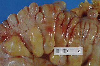
Diverticulitis, also called colonic diverticulitis, is a gastrointestinal disease characterized by inflammation of abnormal pouches—diverticula—that can develop in the wall of the large intestine. Symptoms typically include lower abdominal pain of sudden onset, but the onset may also occur over a few days. There may also be nausea, diarrhea or constipation. Fever or blood in the stool suggests a complication. People may experience a single attack, repeated attacks, or ongoing "smouldering" diverticulitis.

Diverticulosis is the condition of having multiple pouches (diverticula) in the colon that are not inflamed. These are outpockets of the colonic mucosa and submucosa through weaknesses of muscle layers in the colon wall. Diverticula do not cause symptoms in most people. Diverticular disease occurs when diverticula become clinically inflamed, a condition known as diverticulitis.

A Meckel's diverticulum, a true congenital diverticulum, is a slight bulge in the small intestine present at birth and a vestigial remnant of the vitelline duct. It is the most common malformation of the gastrointestinal tract and is present in approximately 2% of the population, with males more frequently experiencing symptoms.

Gastric bypass surgery refers to a technique in which the stomach is divided into a small upper pouch and a much larger lower "remnant" pouch and then the small intestine is rearranged to connect to both. Surgeons have developed several different ways to reconnect the intestine, thus leading to several different gastric bypass procedures (GBP). Any GBP leads to a marked reduction in the functional volume of the stomach, accompanied by an altered physiological and physical response to food.

Ileus is a disruption of the normal propulsive ability of the intestine. It can be caused by lack of peristalsis or by mechanical obstruction. The word 'ileus' is from Ancient Greek eileós. The term 'subileus' refers to a partial obstruction.

In medicine or biology, a diverticulum is an outpouching of a hollow structure in the body. Depending upon which layers of the structure are involved, diverticula are described as being either true or false.
Colic in horses is defined as abdominal pain, but it is a clinical symptom rather than a diagnosis. The term colic can encompass all forms of gastrointestinal conditions which cause pain as well as other causes of abdominal pain not involving the gastrointestinal tract. What makes it tricky is that different causes can manifest with similar signs of distress in the animal. Recognizing and understanding these signs is pivotal, as timely action can spell the difference between a brief moment of discomfort and a life-threatening situation. The most common forms of colic are gastrointestinal in nature and are most often related to colonic disturbance. There are a variety of different causes of colic, some of which can prove fatal without surgical intervention. Colic surgery is usually an expensive procedure as it is major abdominal surgery, often with intensive aftercare. Among domesticated horses, colic is the leading cause of premature death. The incidence of colic in the general horse population has been estimated between 4 and 10 percent over the course of the average lifespan. Clinical signs of colic generally require treatment by a veterinarian. The conditions that cause colic can become life-threatening in a short period of time.
Diverticular disease is when problems occur due to diverticulosis, a benign condition defined by the formation of pouches (diverticula) from the weak spots in the wall of the large intestine. This disease spectrum includes diverticulitis, symptomatic uncomplicated diverticular disease (SUDD), and segmental colitis associated with diverticulosis (SCAD). The most common symptoms across the disease spectrum are abdominal pain and bowel habit changes such as diarrhea or constipation. Otherwise, diverticulitis presents with systemic symptoms such as fever and elevated white blood cell count whereas SUDD and SCAD don’t. Treatment ranges from conservative bowel rest to medications such as antibiotics, antispasmodics, acetaminophen, mesalamine, rifaximin, and corticosteroids depending on the specific conditions.

A calculus, often called a stone, is a concretion of material, usually mineral salts, that forms in an organ or duct of the body. Formation of calculi is known as lithiasis. Stones can cause a number of medical conditions.

Struvite (magnesium ammonium phosphate) is a phosphate mineral with formula: NH4MgPO4·6H2O. Struvite crystallizes in the orthorhombic system as white to yellowish or brownish-white pyramidal crystals or in platy mica-like forms. It is a soft mineral with Mohs hardness of 1.5 to 2 and has a low specific gravity of 1.7. It is sparingly soluble in neutral and alkaline conditions, but readily soluble in acid.
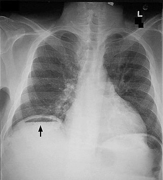
Pneumoperitoneum is pneumatosis in the peritoneal cavity, a potential space within the abdominal cavity. The most common cause is a perforated abdominal organ, generally from a perforated peptic ulcer, although any part of the bowel may perforate from a benign ulcer, tumor or abdominal trauma. A perforated appendix seldom causes a pneumoperitoneum.
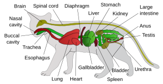
Feline lower urinary tract disease (FLUTD) is a generic category term to describe any disorder affecting the bladder or urethra of cats.

Bladder stones or uroliths are a common occurrence in animals, especially in domestic animals such as dogs and cats. Occurrence in other species, including tortoises, has been reported as well. The stones form in the urinary bladder in varying size and numbers secondary to infection, dietary influences, and genetics. Stones can form in any part of the urinary tract in dogs and cats, but unlike in humans, stones of the kidney are less common and do not often cause significant disease, although they can contribute to pyelonephritis and chronic kidney disease. Types of stones include struvite, calcium oxalate, urate, cystine, calcium phosphate, and silicate. Struvite and calcium oxalate stones are by far the most common. Bladder stones are not the same as bladder crystals but if the crystals coalesce unchecked in the bladder they can become stones.

Intestinal atresia is any congenital malformation of the structure of the intestine that causes bowel obstruction. The malformation can be a narrowing (stenosis), absence or malrotation of a portion of the intestine. These defects can either occur in the small or large intestine.
Jejunoileal bypass (JIB) was a surgical weight-loss procedure performed for the relief of morbid obesity from the 1950s through the 1970s in which all but 30 cm (12 in) to 45 cm (18 in) of the small bowel were detached and set to the side.

A bezoar is a mass often found trapped in the gastrointestinal system, though it can occur in other locations. A pseudobezoar is an indigestible object introduced intentionally into the digestive system.
Proximal enteritis, also known as anterior enteritis or duodenitis-proximal jejunitis (DPJ), is inflammation of the duodenum and upper jejunum. It produces a functional stasis of the affected intestine (ileus) and hypersecretion of fluid into the lumen of that intestine. This leads to large volumes of gastric reflux, dehydration, low blood pressure, and potentially shock. Although the exact cause is not yet definitively known, proximal enteritis requires considerable supportive care.
Carbonated soda treatment of phytobezoars is the use of carbonated soda to try to dissolve a phytobezoar. Bezoars consist of a solid and formed mass trapped in the gastrointestinal system, usually in the stomach. These can also form in other locations.