Eustrongylidosis

Last updated

Eustrongylidosis is a parasitic disease that mainly affects wading birds worldwide; however, the parasite's complex, indirect lifecycle involves other species, such as aquatic worms and fish. Moreover, this disease is zoonotic, which means the parasite can transmit disease from animals to humans. Eustrongylidosis is named after the causative agent Eustrongylides , and typically occurs in eutrophicated waters where concentrations of nutrients and minerals are high enough to provide ideal conditions for the parasite to thrive and persist. Because eutrophication has become a common issue due to agricultural runoff and urban development, cases of eustrongylidosis are becoming prevalent and hard to control. Eustrongylidosis can be diagnosed before or after death by observing behavior and clinical signs, and performing fecal flotations and necropsies. Methods to control it include preventing eutrophication and providing hosts with uninfected food sources in aquaculture farms. Parasites are known to be indicators of environmental health and stability, so should be studied further to better understand the parasite's lifecycle and how it affects predator-prey interactions and improve conservation efforts.

Contents

Cause

Eustrongylidosis is caused by several species of roundworms. The nematodes are red, large, and easily noticeable, and are characterized by the absence of a posterior sucker. [1] These roundworms cause a high mortality rate in nesting egrets and other wading birds, including other coastal populations. [2] The parasite was first detected in fish. The parasite is then transferred from the fish to the waterfowl when the bird is eating the fish. After consumption, the parasite perforates through the stomach lining, often resulting in the death of the host. [3]

Lifecycle

Eustrongylidosis is caused by three species of Eustrongylides: E. ignotus, E. tubifex, and E. excisus. [4] Eustrongylides spp. are macroscopic roundworms or nematodes that reproduce sexually year-round. The parasite has a complex lifecycle that involves two intermediate hosts, a paratenic host, and a definitive host. Therefore, the lifecycle is indirect because the parasite has to infect other species to reach its final host and become sexually mature and reproduce. Intermediate hosts and definitive hosts provide the parasite transport to other hosts and a place to develop, whereas paratenic hosts only provide transport to other hosts. The cycle begins when the wading bird excretes feces in or nearby a water body. [4] In the feces, the eggs are shed, and once they reach the water, an oligochaete, or aquatic worm, feeds on the eggs and becomes the first intermediate host. Within the oligochaete, the parasite eggs hatch and develop into second- and third-stage larvae. When a fish feeds on the infected oligochaete, it becomes the second intermediate host and the parasite develops into a fourth-stage larva. Finally, when a wading bird feeds on the infected fish, it becomes the definitive or final host and Eustrongylides becomes sexually mature. The parasite becomes an adult within 3 to 5 hours after infection and sheds eggs within 14 to 23 days after infection. [3] The cycle repeats itself when the parasite becomes sexually mature and sheds eggs through the feces of the bird to the external environment. If a larger fish, amphibian, or reptile feeds on the infected fish before the wading bird does, it becomes a paratenic hosts until fed on by the definitive host.[ citation needed ]

Species affected

Eustrongylides spp. have been found in a variety of wildlife species, including freshwater fishes, [5] such as perch (Perca fluviatilis), sheat fish (Silurus glanis), pike (Esox lucius), pikeperch (Sander lucioperca), and sun perch (Lepomis eupomotis gibbosus). [5] Eustrongylides spp. have also been observed in wading birds (Ciconiiformes), including great blue herons (Ardea herodias), great egrets (Casmerodius albus), and snowy egrets (Egretta thula). [6]

Distribution

Eustrongylides spp. have been reported throughout most of the world. Within the United States, E. tubifex and E. ignotus have been observed in wildlife. [4] This disease can be found in rookeries, especially in areas consisting of dense bird populations. [4] These species are also found in nesting habitats of birds, including areas with a low tree understory. [4]

Clinical signs

Monkey goby infected with the larvae of nematodes Eustrongylides excisus, Dniester Estuary, Ukraine Infected goby.jpg
Monkey goby infected with the larvae of nematodes Eustrongylides excisus, Dniester Estuary, Ukraine

Eustrongylides has evolved to infect the outside surface of the ventriculus of birds or the stomach in fish. This is due to the bird's response to parasitic infections, which involves regurgitating food to remove the parasite from their bodies. By inhabiting the outside of the stomach, though, the bird cannot get rid of the parasite and continually regurgitates food until it becomes emaciated. [3] Furthermore, the presence of the parasite in the ventriculus causes secondary infections such as bacterial peritonitis, the inflammation of the abdominal cavity walls, and sepsis, the infiltration of bacteria in the blood, when the parasite penetrates the stomach. [4] Birds less than a week old are more susceptible to eustrongylidosis than older birds for several reasons. [3] First, their immune system is still developing and weak. Second, sibling competition for food is common in young birds. Finally, their growth rates become suppressed, so they tend to die before the parasite becomes sexually mature and sheds eggs. Unlike young birds, older birds develop chronic eustrongylidosis because of their stronger immune systems. Similar to young birds, old birds have lesions in their ventriculi and develop secondary infections. A common behavior in both young and old birds includes regurgitation of food, which leads to lack of appetite and eventually anorexia or emaciation. In some cases, the parasite can inhabit the lung and cause respiratory problems that increase stress levels and severity of the disease. [7] In fish, the parasite affects its body condition, making it more susceptible to predation rather than to the disease. [8] Since the parasite depends on the intermediate host to reach the definitive host, it cannot harm the fish without first luring it to its predator, the wading bird.

Diagnosis

Diagnosis in a live specimen is possible in the field by palpating the abdomen. As with birds, prominence of the keel could be a determinant in diagnosis, but natural history of the species needs to be understood to avoid potential misdiagnoses. The best form of diagnosis, though is as necropsy. [1] During the necropsy, the best diagnosis can be determined by scanning the adult nematodes with electron microscopy. [1] Different species Eustrongylides nematodes can be differentiated by specific gender characteristics, i.e. "Male specimens of E. ignotus have a caudal sucker that lacks cuticular cleft, while a cuticular cleft is present in the caudal sucker of male specimens of E. excisus". [1] "Eustrongylidosis can often be misdiagnosed as starvation in nestlings because they are often emaciated at the time of death". [6]

Before necropsy takes place, diagnosis by palpation can be used to find tubular lesions, which are firm in texture, firmly attached to organs, and felt in the subcutaneous tissue. While palpation is practical and simple, errors can be made in nestlings' examinations because their ribs have the potential to present as lesions. Diagnosis is also attainable by examining fecal samples, but has the high potential of false negatives. That possibility is increased in fledging feces "where severe disease may precede appearance of eggs in the feces". [6]

Prevention and control

Because of Eustrongylides species' complex lifecycle with various host species, preventing infection and controlling outbreaks is difficult. Outbreaks of this disease are closely linked to agricultural runoff and urban development. [9] Eutrophication of water bodies supports high population levels of oligochaete worms, which causes increased numbers of infected fish that eat the worms, and then the birds that eat the fish. [10]

One way to prevent eustrongylidosis is to control oligochaete populations. Outbreaks of this parasite are closely linked to high numbers of oligochaete worms in an area's waterways, because the worms are essential for the species to reproduce. Oligochaete populations can be controlled by monitoring nutrient levels in the water, because high nutrient levels support oligochaete populations. [10] They can also be controlled by decreasing the level of oxygen in the water. Encouraging responsible farming practices to reduce chemical run-off can help prevent this disease from occurring.[ citation needed ]

Managers need to be diligent in catching the symptoms of the parasite before it can become an outbreak. [9] Once an outbreak of eustrongylidosis has occurred, ecosystem managers can do little to stop the spread in oligochaetes, fish, and birds. Traditional anthelminthics (dewormers) are not effective in fish because they kill parasites that live inside the gastrointestinal tract, whereas Eustrongylides species live outside the stomach in the body cavity. [11] The parasites can only be removed from fish surgically, which is not feasible. To completely stop the lifecycle in fish, all fish in an affected area must be culled. [11]

Surgical removal of the parasite from wading birds is a viable option, but this would also not be feasible for a large number of birds, and it would not stop the cycle of infection.

Treatment

Treatment for eustrongylidosis is limited in the wading-bird population due to the extensive amount of perforation in the stomach lining and limited funds available for treatment. In humans who are infected with the parasites, surgery is required to remove it from the intestinal wall. [12] As surgery is not a feasible treatment option for wading fowl in the wild en masse, treatment of the infected birds (a large portion of wild populations) has not been found, nor will likely be practical. [1] A possibility exists that killing or removing the nematodes could do more harm to the host specimen than actual good.[ citation needed ]

Related Research Articles

<i>Ascaris lumbricoides</i> One of several species of Ascaris

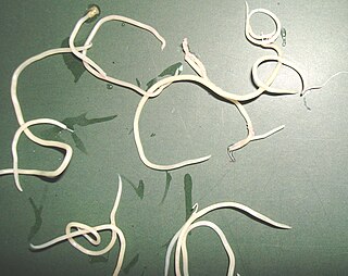
Ascaris lumbricoides is the "large roundworm" of humans, growing to a length of up to 35 cm (14 in). It is one of several species of Ascaris. An ascarid nematode of the phylum Nematoda, it is the most common parasitic worm in humans. This organism is responsible for the disease ascariasis, a type of helminthiasis and one of the group of neglected tropical diseases. An estimated one-sixth of the human population is infected by A. lumbricoides or another roundworm. Ascariasis is prevalent worldwide, especially in tropical and subtropical countries.

Host (biology) Organism that harbours another organism

In biology and medicine, a host is a larger organism that harbours a smaller organism; whether a parasitic, a mutualistic, or a commensalist guest (symbiont). The guest is typically provided with nourishment and shelter. Examples include animals playing host to parasitic worms, cells harbouring pathogenic (disease-causing) viruses, a bean plant hosting mutualistic (helpful) nitrogen-fixing bacteria. More specifically in botany, a host plant supplies food resources to micropredators, which have an evolutionarily stable relationship with their hosts similar to ectoparasitism. The host range is the collection of hosts that an organism can use as a partner.

Toxocariasis is an illness of humans caused by larvae of either the dog roundworm, or cat roundworm. Toxocariasis is often called visceral larva migrans (VLM). Depending on geographic location, degree of eosinophilia, eye and/or pulmonary signs, the terms ocular larva migrans (OLM), Weingarten's disease, Frimodt-Møller's syndrome, and eosinophilic pseudoleukemia are applied to toxocariasis. Other terms sometimes or rarely used include nematode ophthalmitis, toxocaral disease, toxocarose, and covert toxocariasis. This zoonotic, helminthic infection is a rare cause of blindness and may provoke rheumatic, neurologic, or asthmatic symptoms. Humans normally become infected by ingestion of embryonated eggs from contaminated sources.

Gnathostomiasis is the human infection caused by the nematode (roundworm) Gnathostoma spinigerum and/or Gnathostoma hispidum, which infects vertebrates.

<i>Baylisascaris</i> Genus of worms

Baylisascaris is a genus of roundworms that infect more than fifty animal species.

Parelaphostrongylus tenuis is a neurotropic nematode parasite common to white-tailed deer, Odocoileus virginianus, which causes damage to the central nervous system. Moose, elk, caribou, mule deer, and others are also susceptible to the parasite, but are aberrant hosts and are infected in neurological instead of meningeal tissue. The frequency of infection in these species increases dramatically when their ranges overlap high densities of white-tailed deer.

<i>Ascaridia galli</i> Species of roundworm

Ascaridia galli is a parasitic roundworm belonging to the phylum Nematoda. Nematodes of the genus Ascaridia are essentially intestinal parasites of birds. A. galli is the most prevalent and pathogenic species, especially in domestic fowl, Gallus domesticus. It causes ascaridiasis, a disease of poultry due to heavy worm infection, particularly in chickens and turkeys. It inhabits the small intestine, and can be occasionally seen in commercial eggs.

Spirometra erinaceieuropaei is a parasitic tapeworm that infects domestic animals and humans. The medical term for this infection in humans and other animals is sparganosis. Morphologically, these worms are similar to other worms in the genus Spirometra. They have a long body consisting of three sections: the scolex, the neck, and the strobilia. They have a complex life cycle that consists of three hosts, and can live in varying environments and bodily tissues. Humans can contract this parasite in three main ways. Historically, humans are considered a paratenic host; however, the first case of an adult S. erinaceieuropaei infection in humans was reported in 2017. Spirometra tapeworms exist worldwide and infection is common in animals, but S. erinaceieuropaei infections are rare in humans. Treatment for infection typically includes surgical removal and anti-worm medication.

Mammomonogamus is a genus of parasitic nematodes of the family Syngamidae that parasitise the respiratory tracts of cattle, sheep, goats, deer, cats, orangutans, and elephants. The nematodes can also infect humans and cause the disease called mammomonogamiasis. Several known species fall under the genus Mammomonogamus, but the most common species found to infest humans is M. laryngeus. Infection in humans is very rare, with only about 100 reported cases worldwide, and is assumed to be largely accidental. Cases have been reported from the Caribbean, China, Korea, Thailand, and Philippines.

Gapeworm Species of roundworm

A gapeworm, also known as a red worm and forked worm, is a parasitic nematode worm that infects the tracheas of certain birds. The resulting disease, known as "gape" occurs when the worms clog and obstruct the airway. The worms are also known as "red worms" or "forked worms" due to their red color and the permanent procreative conjunction of males and females. Gapeworms are common in young, domesticated chickens and turkeys.

<i>Dioctophyme renale</i> Species of roundworm

Dioctophyme renale, commonly referred to as the giant kidney worm, is a parasitic nematode (roundworm) whose mature form is found in the kidneys of mammals. D. renale is distributed worldwide, but is less common in Africa and Oceania. It affects fish eating mammals, particularly mink and dogs. Human infestation is rare, but results in kidney destruction, usually of one kidney and hence not fatal. A 2019 review listed a total of 37 known human cases of dioctophymiasis in 10 countries with the highest number (22) in China. Upon diagnosis through tissue sampling, the only treatment is surgical excision.

<i>Toxocara canis</i> Species of roundworm

Toxocara canis is a worldwide-distributed helminth parasite of dogs and other canids. The name is derived from the Greek word "toxon," meaning bow or quiver, and the Latin word "caro," meaning flesh. They live in the small intestine of the definitive host. In adult dogs, the infection is usually asymptomatic but may be characterized by diarrhea. By contrast, massive infection with Toxocara canis can be fatal in puppies, causing diarrhea, vomiting, an enlarged abdomen, flatulence, and poor growth rate.

Capillaria philippinensis is a parasitic nematode which causes intestinal capillariasis. This sometimes fatal disease was first discovered in Northern Luzon, Philippines in 1964. Cases have also been reported from China, Egypt, Indonesia, Iran, Japan, Korea, Lao PDR, Taiwan and Thailand. Cases diagnosed in Italy and Spain were believed to be acquired abroad, with one case possibly contracted in Colombia. The natural life cycle of C. philippinensis is believed to involve fish as intermediate hosts, and fish-eating birds as definitive hosts. Humans acquire C. philippinensis by eating small species of infested fish whole and raw.

<i>Heterakis gallinarum</i> Species of roundworm

Heterakis gallinarum is a nematode parasite that lives in the cecum of some galliform birds, particularly in ground feeders such as domestic chickens and turkeys. It causes infection that is mildly pathogenic. However, it often carries a protozoan parasite Histomonas meleagridis which causes of histomoniasis. Transmission of H. meleagridis is through the H. gallinarum egg. H. gallinarum is about 1–2 cm in length with a sharply pointed tail and a preanal sucker. The parasite is a diecious species with marked sexual dimorphism. Males are smaller and shorter, measuring around 9 mm in length, with a unique bent tail. Females are stouter and longer, measuring roughly 13 mm in length, with a straight tail end.

Baylisascaris procyonis, common name raccoon roundworm, is a roundworm nematode, found ubiquitously in raccoons, the definitive hosts. It is named after H. A. Baylis, who studied them in the 1920s–30s, and Greek askaris. Baylisascaris larvae in paratenic hosts can migrate, causing visceral larva migrans (VLM). Baylisascariasis as the zoonotic infection of humans is rare, though extremely dangerous due to the ability of the parasite's larvae to migrate into brain tissue and cause damage. Concern for human infection has been increasing over the years due to urbanization of rural areas resulting in the increase in proximity and potential human interaction with raccoons.

Gnathostoma hispidum is a nematode (roundworm) that infects many vertebrate animals including humans. Infection of Gnathostoma hispidum, like many species of Gnathostoma causes the disease gnathostomiasis due to the migration of immature worms in the tissues.

Histomoniasis Medical condition

Histomoniasis is a commercially significant disease of poultry, particularly of chickens and turkeys, due to parasitic infection of a protozoan, Histomonas meleagridis. The protozoan is transmitted to the bird by the nematode parasite Heterakis gallinarum. H. meleagridis resides within the eggs of H. gallinarum, so birds ingest the parasites along with contaminated soil or food. Earthworms can also act as a paratenic host.

<i>Anisakis simplex</i>

Anisakis simplex, known as the herring worm, is a species of nematode in the genus Anisakis. Like other nematodes, it infects and settles in the organs of marine animals, such as salmon, mackerels and squids. It is commonly found in cold marine waters, such as the Pacific Ocean and Atlantic Ocean.

<i>Baylisascaris shroederi</i> Species of roundworm

Baylisascaris shroederi, common name giant panda roundworm, is a roundworm (nematode), found ubiquitously in giant pandas of central China, the definitive hosts. Baylisascaris larvae in paratenic hosts can migrate, causing visceral larva migrans (VLM). It is extremely dangerous to the host due to the ability of the parasite's larvae to migrate into brain tissue and cause damage. Concern for human infection is minimal as there are very few giant pandas living today and most people do not encounter giant pandas in their everyday activities. There is growing recognition that the infection of Baylisascaris shroederi is one of the major causes of death in the species. This is confirmed by a report stating that during the period of 2001 to 2005; about 50% of deaths in wild giant pandas were caused by the parasite infection.

<i>Corynosoma australe</i> Species of thorny-headed worm

Corynosoma autrale is a species of acanthocephalan. This species usually infects pinnipeds; the semi-aquatic fin-footed marine mammals most commonly known as seals and sea lions. Pinniped infections are not exclusive, recently C. australe has been discovered in Magellanic penguins.

References

  1. 1 2 3 4 5 Spalding, M.; Forrester, D. (2008). "Eustrongylidosis". Parasitic Diseases of Wild Birds. pp. 289–315. doi:10.1002/9780813804620.ch16. ISBN   9780813804620.
  2. National Wildlife Health Center, 1999
  3. 1 2 3 4 Spalding, M. G.; Forrester, D. J. (1993). "Pathogenesis of Eustrongylides ignotus (Nematoda: Dioctophymatoidea) in Ciconiiformes". Journal of Wildlife Diseases. 29 (2): 250–260. doi:10.7589/0090-3558-29.2.250. PMID   8487374. S2CID   11257605.
  4. 1 2 3 4 5 6 Ciganovich, E. A., Redman, P. J., & Stenback, R. S. (2013). USGS National Wildlife Health Center Field Manual of Wildlife Diseases: Birds, 29: 223-228.
  5. 1 2 Urdeş, L.; Hangan, M.; Diaconescu, C.; Ianiţchi, D.; Serafim, V. (2008). "Eustrongylidosis' occurrence in freshwater fish from the Danubian Delta area". Scientific Papers: Animal Science and Biotechnologies. 41 (2): 182–186. doi:10.1016/j.aaspro.2015.08.072.
  6. 1 2 3 Spalding, M. G. (1990). "Antemortem diagnosis of Eustrongylidosis in wading birds (Ciconiiformes)". Colonial Waterbirds. 13 (1): 75–77. doi:10.2307/1521425. JSTOR   1521425.
  7. Ballweber, L. R. (2004). "Waterfowl parasites". Seminars in Avian and Exotic Pet Medicine. 13 (4): 197–205. doi:10.1053/j.saep.2004.04.005.
  8. Scholz, T. (1999). "Parasites in cultured and feral fish". Veterinary Parasitology. 84 (3–4): 317–335. doi:10.1016/s0304-4017(99)00039-4. PMID   10456421.
  9. 1 2 Coyner, D. F.; Spalding, M. G.; Forrester, D. J. (2002). "Epizootiology of Eustronglyides Ignotus in Florida: Distribution, density, and natural infections in intermediate hosts". Journal of Wildlife Diseases. 38 (3): 483–499. doi: 10.7589/0090-3558-38.3.483 . PMID   12238365. S2CID   7853013.
  10. 1 2 Franson, J. C.; Cluster, T. W. (1994). "Eustronglyidosis in wading birds from colonies in California, Texas, and Rhode Island, USA". Colonial Waterbirds. 17 (2): 168–172. doi:10.2307/1521295. JSTOR   1521295.
  11. 1 2 Yanong, R. P. E. (2006). Nematode (roundworm) infections in fish. Circular 91, Department of Fisheries and Aquatic Sciences, Florida Cooperative Extension Service, Institute of Food and Agricultural Sciences, University of Florida. https://edis.ifas.ufl.edu/fa091
  12. (Martin et al. 2000)