FtsZ

Last updated
Cell division protein FtsZ
Identifiers
SymbolFtsZ
InterPro IPR000158
CATH 1fsz
SCOP2 1fsz / SCOPe / SUPFAM
CDD cd02201
FtsZ, C-terminal sandwich
Identifiers
SymbolFtsZ_C
Pfam PF12327
InterPro IPR024757
Available protein structures:
Pfam   structures / ECOD  
PDB RCSB PDB; PDBe; PDBj
PDBsum structure summary
Cell division protein FtsZ
PDB 1fsz EBI.jpg
Molecular Structure of FtsZ (PDB 1fsz).
Identifiers
Organism Escherichia coli
SymbolftsZ
UniProt P0A9A6
Search for
Structures Swiss-model
Domains InterPro

FtsZ is a protein encoded by the ftsZ gene that assembles into a ring at the future site of bacterial cell division (also called the Z ring). FtsZ is a prokaryotic homologue of the eukaryotic protein tubulin. The initials FtsZ mean "Filamenting temperature-sensitive mutant Z." The hypothesis was that cell division mutants of E. coli would grow as filaments due to the inability of the daughter cells to separate from one another. FtsZ is found in almost all bacteria, many archaea, all chloroplasts and some mitochondria, where it is essential for cell division. FtsZ assembles the cytoskeletal scaffold of the Z ring that, along with additional proteins, constricts to divide the cell in two.

Contents

History

In the 1960s scientists screened for temperature sensitive mutations that blocked cell division at 42 °C. The mutant cells divided normally at 30°, but failed to divide at 42°. Continued growth without division produced long filamentous cells (Filamenting temperature sensitive). Several such mutants were discovered and mapped to a locus originally named ftsA, which could be one or more genes. In 1980 Lutkenhaus and Donachie [1] showed that several of these mutations mapped to one gene, ftsA, but one well-characterized mutant, PAT84, originally discovered by Hirota et al, [2] mapped to a separate, adjacent gene. They named this cell division gene ftsZ. In 1991 Bi and Lutkenhaus used immunogold electron microscopy to show that FtsZ localized to the invaginating septum at midcell. [3] Subsequently, the Losick and Margolin groups used immuno-fluorescence microscopy [4] and GFP fusions [5] to show that FtsZ assembled Z rings early in the cell cycle, well before the septum began to constrict. Other division proteins then assemble onto the Z ring and constriction occurs in the last part of the cell cycle.

In 1992-3 three labs independently discovered that FtsZ was related to eukaryotic tubulin, which is the protein subunit that assembles into microtubules. [6] [7] [8] This was the first discovery that bacteria have homologs of eukaryotic cytoskeletal proteins. Later work showed that FtsZ was present in, and essential for, cell division in almost all bacteria and in many but not all archaea.

Mitochondria and chloroplasts are eukaryotic organelles that originated as bacterial endosymbionts, so there was much interest in whether they use FtsZ for division. Chloroplast FtsZ was first discovered by Osteryoung, [9] and it is now known that all chloroplasts use FtsZ for division. Mitochondrial FtsZ was discovered by Beech [10] in an alga; FtsZ is used for mitochondrial division in some eukaryotes, while others have replaced it with a dynamin-based machinery.

In 2014, scientists identified two FtsZ homologs in archaea, FtsZ1 and FtsZ2. [11]

Function

Inhibition of FtsZ disrupts septum formation, resulting in filamentation of bacterial cells (top right of electron micrograph). Filamentation 1.jpg
Inhibition of FtsZ disrupts septum formation, resulting in filamentation of bacterial cells (top right of electron micrograph).

During cell division, FtsZ is the first protein to move to the division site, and is essential for recruiting other proteins that produce a new cell wall (septum) between the dividing cells. FtsZ's role in cell division is analogous to that of actin in eukaryotic cell division, but, unlike the actin-myosin ring in eukaryotes, FtsZ has no known motor protein associated with it. Cell wall synthesis may externally push the cell membrane, providing the force for cytokinesis. Supporting this, in E. coli the rate of division is affected by mutations in cell wall synthesis. [12] Alternatively, FtsZ may pull the membrane from the inside based on Osawa (2009) showing the protein's contractile force on liposomes with no other proteins present. [13]

Erickson (2009) proposed how the roles of tubulin-like proteins and actin-like proteins in cell division became reversed in an evolutionary mystery. [14] The use of the FtsZ ring in dividing chloroplasts and some mitochondria further establishes their prokaryotic ancestry. [15] L-form bacteria that lack a cell wall do not require FtsZ for division, which implies that bacteria may have retained components of an ancestral mode of cell division. [16]

Much is known about the dynamic polymerization activities of tubulin and microtubules, but little is known about these activities in FtsZ. While it is known that single-stranded tubulin protofilaments form into 13 stranded microtubules, the multistranded structure of the FtsZ-containing Z-ring is not known. It is only speculated that the structure consists of overlapping protofilaments. Nevertheless, recent work with purified FtsZ on supported lipid bilayers as well as imaging FtsZ in living bacterial cells revealed that FtsZ protofilaments have polarity and move in one direction by treadmilling [17] (see also below).

Recently, proteins similar to tubulin and FtsZ have been discovered in large plasmids found in Bacillus species. They are believed to function as components of segrosomes, which are multiprotein complexes that partition chromosomes/plasmids in bacteria. The plasmid homologs of tubulin/FtsZ seem to have conserved the ability to polymerize into filaments.

The contractile ring (the "Z ring")

The Z-ring forms from smaller subunits of FtsZ filaments. These filaments may pull on each other and tighten to divide the cell. FtsZ Filaments.svg
The Z-ring forms from smaller subunits of FtsZ filaments. These filaments may pull on each other and tighten to divide the cell.
Super-resolution image of Z-rings (green) at different stages of constriction in two E. coli cells. Zrings.png
Super-resolution image of Z-rings (green) at different stages of constriction in two E. coli cells.

FtsZ has the ability to bind to GTP and also exhibits a GTPase domain that allows it to hydrolyze GTP to GDP and a phosphate group. In vivo, FtsZ forms filaments with a repeating arrangement of subunits, all arranged head-to-tail. [18] These filaments form a ring around the longitudinal midpoint, or septum, of the cell. This ring is called the Z-ring.

The GTP hydrolyzing activity of the protein is not essential to the formation of filaments or cell division. Mutants defective in GTPase activity often still divide, but sometimes form twisted and disordered septa. It is unclear as to whether FtsZ actually provides the physical force that results in division or serves as a scaffold for other proteins to execute division.

There are two models for how FtsZ might generate a constriction force. One model is based on the observation that FtsZ protfilaments can be straight or curved. The transition from straight to curved is suggested to generate a bending force on the membrane. [19] Another model is based on sliding protofilaments. Computer models and in vivo measurements suggest that single FtsZ filaments cannot sustain a length more than 30 subunits long. In this model, FtsZ scission force comes from the relative lateral movement of subunits. [20] Lines of FtsZ would line up together parallel and pull on each other creating a "cord" of many strings that tightens itself.

In other models, FtsZ does not provide the contractile force but provides the cell a spatial scaffold for other proteins to execute the division of the cell. This is akin to the creating of a temporary structure by construction workers to access hard-to-reach places of a building. The temporary structure allows unfettered access and ensures that the workers can reach all places. If the temporary structure is not correctly built, the workers will not be able to reach certain places, and the building will be deficient.

The scaffold theory is supported by information that shows that the formation of the ring and localization to the membrane requires the concerted action of a number of accessory proteins. ZipA or the actin homologue FtsA permit initial FtsZ localization to the membrane. [21] Following localization to the membrane, division proteins of the Fts family are recruited for ring assembly. [22] Many of these proteins direct the synthesis of the new division septum at midcell (FtsI, FtsW), or regulate the activity of this synthesis (FtsQ, FtsL, FtsB, FtsN). The timing of Z-ring formation suggests the possibility of a spatial or temporal signal that permits the formation of FtsZ filaments.

Recent super-resolution imaging in several species supports a dynamic scaffold model, in which small clusters of FtsZ protofilaments or protofilament bundles move unidirectionally around the ring's circumference by treadmilling, anchored to the membrane by FtsA and other FtsZ-specific membrane tethers. [23] [24] The speed of treadmilling depends on the rate of GTP hydrolysis within the FtsZ protofilaments, but in Escherichia coli , synthesis of the division septum remains the rate limiting step for cytokinesis. [25] The treadmilling action of FtsZ is required for proper synthesis of the division septum by septal peptidoglycan synthesis enzymes, suggesting that these enzymes can track the growing ends of the filaments.

Septal localization and intracellular signaling

The formation of the Z-ring closely coincides with cellular processes associated with replication. Z-ring formation coincides with the termination of genome replication in E. coli and 70% of chromosomal replication in B. subtilis . [26] The timing of Z-ring formation suggests the possibility of a spatial or temporal signal that permits the formation of FtsZ filaments. In Escherichia coli , at least two negative regulators of FtsZ assembly form a bipolar gradient, such that the concentration of active FtsZ required for FtsZ assembly is highest at mid-cell between the two segregating chromosomes, and lowest at the poles and over the chromosomes. This type of regulation seems to occur in other species such as Bacillus subtilis and Caulobacter crescentus . However, other species including Streptococcus pneumoniae and Myxococcus xanthus seem to use positive regulators that stimulate FtsZ assembly at mid-cell. [27]

Communicating distress

FtsZ polymerization is also linked to stressors like DNA damage. DNA damage induces a variety of proteins to be manufactured, one of them called SulA. [28] SulA prevents the polymerization and GTPase activity of FtsZ. SulA accomplishes this task by binding to self-recognizing FtsZ sites. By sequestering FtsZ, the cell can directly link DNA damage to inhibiting cell division. [29]

Preventing DNA damage

Like SulA, there are other mechanisms that prevent cell division that would result in disrupted genetic information sent to daughter cells. So far, two proteins have been identified in E. coli and B. subtilis that prevent division over the nucleoid region: Noc and SlmA. Noc gene knockouts result in cells that divide without respect to the nucleoid region, resulting in its asymmetrical partitioning between the daughter cells. The mechanism is not well understood, but thought to involve sequestration of FtsZ, preventing polymerization over the nucleoid region. [30] The mechanism used by SlmA to inhibit FtsZ polymerization over the nucleoid [31] is better understood, and uses two separate steps. One domain of SlmA binds to a FtsZ polymer, then a separate domain of SlmA severs the polymer. [32] A similar mechanism is thought to be used by MinC, another inhibitor of FtsZ polymerization involved in positioning of the FtsZ ring. [33]

Clinical significance

The number of multidrug-resistant bacterial strains is currently increasing; thus, the determination of drug targets for the development of novel antimicrobial drugs is urgently needed. The potential role of FtsZ in the blockage of cell division, together with its high degree of conservation across bacterial species, makes FtsZ a highly attractive target for developing novel antibiotics. [34] Researchers have been working on synthetic molecules and natural products as inhibitors of FtsZ. [35]

The spontaneous self-assembly of FtsZ can also be used in nanotechnology to fabricate metal nanowires. [36] [37]

See also

Related Research Articles

<span class="mw-page-title-main">Microtubule</span> Polymer of tubulin that forms part of the cytoskeleton

Microtubules are polymers of tubulin that form part of the cytoskeleton and provide structure and shape to eukaryotic cells. Microtubules can be as long as 50 micrometres, as wide as 23 to 27 nm and have an inner diameter between 11 and 15 nm. They are formed by the polymerization of a dimer of two globular proteins, alpha and beta tubulin into protofilaments that can then associate laterally to form a hollow tube, the microtubule. The most common form of a microtubule consists of 13 protofilaments in the tubular arrangement.

<span class="mw-page-title-main">Cytoskeleton</span> Network of filamentous proteins that forms the internal framework of cells

The cytoskeleton is a complex, dynamic network of interlinking protein filaments present in the cytoplasm of all cells, including those of bacteria and archaea. In eukaryotes, it extends from the cell nucleus to the cell membrane and is composed of similar proteins in the various organisms. It is composed of three main components:microfilaments, intermediate filaments, and microtubules, and these are all capable of rapid growth or disassembly depending on the cell's requirements.

<span class="mw-page-title-main">MreB</span>

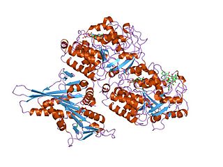
MreB is a protein found in bacteria that has been identified as a homologue of actin, as indicated by similarities in tertiary structure and conservation of active site peptide sequence. The conservation of protein structure suggests the common ancestry of the cytoskeletal elements formed by actin, found in eukaryotes, and MreB, found in prokaryotes. Indeed, recent studies have found that MreB proteins polymerize to form filaments that are similar to actin microfilaments. It has been shown to form multilayer sheets comprising diagonally interwoven filaments in the presence of ATP or GTP.

<span class="mw-page-title-main">Tubulin</span> Superfamily of proteins that make up microtubules

Tubulin in molecular biology can refer either to the tubulin protein superfamily of globular proteins, or one of the member proteins of that superfamily. α- and β-tubulins polymerize into microtubules, a major component of the eukaryotic cytoskeleton. Microtubules function in many essential cellular processes, including mitosis. Tubulin-binding drugs kill cancerous cells by inhibiting microtubule dynamics, which are required for DNA segregation and therefore cell division.

<span class="mw-page-title-main">Nucleoid</span> Region within a prokaryotic cell containing genetic material

The nucleoid is an irregularly shaped region within the prokaryotic cell that contains all or most of the genetic material. The chromosome of a typical prokaryote is circular, and its length is very large compared to the cell dimensions, so it needs to be compacted in order to fit. In contrast to the nucleus of a eukaryotic cell, it is not surrounded by a nuclear membrane. Instead, the nucleoid forms by condensation and functional arrangement with the help of chromosomal architectural proteins and RNA molecules as well as DNA supercoiling. The length of a genome widely varies and a cell may contain multiple copies of it.

<span class="mw-page-title-main">Filamentation</span>

Filamentation is the anomalous growth of certain bacteria, such as Escherichia coli, in which cells continue to elongate but do not divide. The cells that result from elongation without division have multiple chromosomal copies.

Crescentin is a protein which is a bacterial relative of the intermediate filaments found in eukaryotic cells. Just as tubulins and actins, the other major cytoskeletal proteins, have prokaryotic homologs in, respectively, the FtsZ and MreB proteins, intermediate filaments are linked to the crescentin protein. Some of its homologs are erroneously labelled Chromosome segregation protein ParA. This protein family is found in Caulobacter and Methylobacterium.

The bacterium, despite its simplicity, contains a well-developed cell structure which is responsible for some of its unique biological structures and pathogenicity. Many structural features are unique to bacteria and are not found among archaea or eukaryotes. Because of the simplicity of bacteria relative to larger organisms and the ease with which they can be manipulated experimentally, the cell structure of bacteria has been well studied, revealing many biochemical principles that have been subsequently applied to other organisms.

<span class="mw-page-title-main">Treadmilling</span> Simultaneous growth and breakdown on opposite ends of a protein filament

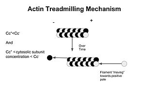
In molecular biology, treadmilling is a phenomenon observed within protein filaments of the cytoskeletons of many cells, especially in actin filaments and microtubules. It occurs when one end of a filament grows in length while the other end shrinks, resulting in a section of filament seemingly "moving" across a stratum or the cytosol. This is due to the constant removal of the protein subunits from these filaments at one end of the filament, while protein subunits are constantly added at the other end. Treadmilling was discovered by Wegner, who defined the thermodynamic and kinetic constraints. Wegner recognized that: “The equilibrium constant (K) for association of a monomer with a polymer is the same at both ends, since the addition of a monomer to each end leads to the same polymer.”; a simple reversible polymer can’t treadmill; ATP hydrolysis is required. GTP is hydrolyzed for microtubule treadmilling.

<span class="mw-page-title-main">Prokaryotic cytoskeleton</span> Structural filaments in prokaryotes

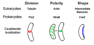
The prokaryotic cytoskeleton is the collective name for all structural filaments in prokaryotes. It was once thought that prokaryotic cells did not possess cytoskeletons, but advances in visualization technology and structure determination led to the discovery of filaments in these cells in the early 1990s. Not only have analogues for all major cytoskeletal proteins in eukaryotes been found in prokaryotes, cytoskeletal proteins with no known eukaryotic homologues have also been discovered. Cytoskeletal elements play essential roles in cell division, protection, shape determination, and polarity determination in various prokaryotes.

Fission, in biology, is the division of a single entity into two or more parts and the regeneration of those parts to separate entities resembling the original. The object experiencing fission is usually a cell, but the term may also refer to how organisms, bodies, populations, or species split into discrete parts. The fission may be binary fission, in which a single organism produces two parts, or multiple fission, in which a single entity produces multiple parts.

Bacterial morphological plasticity refers to changes in the shape and size that bacterial cells undergo when they encounter stressful environments. Although bacteria have evolved complex molecular strategies to maintain their shape, many are able to alter their shape as a survival strategy in response to protist predators, antibiotics, the immune response, and other threats.

<span class="mw-page-title-main">Tubulin domain</span>

Tubulin/FtsZ family, GTPase domain is an evolutionary conserved protein domain.

<span class="mw-page-title-main">Min System</span> Mechanism used by E. coli in cell division

The Min System is a mechanism composed of three proteins MinC, MinD, and MinE used by E. coli as a means of properly localizing the septum prior to cell division. Each component participates in generating a dynamic oscillation of FtsZ protein inhibition between the two bacterial poles to precisely specify the mid-zone of the cell, allowing the cell to accurately divide in two. This system is known to function in conjunction with a second negative regulatory system, the nucleoid occlusion system (NO), to ensure proper spatial and temporal regulation of chromosomal segregation and division.

The MinC protein is one of three proteins in the Min system encoded by the minB operon and which is required to generate pole to pole oscillations prior to bacterial cell division as a means of specifying the midzone of the cell. This function is achieved by preventing the formation of the divisome Z-ring around the poles.

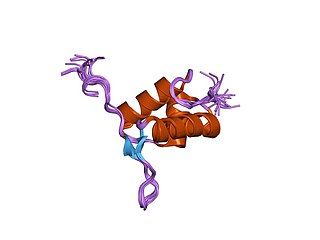
The MinE protein is one of three proteins of the Min system encoded by the minB operon required to generate pole to pole oscillations prior to bacterial cell division as a means of specifying the midzone of the cell, as seen in E.coli.

A plasmid partition system is a mechanism that ensures the stable inheritance of plasmids during bacterial cell division. Each plasmid has its independent replication system which controls the number of copies of the plasmid in a cell. The higher the copy number, the more likely the two daughter cells will contain the plasmid. Generally, each molecule of plasmid diffuses randomly, so the probability of having a plasmid-less daughter cell is 21−N, where N is the number of copies. For instance, if there are 2 copies of a plasmid in a cell, there is 50% chance of having one plasmid-less daughter cell. However, high-copy number plasmids have a cost for the hosting cell. This metabolic burden is lower for low-copy plasmids, but those have a higher probability of plasmid loss after a few generations. To control vertical transmission of plasmids, in addition to controlled-replication systems, bacterial plasmids use different maintenance strategies, such as multimer resolution systems, post-segregational killing systems, and partition systems.

<span class="mw-page-title-main">FtsA</span> Bacterial protein that is related to actin

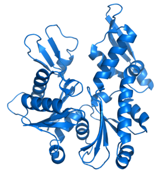
FtsA is a bacterial protein that is related to actin by overall structural similarity and in its ATP binding pocket.

<span class="mw-page-title-main">Divisome</span> A protein complex in bacteria responsible for cell division

The divisome is a protein complex in bacteria that is responsible for cell division, constriction of inner and outer membranes during division, and peptidoglycan (PG) synthesis at the division site. The divisome is a membrane protein complex with proteins on both sides of the cytoplasmic membrane. In gram-negative cells it is located in the inner membrane. The divisome is nearly ubiquitous in bacteria although its composition may vary between species.

<span class="mw-page-title-main">FtsK</span> Protein involved in bacterial cell division

FtsK is a protein in E.Coli involved in bacterial cell division and chromosome segregation. It is one of the largest proteins, consisting of 1329 amino acids. FtsK stands for "Filament temperature sensitive mutant K" because cells expressing a mutant ftsK allele called ftsK44, which encodes an FtsK variant containing an G80A residue change in the second transmembrane segment, fail to divide at high temperatures and form long filaments instead. FtsK, specifically its C-terminal domain, functions as a DNA translocase, interacts with other cell division proteins, and regulates Xer-mediated recombination. FtsK belongs to the AAA superfamily and is present in most bacteria.

References

  1. Lutkenhaus JF, Wolf-Watz H, Donachie WD (May 1980). "Organization of genes in the ftsA-envA region of the Escherichia coli genetic map and identification of a new fts locus (ftsZ)". Journal of Bacteriology. 142 (2): 615–620. doi:10.1128/JB.142.2.615-620.1980. OCLC   678550294. PMC   294035 . PMID   6991482.
  2. Hirota Y, Ryter A, Jacob F (1968-01-01). "Thermosensitive mutants of E. coli affected in the processes of DNA synthesis and cellular division". Cold Spring Harbor Symposia on Quantitative Biology. 33: 677–693. doi:10.1101/sqb.1968.033.01.077. PMID   4892005.
  3. Bi EF, Lutkenhaus J (November 1991). "FtsZ ring structure associated with division in Escherichia coli". Nature. 354 (6349): 161–164. Bibcode:1991Natur.354..161B. doi:10.1038/354161a0. PMID   1944597. S2CID   4329947.
  4. Levin PA, Losick R (February 1996). "Transcription factor Spo0A switches the localization of the cell division protein FtsZ from a medial to a bipolar pattern in Bacillus subtilis". Genes & Development. 10 (4): 478–488. doi: 10.1101/gad.10.4.478 . PMID   8600030.
  5. Ma X, Ehrhardt DW, Margolin W (November 1996). "Colocalization of cell division proteins FtsZ and FtsA to cytoskeletal structures in living Escherichia coli cells by using green fluorescent protein". Proceedings of the National Academy of Sciences of the United States of America. 93 (23): 12998–13003. Bibcode:1996PNAS...9312998M. doi: 10.1073/pnas.93.23.12998 . PMC   24035 . PMID   8917533.
  6. RayChaudhuri D, Park JT (September 1992). "Escherichia coli cell-division gene ftsZ encodes a novel GTP-binding protein". Nature. 359 (6392): 251–254. Bibcode:1992Natur.359..251R. doi:10.1038/359251a0. PMID   1528267. S2CID   4355143.
  7. de Boer P, Crossley R, Rothfield L (September 1992). "The essential bacterial cell-division protein FtsZ is a GTPase". Nature. 359 (6392): 254–256. Bibcode:1992Natur.359..254D. doi:10.1038/359254a0. PMID   1528268. S2CID   2748757.
  8. Mukherjee A, Dai K, Lutkenhaus J (February 1993). "Escherichia coli cell division protein FtsZ is a guanine nucleotide binding protein". Proceedings of the National Academy of Sciences of the United States of America. 90 (3): 1053–1057. Bibcode:1993PNAS...90.1053M. doi: 10.1073/pnas.90.3.1053 . PMC   45809 . PMID   8430073.
  9. Osteryoung KW, Vierling E (August 1995). "Conserved cell and organelle division". Nature. 376 (6540): 473–474. Bibcode:1995Natur.376..473O. doi:10.1038/376473b0. PMID   7637778. S2CID   5399155.
  10. Beech PL, Nheu T, Schultz T, Herbert S, Lithgow T, Gilson PR, McFadden GI (February 2000). "Mitochondrial FtsZ in a chromophyte alga". Science. 287 (5456): 1276–1279. Bibcode:2000Sci...287.1276B. doi:10.1126/science.287.5456.1276. PMID   10678836. S2CID   26587576.
  11. Duggin, Iain G.; Aylett, Christopher H. S.; Walsh, James C.; Michie, Katharine A.; Wang, Qing; Turnbull, Lynne; Dawson, Emma M.; Harry, Elizabeth J.; Whitchurch, Cynthia B.; Amos, Linda A.; Löwe, Jan (2014-12-22). "CetZ tubulin-like proteins control archaeal cell shape". Nature. Springer Science and Business Media LLC. 519 (7543): 362–365. doi:10.1038/nature13983. ISSN   0028-0836. PMC   4369195 . PMID   25533961.
  12. Coltharp C, Buss J, Plumer TM, Xiao J (February 2016). "Defining the rate-limiting processes of bacterial cytokinesis". Proceedings of the National Academy of Sciences of the United States of America. 113 (8): E1044–E1053. Bibcode:2016PNAS..113E1044C. doi: 10.1073/pnas.1514296113 . PMC   4776500 . PMID   26831086.
  13. Osawa M, Anderson DE, Erickson HP (May 2008). "Reconstitution of contractile FtsZ rings in liposomes". Science. 320 (5877): 792–794. Bibcode:2008Sci...320..792O. doi:10.1126/science.1154520. PMC   2645864 . PMID   18420899.
  14. Erickson HP (July 2007). "Evolution of the cytoskeleton". BioEssays. 29 (7): 668–677. doi:10.1002/bies.20601. PMC   2630885 . PMID   17563102.
  15. Leger MM, Petrů M, Žárský V, Eme L, Vlček Č, Harding T, et al. (August 2015). "An ancestral bacterial division system is widespread in eukaryotic mitochondria". Proceedings of the National Academy of Sciences of the United States of America. 112 (33): 10239–10246. Bibcode:2015PNAS..11210239L. doi: 10.1073/pnas.1421392112 . PMC   4547283 . PMID   25831547.
  16. Leaver M, Domínguez-Cuevas P, Coxhead JM, Daniel RA, Errington J (February 2009). "Life without a wall or division machine in Bacillus subtilis". Nature. 457 (7231): 849–853. Bibcode:2009Natur.457..849L. doi:10.1038/nature07742. PMID   19212404. S2CID   4413852.
  17. Loose M, Mitchison TJ (January 2014). "The bacterial cell division proteins FtsA and FtsZ self-organize into dynamic cytoskeletal patterns". Nature Cell Biology. 16 (1): 38–46. doi:10.1038/ncb2885. PMC   4019675 . PMID   24316672.
  18. Desai A, Mitchison TJ (1997). "Microtubule polymerization dynamics". Annual Review of Cell and Developmental Biology. 13: 83–117. doi:10.1146/annurev.cellbio.13.1.83. PMID   9442869.
  19. Erickson HP, Anderson DE, Osawa M (December 2010). "FtsZ in bacterial cytokinesis: cytoskeleton and force generator all in one". Microbiology and Molecular Biology Reviews. 74 (4): 504–528. doi:10.1128/MMBR.00021-10. PMC   3008173 . PMID   21119015.
  20. Lan G, Daniels BR, Dobrowsky TM, Wirtz D, Sun SX (January 2009). "Condensation of FtsZ filaments can drive bacterial cell division". Proceedings of the National Academy of Sciences of the United States of America. 106 (1): 121–126. Bibcode:2009PNAS..106..121L. doi: 10.1073/pnas.0807963106 . PMC   2629247 . PMID   19116281.
  21. Pichoff S, Lutkenhaus J (March 2005). "Tethering the Z ring to the membrane through a conserved membrane targeting sequence in FtsA". Molecular Microbiology. 55 (6): 1722–1734. doi: 10.1111/j.1365-2958.2005.04522.x . PMID   15752196.
  22. Buddelmeijer N, Beckwith J (December 2002). "Assembly of cell division proteins at the E. coli cell center". Current Opinion in Microbiology. 5 (6): 553–557. doi:10.1016/S1369-5274(02)00374-0. PMID   12457697.
  23. Yang X, Lyu Z, Miguel A, McQuillen R, Huang KC, Xiao J (February 2017). "GTPase activity-coupled treadmilling of the bacterial tubulin FtsZ organizes septal cell wall synthesis". Science. 355 (6326): 744–747. Bibcode:2017Sci...355..744Y. doi:10.1126/science.aak9995. PMC   5851775 . PMID   28209899.
  24. Bisson-Filho AW, Hsu YP, Squyres GR, Kuru E, Wu F, Jukes C, et al. (February 2017). "Treadmilling by FtsZ filaments drives peptidoglycan synthesis and bacterial cell division". Science. 355 (6326): 739–743. Bibcode:2017Sci...355..739B. doi:10.1126/science.aak9973. PMC   5485650 . PMID   28209898.
  25. Coltharp C, Buss J, Plumer TM, Xiao J (February 2016). "Defining the rate-limiting processes of bacterial cytokinesis". Proceedings of the National Academy of Sciences of the United States of America. 113 (8): E1044–E1053. Bibcode:2016PNAS..113E1044C. doi: 10.1073/pnas.1514296113 . PMC   4776500 . PMID   26831086.
  26. Harry EJ (January 2001). "Coordinating DNA replication with cell division: lessons from outgrowing spores". Biochimie. 83 (1): 75–81. doi:10.1016/S0300-9084(00)01220-7. PMID   11254978.
  27. Rowlett VW, Margolin W (2015). "The Min system and other nucleoid-independent regulators of Z ring positioning". Frontiers in Microbiology. 6: 478. doi: 10.3389/fmicb.2015.00478 . PMC   4429545 . PMID   26029202.
  28. He AS, Rohatgi PR, Hersh MN, Rosenberg SM (February 2006). "Roles of E. coli double-strand-break-repair proteins in stress-induced mutation". DNA Repair. 5 (2): 258–273. doi:10.1016/j.dnarep.2005.10.006. PMC   3685484 . PMID   16310415.
  29. Mukherjee A, Lutkenhaus J (January 1998). "Dynamic assembly of FtsZ regulated by GTP hydrolysis". The EMBO Journal. 17 (2): 462–469. doi:10.1093/emboj/17.2.462. PMC   1170397 . PMID   9430638.
  30. Wu LJ, Errington J (June 2004). "Coordination of cell division and chromosome segregation by a nucleoid occlusion protein in Bacillus subtilis". Cell. 117 (7): 915–925. doi: 10.1016/j.cell.2004.06.002 . PMID   15210112.
  31. Bernhardt TG, de Boer PA (May 2005). "SlmA, a nucleoid-associated, FtsZ binding protein required for blocking septal ring assembly over Chromosomes in E. coli". Molecular Cell. 18 (5): 555–564. doi:10.1016/j.molcel.2005.04.012. PMC   4428309 . PMID   15916962.
  32. Du S, Lutkenhaus J (July 2014). "SlmA antagonism of FtsZ assembly employs a two-pronged mechanism like MinCD". PLOS Genetics. 10 (7): e1004460. doi: 10.1371/journal.pgen.1004460 . PMC   4117426 . PMID   25078077.
  33. Shen B, Lutkenhaus J (March 2010). "Examination of the interaction between FtsZ and MinCN in E. coli suggests how MinC disrupts Z rings". Molecular Microbiology. 75 (5): 1285–1298. doi: 10.1111/j.1365-2958.2010.07055.x . PMID   20132438.
  34. Casiraghi A, Suigo L, Valoti E, Straniero V (February 2020). "Targeting Bacterial Cell Division: A Binding Site-Centered Approach to the Most Promising Inhibitors of the Essential Protein FtsZ". Antibiotics. 9 (2): 69. doi: 10.3390/antibiotics9020069 . PMC   7167804 . PMID   32046082.
  35. Ur Rahman M, Wang P, Wang N, Chen Y (August 2020). "A key bacterial cytoskeletal cell division protein FtsZ as a novel therapeutic antibacterial drug target". Bosnian Journal of Basic Medical Sciences. 20 (3): 310–318. doi: 10.17305/bjbms.2020.4597 . PMC   7416170 . PMID   32020845.
  36. Ostrov N, Gazit E (April 2010). "Genetic engineering of biomolecular scaffolds for the fabrication of organic and metallic nanowires". Angewandte Chemie. 49 (17): 3018–3021. doi:10.1002/anie.200906831. PMID   20349481.
  37. Ostrov N, Fichman G, Adler-Abramovich L, Gazit E (January 2015). "FtsZ Cytoskeletal Filaments as a Template for Metallic Nanowire Fabrication". Journal of Nanoscience and Nanotechnology. 15 (1): 556–561. doi:10.1166/jnn.2015.9203. PMID   26328401.