Glial scar

Last updated
Glial scar
Reactive astrocytes - lfb - high mag.jpg
Micrograph of the superficial cerebral cortex showing neuron loss and reactive astrocytes in a person who had a stroke. H&E-LFB stain.
Specialty Pathology
Causes Trauma

A glial scar formation (gliosis) is a reactive cellular process involving astrogliosis that occurs after injury to the central nervous system. As with scarring in other organs and tissues, the glial scar is the body's mechanism to protect and begin the healing process in the nervous system.

Contents

In the context of neurodegeneration, formation of the glial scar has been shown to have both beneficial and detrimental effects. Particularly, many neuro-developmental inhibitor molecules are secreted by the cells within the scar that prevent complete physical and functional recovery of the central nervous system after injury or disease.[ citation needed ] On the other hand, absence of the glial scar has been associated with impairments in the repair of the blood brain barrier. [1]

Scar components

The glial scar is composed of several components briefly discussed below.

Reactive astrocytes

Reactive astrocytes are the main cellular component of the glial scar. [2] After injury, astrocytes undergo morphological changes, extend their processes, and increase synthesis of glial fibrillary acidic protein (GFAP). GFAP is an important intermediate filament protein that allows the astrocytes to begin synthesizing more cytoskeletal supportive structures and extend pseudopodia. Ultimately, the astrocytes form a dense web of their plasma membrane extensions that fills the empty space generated by the dead or dying neuronal cells (a process called astrogliosis). The heavy proliferation of astrocytes also modifies the extracellular matrix surrounding the damaged region by secreting many molecules including laminin, fibronectin, tenascin C, and proteoglycans. [3] [4] These molecules are important modulators of neuronal outgrowth. Accordingly, their presence after injury contributes to inhibition of regeneration. [5] [6]

Another important caveat of the astrocytic response to CNS injuries is its heterogeneity. Particularly, the response of the astrocytes to the injury varies depending on factors such as the nature of the injury and the microenvironment at the injury location. [7] [8] Further, the reactive astrocytes in the immediate vicinity of the injury increase gene expression, thus compounding the response of other astrocytes and contributing to the heterogeneity. Particularly, astrocytes closest to the lesion generally secrete more inhibitory molecules into the extracellular matrix. [2]

Microglia

Microglia are the second most prominent cell type present within the glial scar. They are the nervous system analog of immune system macrophages. Microglia rapidly activate near the injury and secrete several cytokines, bioactive lipids, coagulation factors, reactive oxygen intermediates, and neurotrophic factors. [9] The expression of these molecules depends on the location of the microglial cells relative to the injury, with the cells closest to the injury secreting the largest amount of such biologically active molecules.[ citation needed ]

Endothelial cells and fibroblasts

The various biologically active molecules secreted by microglia stimulate and recruit endothelial cells and fibroblasts. These cells help stimulate angiogenesis and collagen secretion into the injured area. Ultimately, the amount of capillaries extended into the injured area is twice that of uninjured central nervous system regions. [10]

Basal membrane

The basal membrane is a histopathological extracellular matrix feature that forms at the center of injury and partially covers the astrocytic processes. It is composed of three layers with the basal lamina as the prominent layer. Molecularly, the basal membrane is created by glycoprotein and proteoglycan protomers. Further, two independent networks are formed within the basal membrane by collagen IV and laminin for structural support. Other molecular components of the basal membrane include fibulin-1, fibronectin, entactin, and heparin sulfate proteoglycan perlecan. Ultimately, the astrocytes attach to the basal membrane, and the complex surrounds the blood vessels and nervous tissue to form the initial wound covering. [2]

Beneficial effects of the scar

The ultimate function of the glial scar is to reestablish the physical and chemical integrity of the CNS. This is done by generating a barrier across the injured area that seals the nervous/non-nervous tissue boundary. This also allows for the regeneration of the selective barrier to prevent further microbial infections and spread of cellular damage. Moreover, the glial scar stimulates revascularization of blood capillaries to increase the nutritional, trophic, and metabolic support of the nervous tissue. [2]

Detrimental effects of the scar

The glial scar also prevents neuronal regrowth. Following trauma to the CNS, axons begin to sprout and attempt to extend across the injury site in order to repair the damaged regions. However, the scar prevents axonal extensions via physical and chemical means. Astrocytes form a dense network of gap junctions that generates a physical barrier to axonal regrowth. Further, the astrocytes secrete several growth-inhibitory molecules that chemically prevent axonal extensions. Moreover, the basal membrane component is expected to generate an additional physical and chemical barrier to axonal extensions. [2]

Primary scar molecular inducers

The formation of the glial scar is a complex process. Several main classes of molecular mediators of gliosis have been identified and are briefly discussed below.

Transforming growth factor β

Two neuronally-important subclasses of transforming growth factor family of molecules are TGFβ-1 and TGFβ-2 that directly stimulate astrocytes, endothelial cells, and macrophages. TGFβ-1 has been observed to increase immediately after injury to the central nervous system, whereas TGFβ-2 expression occurs more slowly near the injury site. Further, TGFβ-2 has been shown to stimulate growth-inhibitory proteoglycans by astrocytes. [11] Experimental reduction of TGFβ-1 and TGFβ-2 has been shown to partially reduce glial scarring. [12]

Interleukins

Interleukins are another potential family of scar-inducing cellular messengers. Particularly, interleukin-1, a protein produced by mononuclear phagocytes, helps to initiate the inflammatory response in astrocytes, leading to reactive astrogliosis and the formation of the glial scar. [13] [14]

Cytokines

The cytokine family of glial scar inducers include interferon-γ (IFNγ) and fibroblast growth factor 2 (FGF2). IFNγ has been shown to induce astrocyte proliferation and increase the extent of glial scarring in injured brain models. [15] Further, FGF2 production increases after injury to the brain and spinal cord. FGF2 has also been shown to increase astrocyte proliferation in vitro. [16] [17]

Ciliary neurotrophic factor

Ciliary neurotrophic factor (CNTF) is a cytosolic protein that is not secreted. CNTF has been shown to promote the survival of neuronal cultures in vitro, and it can also act as a differentiator and trophic factor on glial cells. Further, CNTF has been previously shown to affect the differentiation of glial precursor cells in vitro; however, the influence of CNTF in the in vivo setting has only recently been determined. Winter et al. used CNTF over-expressing transgenic mice as well as wildtype controls that had CNTF levels artificially elevated via injection, were subjected to neuronal damage using ZnSO4 (a known neuronal degenerative factor), which was injected intranasally in the olfactory epithelium. The olfactory bulb was then assessed for the expression of GFAP mRNA- a common marker for the glial scar. It was determined that mice with elevated levels of CNTF increased their GFAP mRNA expression two-fold. This data suggests that CNTF may mediate glial scar formation following CNS damage. [18]

Upregulation of nestin intermediate filament protein

Nestin is an intermediate filament (IF) protein that assists with IF polymerization and macromolecule stability. Intermediate filaments are an integral part of cell motility, a requirement for any large migration or cellular reaction. Nestin is normally present during (CNS) development and reactivates after minor stresses to the nervous system. However, Frisen et al. determined that nestin is also upregulated during severe stresses such as lesions which involve the formation of the glial scar. Mid-thoracic spinal cord lesions, optic nerve lesions, but not lesions to the sciatic nerve, have shown marked increases in nestin expression within the first 48 hours after trauma. Further, nestin upregulation was shown to last for up to 13 months post-injury. This data suggests that nestin upregulation may be associated with CNS glial scarring. [19]

Suppression of glial scar formation

Several techniques have been devised to impede scar formation. Such techniques can be combined with other neuroregeneration techniques to help with functional recovery.

Olomoucine

Olomoucine, a purine derivative, is a cyclin-dependent kinase (CDK) inhibitor. CDK is a cell-cycle promoting protein, which along with other pro-growth proteins is abnormally activated during glial scar formation.[ citation needed ] Such proteins can increase astrocyte proliferation and can also lead to cell death, thus exacerbating cellular damage at the lesion site. Administration of olomoucine peritoneally has been shown to suppress CDK function. Further, olomoucine has been shown to reduce neuronal cell death, reduce astroglial proliferation (and therefore reduce astrogliosis), and increase GAP-43 expression, a useful protein marker for neurite growth. Moreover, reduced astrocyte proliferation decreases expression of chondroitin sulfate proteoglycans (CSPGs), major extracellular matrix molecules associated with inhibition of neuroregneration after trauma to the CNS. [20]

Recent work has also shown that olomoucine suppresses microglial proliferation within the glial scar. This is particularly important because microglia play an important role in the secondary damage following lesion to the CNS, during the time of scar formation. Microglial cells are activated via various pro-inflammatory cytokines (some discussed above). Rat spinal cord injury models have shown remarkable improvements after the administration of olomoucine. One hour-post administration, olomoucine suppressed microglial proliferation, as well as reduced the tissue edema normally present during the early stages of glial scar formation. Further, 24 hours post-administration, a reduction in concentration of interleukin-1β was observed. Additionally, the administration of olomoucine has also been shown to decrease neuronal cell death. [21]

Inhibition of phosphodiesterase 4 (PDE4)

Phosphodiesterase 4 is a member of the phosphodiesterase family of proteins that cleave phosphodiester bonds. This is an important step in degrading cyclic adenosine monophosphate (cAMP), a major intracellular signaling molecule; conversely, blocking PDE4 will increase cAMP. Increased intracellular cAMP levels in neurons has been previously shown to induce axonal growth. [22] In 2004, Nikulina et al. showed that administration of rolipram , a PDE4 inhibitor, can increase cAMP levels in neurons after spinal cord injury. This is partially possible because rolipram is sufficiently small to pass through the blood–brain barrier and immediately begin to catalyze reactions in neurons. 10 day administration of rolipram in spinal cord injured rodents resulted in considerable axonal growth associated with a reduction in glial scarring at 2 weeks post-injury. The mechanism for this reduction in glial scarring is currently unknown, but possible mechanisms include axonal extensions that physically prevent reactive astrocytes from proliferating, as well as chemical signaling events to reduce reactive astrogliosis. [23]

Ribavirin

Ribavirin is a purine nucleoside analogue that is generally used as an anti-viral medication. However, it has also been shown to decrease the amount of reactive astrocytes. Daily administration for at least five days following brain trauma was shown to significantly decrease the number of reactive astrocytes. [24]

Antisense GFAP retrovirus

An antisense GFAP retrovirus (PLBskG) to reduce GFAP mRNA expression, has been implemented in suppressing growth and arresting astrocytes in the G1 phase of the cell cycle. However, a main caveat to the clinical application of retroviral use is the non-discriminatory effects of PLBskG on normal as well as injured astrocytes. Further in vivo studies are needed to determine the systemic effects of PLBskG administration. [25]

Recombinant monoclonal antibody to transforming growth factor-β2

As noted in the above section, transforming growth factor-β2 (TGFβ2) is an important glial scar stimulant that directly affects astrocyte proliferation. Logan et al. developed monoclonal antibodies to TGFβ2, cerebral wounds were generated in rat brains, and the antibodies were administered via the ventricles, daily for 10 days. Subsequent analysis showed a marked reduction in glial scarring. Particularly, extracellular matrix protein deposition (laminin, fibronectin, and chondroitin sulfate proteoglycans) was closer to baseline (levels of protein expression in an uninjured animal). Further, a reduction in astrocytes and microglia, as well as a reduction in inflammation and angiogenesis, were observed. [26]

Recombinant monoclonal antibody to interleukin-6 receptor

Interleukin-6 (IL-6) is thought to be a molecular mediator of glial scar formation. It has been shown to promote differentiation of neural stem cells into astrocytes.[ citation needed ] A monoclonal antibody, MR16-1, has been used to target and block the IL-6 receptors in rat spinal cord injury models. In a study by Okada et al., mice were intraperitoneally injected with a single dose of MR16-1 immediately after generating a spinal cord injury. Blockade of IL-6 receptors decreased the number of astrocytes present at the spinal cord lesion and this decrease was associated with a reduction in glial scarring. [27]

Glial scar treatment or removal

Chondroitinase ABC has been shown to degrade glial scars. [28] [29] Degrading the glial scar with chondroitinase has been shown to promote recovery from spinal cord injury, [30] especially when combined with other techniques such as nerve guidance conduits, schwann cell transplants, [31] and peripheral nerve autografts. [32]

See also

Related Research Articles

<span class="mw-page-title-main">Glia</span> Support cells in the nervous system

Glia, also called glial cells (gliocytes) or neuroglia, are non-neuronal cells in the central nervous system and the peripheral nervous system that do not produce electrical impulses. The neuroglia make up more than one half the volume of neural tissue in the human body. They maintain homeostasis, form myelin in the peripheral nervous system, and provide support and protection for neurons. In the central nervous system, glial cells include oligodendrocytes, astrocytes, ependymal cells and microglia, and in the peripheral nervous system they include Schwann cells and satellite cells.

<span class="mw-page-title-main">Astrocyte</span> Type of brain cell

Astrocytes, also known collectively as astroglia, are characteristic star-shaped glial cells in the brain and spinal cord. They perform many functions, including biochemical control of endothelial cells that form the blood–brain barrier, provision of nutrients to the nervous tissue, maintenance of extracellular ion balance, regulation of cerebral blood flow, and a role in the repair and scarring process of the brain and spinal cord following infection and traumatic injuries. The proportion of astrocytes in the brain is not well defined; depending on the counting technique used, studies have found that the astrocyte proportion varies by region and ranges from 20% to around 40% of all glia. Another study reports that astrocytes are the most numerous cell type in the brain. Astrocytes are the major source of cholesterol in the central nervous system. Apolipoprotein E transports cholesterol from astrocytes to neurons and other glial cells, regulating cell signaling in the brain. Astrocytes in humans are more than twenty times larger than in rodent brains, and make contact with more than ten times the number of synapses.

<span class="mw-page-title-main">Wallerian degeneration</span> Biological process of axonal degeneration

Wallerian degeneration is an active process of degeneration that results when a nerve fiber is cut or crushed and the part of the axon distal to the injury degenerates. A related process of dying back or retrograde degeneration known as 'Wallerian-like degeneration' occurs in many neurodegenerative diseases, especially those where axonal transport is impaired such as ALS and Alzheimer's disease. Primary culture studies suggest that a failure to deliver sufficient quantities of the essential axonal protein NMNAT2 is a key initiating event.

<span class="mw-page-title-main">Astrogliosis</span> Increase in astrocytes in response to brain injury

Astrogliosis is an abnormal increase in the number of astrocytes due to the destruction of nearby neurons from central nervous system (CNS) trauma, infection, ischemia, stroke, autoimmune responses or neurodegenerative disease. In healthy neural tissue, astrocytes play critical roles in energy provision, regulation of blood flow, homeostasis of extracellular fluid, homeostasis of ions and transmitters, regulation of synapse function and synaptic remodeling. Astrogliosis changes the molecular expression and morphology of astrocytes, in response to infection for example, in severe cases causing glial scar formation that may inhibit axon regeneration.

<span class="mw-page-title-main">Glial fibrillary acidic protein</span> Type III intermediate filament protein

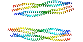
Glial fibrillary acidic protein (GFAP) is a protein that is encoded by the GFAP gene in humans. It is a type III intermediate filament (IF) protein that is expressed by numerous cell types of the central nervous system (CNS), including astrocytes and ependymal cells during development. GFAP has also been found to be expressed in glomeruli and peritubular fibroblasts taken from rat kidneys, Leydig cells of the testis in both hamsters and humans, human keratinocytes, human osteocytes and chondrocytes and stellate cells of the pancreas and liver in rats.

<span class="mw-page-title-main">Microglia</span> Glial cell located throughout the brain and spinal cord

Microglia are a type of neuroglia located throughout the brain and spinal cord. Microglia account for about 10-15% of cells found within the brain. As the resident macrophage cells, they act as the first and main form of active immune defense in the central nervous system (CNS). Microglia originate in the yolk sac under a tightly regulated molecular process. These cells are distributed in large non-overlapping regions throughout the CNS. Microglia are key cells in overall brain maintenance—they are constantly scavenging the CNS for plaques, damaged or unnecessary neurons and synapses, and infectious agents. Since these processes must be efficient to prevent potentially fatal damage, microglia are extremely sensitive to even small pathological changes in the CNS. This sensitivity is achieved in part by the presence of unique potassium channels that respond to even small changes in extracellular potassium. Recent evidence shows that microglia are also key players in the sustainment of normal brain functions under healthy conditions. Microglia also constantly monitor neuronal functions through direct somatic contacts and exert neuroprotective effects when needed.

Oligodendrocyte progenitor cells (OPCs), also known as oligodendrocyte precursor cells, NG2-glia, O2A cells, or polydendrocytes, are a subtype of glia in the central nervous system named for their essential role as precursors to oligodendrocytes. They are typically identified in the human by co-expression of PDGFRA and CSPG4.

<span class="mw-page-title-main">Rostral migratory stream</span> One path neural stem cells take to reach the olfactory bulb


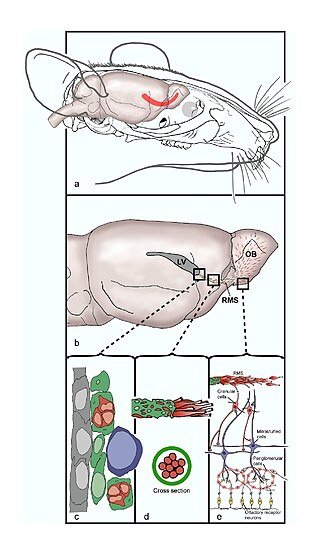
The rostral migratory stream (RMS) is a specialized migratory route found in the brain of some animals along which neuronal precursors that originated in the subventricular zone (SVZ) of the brain migrate to reach the main olfactory bulb (OB). The importance of the RMS lies in its ability to refine and even change an animal's sensitivity to smells, which explains its importance and larger size in the rodent brain as compared to the human brain, as our olfactory sense is not as developed. This pathway has been studied in the rodent, rabbit, and both the squirrel monkey and rhesus monkey. When the neurons reach the OB they differentiate into GABAergic interneurons as they are integrated into either the granule cell layer or periglomerular layer.

Gliosis is a nonspecific reactive change of glial cells in response to damage to the central nervous system (CNS). In most cases, gliosis involves the proliferation or hypertrophy of several different types of glial cells, including astrocytes, microglia, and oligodendrocytes. In its most extreme form, the proliferation associated with gliosis leads to the formation of a glial scar.

<span class="mw-page-title-main">Floor plate</span> Embryonic structure

The floor plate is a structure integral to the developing nervous system of vertebrate organisms. Located on the ventral midline of the embryonic neural tube, the floor plate is a specialized glial structure that spans the anteroposterior axis from the midbrain to the tail regions. It has been shown that the floor plate is conserved among vertebrates, such as zebrafish and mice, with homologous structures in invertebrates such as the fruit fly Drosophila and the nematode C. elegans. Functionally, the structure serves as an organizer to ventralize tissues in the embryo as well as to guide neuronal positioning and differentiation along the dorsoventral axis of the neural tube.

<span class="mw-page-title-main">Radial glial cell</span> Bipolar-shaped progenitor cells of all neurons in the cerebral cortex and some glia

Radial glial cells, or radial glial progenitor cells (RGPs), are bipolar-shaped progenitor cells that are responsible for producing all of the neurons in the cerebral cortex. RGPs also produce certain lineages of glia, including astrocytes and oligodendrocytes. Their cell bodies (somata) reside in the embryonic ventricular zone, which lies next to the developing ventricular system.

<span class="mw-page-title-main">Glia limitans</span> Thin astrocyte membrane surrounding the brain and spinal cord

The glia limitans, or the glial limiting membrane, is a thin barrier of astrocyte foot processes associated with the parenchymal basal lamina surrounding the brain and spinal cord. It is the outermost layer of neural tissue, and among its responsibilities is the prevention of the over-migration of neurons and neuroglia, the supporting cells of the nervous system, into the meninges. The glia limitans also plays an important role in regulating the movement of small molecules and cells into the brain tissue by working in concert with other components of the central nervous system (CNS) such as the blood–brain barrier (BBB).

<span class="mw-page-title-main">Subventricular zone</span> Region outside each lateral ventricle of the brain

The subventricular zone (SVZ) is a region situated on the outside wall of each lateral ventricle of the vertebrate brain. It is present in both the embryonic and adult brain. In embryonic life, the SVZ refers to a secondary proliferative zone containing neural progenitor cells, which divide to produce neurons in the process of neurogenesis. The primary neural stem cells of the brain and spinal cord, termed radial glial cells, instead reside in the ventricular zone (VZ).

Neuroregeneration involves the regrowth or repair of nervous tissues, cells or cell products. Neuroregenerative mechanisms may include generation of new neurons, glia, axons, myelin, or synapses. Neuroregeneration differs between the peripheral nervous system (PNS) and the central nervous system (CNS) by the functional mechanisms involved, especially in the extent and speed of repair. When an axon is damaged, the distal segment undergoes Wallerian degeneration, losing its myelin sheath. The proximal segment can either die by apoptosis or undergo the chromatolytic reaction, which is an attempt at repair. In the CNS, synaptic stripping occurs as glial foot processes invade the dead synapse.

<span class="mw-page-title-main">Perineuronal net</span> Structures of the brain

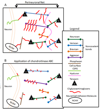
Perineuronal nets (PNNs) are specialized extracellular matrix structures responsible for synaptic stabilization in the adult brain. PNNs are found around certain neuron cell bodies and proximal neurites in the central nervous system. PNNs play a critical role in the closure of the childhood critical period, and their digestion can cause restored critical period-like synaptic plasticity in the adult brain. They are largely negatively charged and composed of chondroitin sulfate proteoglycans, molecules that play a key role in development and plasticity during postnatal development and in the adult.

<span class="mw-page-title-main">Chondroitin sulfate proteoglycan</span>

Chondroitin sulfate proteoglycans (CSPGs) are proteoglycans consisting of a protein core and a chondroitin sulfate side chain. They are known to be structural components of a variety of human tissues, including cartilage, and also play key roles in neural development and glial scar formation. They are known to be involved in certain cell processes, such as cell adhesion, cell growth, receptor binding, cell migration, and interaction with other extracellular matrix constituents. They are also known to interact with laminin, fibronectin, tenascin, and collagen. CSPGs are generally secreted from cells.

<span class="mw-page-title-main">Olfactory ensheathing cell</span> Type of macroglia that ensheath unmyelinated olfactory neurons

Olfactory ensheathing cells (OECs), also known as olfactory ensheathing glia or olfactory ensheathing glial cells, are a type of macroglia found in the nervous system. They are also known as olfactory Schwann cells, because they ensheath the non-myelinated axons of olfactory neurons in a similar way to which Schwann cells ensheath non-myelinated peripheral neurons. They also share the property of assisting axonal regeneration.

Endogenous regeneration in the brain is the ability of cells to engage in the repair and regeneration process. While the brain has a limited capacity for regeneration, endogenous neural stem cells, as well as numerous pro-regenerative molecules, can participate in replacing and repairing damaged or diseased neurons and glial cells. Another benefit that can be achieved by using endogenous regeneration could be avoiding an immune response from the host.

Neuroinflammation is inflammation of the nervous tissue. It may be initiated in response to a variety of cues, including infection, traumatic brain injury, toxic metabolites, or autoimmunity. In the central nervous system (CNS), including the brain and spinal cord, microglia are the resident innate immune cells that are activated in response to these cues. The CNS is typically an immunologically privileged site because peripheral immune cells are generally blocked by the blood–brain barrier (BBB), a specialized structure composed of astrocytes and endothelial cells. However, circulating peripheral immune cells may surpass a compromised BBB and encounter neurons and glial cells expressing major histocompatibility complex molecules, perpetuating the immune response. Although the response is initiated to protect the central nervous system from the infectious agent, the effect may be toxic and widespread inflammation as well as further migration of leukocytes through the blood–brain barrier may occur.

<span class="mw-page-title-main">Neuronal lineage marker</span> Endogenous tag expressed in different cells along neurogenesis and differentiated cells

A neuronal lineage marker is an endogenous tag that is expressed in different cells along neurogenesis and differentiated cells such as neurons. It allows detection and identification of cells by using different techniques. A neuronal lineage marker can be either DNA, mRNA or RNA expressed in a cell of interest. It can also be a protein tag, as a partial protein, a protein or an epitope that discriminates between different cell types or different states of a common cell. An ideal marker is specific to a given cell type in normal conditions and/or during injury. Cell markers are very valuable tools for examining the function of cells in normal conditions as well as during disease. The discovery of various proteins specific to certain cells led to the production of cell-type-specific antibodies that have been used to identify cells.

References

  1. Faulkner JR, Herrmann JE, Woo MJ, Tansey KE, Doan NB, Sofroniew MV (March 2004). "Reactive astrocytes protect tissue and preserve function after spinal cord injury". J. Neurosci. 24 (9): 2143–55. doi: 10.1523/JNEUROSCI.3547-03.2004 . PMC   6730429 . PMID   14999065.
  2. 1 2 3 4 5 Stichel CC, Müller HW (October 1998). "The CNS lesion scar: new vistas on an old regeneration barrier". Cell Tissue Res. 294 (1): 1–9. doi:10.1007/s004410051151. PMID   9724451. S2CID   13652357.
  3. Jones LL, Margolis RU, Tuszynski MH (August 2003). "The chondroitin sulfate proteoglycans neurocan, brevican, phosphacan, and versican are differentially regulated following spinal cord injury". Exp. Neurol. 182 (2): 399–411. doi:10.1016/S0014-4886(03)00087-6. PMID   12895450. S2CID   16748373.
  4. 14561854
  5. Davies SJ, Fitch MT, Memberg SP, Hall AK, Raisman G, Silver J (1997). "Regeneration of adult axons in white matter tracts of the central nervous system". Nature . 390 (6661): 680–3. Bibcode:1997Natur.390..680D. doi:10.1038/37776. PMID   9414159. S2CID   205026020.
  6. Silver, Jerry (2004). "Regeneration beyond the glial scar". Nature Reviews Neuroscience. 5 (2): 146–156. doi: 10.1038/nrn1326 . PMID   14735117.
  7. David S, Ness R. (1993). "Heterogeneity of reactive astrocytes." In: Fedoroff S (ed) Biology and pathology of astrocyte-neuron interactions. Plenum Press, New York, pp. 303-312.
  8. Fernaud-Espinosa I, Nieto-Sampedro N, Bovolenta P. (1993). "Differential activation of microglia and astrocytes in aniso- and isomorphic gliotic tissue." Glia 8: 277-291.
  9. Elkabes S, DiCicco-Bloom EM, Black IB (1996). "Brain microglia/ macrophages express neurotrophins that selectively regulate microglial proliferation and function", Journal of Neuroscience 16: 2508–2521
  10. Jaeger CB, Blight AR (1997). "Spinal compression injury in guinea pigs: structural changes of endothelium and its perivascular cell associations after blood–brain barrier breakdown and repair." Experimental Neurology 144: 381-399.
  11. Asher RA, et al. (2000). "Neurocan is upregulated in injured brain and in cytokine-treated astrocytes." Journal of Neurosciemce 20, 2427–2438.
  12. Moon LDF, Fawcett JW. (2001). "Reduction in CNS scar formation without concomitant increase in axon regeneration following treatment of adult rat brain with a combination of antibodies to TGFβ1 and β2." European Journal of Neuroscience 14, 1667–1677.
  13. Giulian D, et al. (1988). "Interleukin-1 injected into mammalian brain stimulates astrogliosis and neovascularization." Journal of Neuroscience 8, 2485–2490.
  14. Silver J, Miller J. (2004). "Regeneration beyond the glial scar." Nature Reviews Neuroscience. 5(2): 146-156.
  15. Yong VW et al. (1991). "γ-Interferon promotes proliferation of adult human astrocytes in vitro and reactive gliosis in the adult mouse brain in vivo." PNAS USA 88, 7016–7020.
  16. Lander C, et al. (1997). "A family of activity-dependent neuronal cell-surface chondroitin sulfate proteoglycans in cat visual cortex." Journal of Neuroscience 17, 1928–1939.
  17. Mocchetti I, et al. (1996). "Increased basic fibroblast growth factor expression following contusive spinal cord injury." Experimental Neurology 141, 154–164.
  18. Winger, CG, et al. (1995). "A role for ciliary neurotrophic factor as an inducer of reactive gliosis, the glial response to central nervous system injury", Proc. Natl. Acad. Sci, USA, 92, 5865 - 5869.
  19. Frisen, J. (1995). "Rapid, widespread, and long lasting induction of nestin contributes to the generation of glial scar tissue after CNS injury", The Journal of Cell Biology 131(2): 453-464.
  20. Tian D, et al. (2006). "Suppression of Astroglial Scar Formation and Enhanced Axonal Regeneration Associated with Functional Recovery in a Spinal Cord Injury Rat Model by the Cell Cycle Inhibitor Olomoucine", Journal of Neuroscience Research 84: 1053-1063.
  21. Tian D., et al. (2007). "Cell cycle inhibition attenuates microglia induced inflammatory response and alleviates neuronal cell death after spinal cord injury in rats." Brain Research 1135: 177-185.
  22. Neumann, S., et al. (2002). "Regeneration of Sensory Axons within the Injured Spinal Cord Induced by Intraganglionic cAMP Elevation." Neuron 34, 885–893.
  23. Nikulina, E. et al. (2004). "The phosphodiesterase inhibitor rolipram delivered after a spinal cord lesion promotes axonal regeneration and functional recovery", Proc Natl Acad Sci USA 101(23): 8786–8790.
  24. Pekovic, S., et al. (2006). "Downregulation of glial scarring after brain injury", Annals of the New York Academy of Sciences 1048(1): 296-310.
  25. Huang QL, Cai WQ, Zhang KC. (2000). "Effect of the control proliferation of astrocyte on the formation of glial scars by antisense GFAP retrovirus", Chinese Science Bulletin 45(1): 38-44.
  26. Logan A, et al. (1999). "Inhibition of glial scarring in the injured rat brain by a recombinant human monoclonal antibody to transforming growth factor-β2", European Journal of Neuroscience 11: 2367-2374.
  27. Okada S, et al. (2004). "Blockade of Interleukin-6 Receptor Suppresses Reactive Astrogliosis and Ameliorates Functional Recovery in Experimental Spinal Cord Injury", Journal of Neuroscience Research 76: 265-276.
  28. Bradbury, Elizabeth J. (2002). "Chondroitinase ABC promotes functional recovery after spinal cord injury". Nature. 416 (6881): 636–640. Bibcode:2002Natur.416..636B. doi:10.1038/416636a. PMID   11948352. S2CID   4430737.
  29. "Re-engineered enzyme could help reverse damage from spinal cord injury and stroke". August 24, 2020. Archived from the original on August 25, 2020. Retrieved August 26, 2020.
  30. Bradbury, Elizabeth J. (2011). "Manipulating the glial scar: chondroitinase ABC as a therapy for spinal cord injury". Brain Research Bulletin. 84 (4–5): 306–316. doi:10.1016/j.brainresbull.2010.06.015. PMID   20620201. S2CID   10605553.
  31. Fouad, Karim; Lisa Schnell; Mary B. Bunge; Martin E. Schwab; Thomas Liebscher; Damien D. Pearse (2 February 2005). "Combining Schwann Cell Bridges and Olfactory-Ensheathing Glia Grafts with Chondroitinase Promotes Locomotor Recovery after Complete Transection of the Spinal Cord". The Journal of Neuroscience. 25 (5): 1169–1178. doi: 10.1523/JNEUROSCI.3562-04.2005 . PMC   6725952 . PMID   15689553.
  32. Alilain, Warren J. (2011). "Functional regeneration of respiratory pathways after spinal cord injury". Nature. 475 (7355): 196–200. doi:10.1038/nature10199. PMC   3163458 . PMID   21753849.