Hepatic portal system

Last updated
Hepatic portal system
Gray591.png
The hepatic portal vein and its territory
Details
Location Abdomen
Anatomical terminology

In human anatomy, the hepatic portal system or portal venous system is the system of veins comprising the portal vein and its tributaries. The other portal venous systems in the body are the renal portal system, and the hypophyseal portal system. [1]

Contents

Structure

Large veins that are considered part of the portal venous system are the:

The superior mesenteric vein and the splenic vein come together to form the actual hepatic portal vein. The inferior mesenteric vein connects in the majority of people on the splenic vein, but in some people, it is known to connect on the portal vein or the superior mesenteric vein.

Roughly, the portal venous system corresponds to areas supplied by the celiac trunk, the superior mesenteric artery, and the inferior mesenteric artery.

Function

Microscopic section through a small portal vein branch inside the liver (x250 magnified) Gray1093.png
Microscopic section through a small portal vein branch inside the liver (×250 magnified)

The portal venous system is responsible for directing blood from parts of the gastrointestinal tract to the liver. Substances absorbed in the small intestine travel first to the liver for processing before continuing to the heart. Not all of the gastrointestinal tract is part of this system. The system extends from about the lower portion of the esophagus to the upper part of the anal canal. It also includes venous drainage from the spleen, pancreas and visceral fat. [2] [3]

The benefit of first-pass metabolism, whereby substances absorbed from food in the gut pass through the liver before entering the systemic circulation, is to use the liver as a shield (a first line of defense) between (a) the food, its toxins (whatever they may be), and its metabolic intermediates/metabolites (such as ammonia) and (b) the rest of the body's tissues, including the brain. The necessity of such a system is demonstrated by what happens when the system breaks down, as seen when advanced hepatic fibrosis in cirrhosis leads to hepatic encephalopathy in the brain owing to the blood being loaded with ammonia and other substances not conducive to brain function.

Blood flow to the liver is unique in that it receives both oxygenated and (partially) deoxygenated blood. As a result, the partial gas pressure of oxygen (pO2) and perfusion pressure of portal blood are lower than in other organs of the body. Blood passes from branches of the portal vein through cavities between "plates" of hepatocytes called sinusoids. Blood also flows from branches of the hepatic artery and mixes in the sinusoids to supply the hepatocytes with oxygen. This mixture percolates through the sinusoids and collects in a central vein which drains into the hepatic vein. The hepatic vein subsequently drains into the inferior vena cava.

The hepatic artery provides 30 to 40% of the oxygen to the liver, while only accounting for 25% of the total liver blood flow. The rest comes from the partially deoxygenated blood from the portal vein. The liver consumes about 20% of the total body oxygen when at rest. That is why the total liver blood flow is quite high, at about 1 litre a minute and up to two litres a minute. That is on average one fourth of the average cardiac output at rest.

Clinical significance

Portal hypertension is a condition in which the blood pressure of the portal venous system is too high. It is often the result of cirrhosis of the liver. Liver cirrhosis can lead to increased intrahepatic vascular resistance and vasodilation of portal system arteries, both of which increase pressure in the portal vein [4] . Color Doppler Ultrasound is the most useful imaging tool used to identify aneurysms, thrombosis, and branching patterns of the portal venous system, and to determine if treatment or surgery will be necessary [5] . In preparation for surgery, Color Doppler Ultrasound is used to identify portal vein branching patterns to ensure preserved portal perfusion and choose appropriate vessel connections for a liver transplant [6] .


Drug metabolism

Many drugs that are absorbed through the GI tract are substantially metabolized by the liver before reaching general circulation. This is known as the first pass effect. As a consequence, certain drugs can only be taken via certain routes. For example, nitroglycerin cannot be swallowed because the liver would deactivate the medication, but it can be taken under the tongue or transdermally (through the skin) and thus is absorbed in a way that bypasses the portal venous system. Inversely, dextromethorphan, a cough suppressor, is best taken orally because it needs to be metabolised by the liver into dextrorphan in order to be effective. This latter principle is that of most prodrugs.

The use of suppositories is a way to partially bypass the portal vein: the upper 1/3 of the rectum is drained into the portal vein while the lower 2/3 are drained into the internal iliac vein that goes directly in the inferior vena cava (thus bypassing the liver).

See also

Related Research Articles

<span class="mw-page-title-main">Vein</span> Blood vessels that carry blood towards the heart

Veins are blood vessels in the circulatory system of humans and most other animals that carry blood toward the heart. Most veins carry deoxygenated blood from the tissues back to the heart; exceptions are those of the pulmonary and fetal circulations which carry oxygenated blood to the heart. In the systemic circulation arteries carry oxygenated blood away from the heart, and veins return deoxygenated blood to the heart, in the deep veins.

<span class="mw-page-title-main">Circulatory system</span> Organ system for circulating blood in animals

The blood circulatory system is a system of organs that includes the heart, blood vessels, and blood which is circulated throughout the entire body of a human or other vertebrate. It includes the cardiovascular system, or vascular system, that consists of the heart and blood vessels. The circulatory system has two divisions, a systemic circulation or circuit, and a pulmonary circulation or circuit. Some sources use the terms cardiovascular system and vascular system interchangeably with the circulatory system.

<span class="mw-page-title-main">Portal vein</span> Short thick vein formed by the union of the superior mesenteric vein and the splenic vein

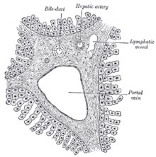
The portal vein or hepatic portal vein (HPV) is a blood vessel that carries blood from the gastrointestinal tract, gallbladder, pancreas and spleen to the liver. This blood contains nutrients and toxins extracted from digested contents. Approximately 75% of total liver blood flow is through the portal vein, with the remainder coming from the hepatic artery proper. The blood leaves the liver to the heart in the hepatic veins.

<span class="mw-page-title-main">Inferior vena cava</span> One of two veinous trunks bringing deoxygenated blood back to the heart

The inferior vena cava is a large vein that carries the deoxygenated blood from the lower and middle body into the right atrium of the heart. It is formed by the joining of the right and the left common iliac veins, usually at the level of the fifth lumbar vertebra.

<span class="mw-page-title-main">Esophageal varices</span> Medical condition

Esophageal varices are extremely dilated sub-mucosal veins in the lower third of the esophagus. They are most often a consequence of portal hypertension, commonly due to cirrhosis. People with esophageal varices have a strong tendency to develop severe bleeding which left untreated can be fatal. Esophageal varices are typically diagnosed through an esophagogastroduodenoscopy.

<span class="mw-page-title-main">Portal hypertension</span> Abnormally increased portal venous pressure

Portal hypertension is increased portal venous pressure – blood pressure in the portal vein and its branches, that drain from most of the intestine to the liver. Portal hypertension is defined as a hepatic venous pressure gradient greater than 5 mmHg. Cirrhosis is the most common cause of portal hypertension; other, less frequent causes are therefore grouped as non-cirrhotic portal hypertension. When it becomes severe enough to cause symptoms or complications, treatment may be given to decrease portal hypertension itself or to manage its complications.

<span class="mw-page-title-main">Abdominal aorta</span> Largest artery in the abdomen

In human anatomy, the abdominal aorta is the largest artery in the abdominal cavity. As part of the aorta, it is a direct continuation of the descending aorta.

<span class="mw-page-title-main">Celiac artery</span> First major branch of the abdominal aorta

The celiacartery, also known as the celiac trunk or truncus coeliacus, is the first major branch of the abdominal aorta. It is about 1.25 cm in length. Branching from the aorta at thoracic vertebra 12 (T12) in humans, it is one of three anterior/ midline branches of the abdominal aorta.

<span class="mw-page-title-main">Superior mesenteric vein</span> Vein which drains blood from the small intestine

In human anatomy, the superior mesenteric vein (SMV) is a blood vessel that drains blood from the small intestine. Behind the neck of the pancreas, the superior mesenteric vein combines with the splenic vein to form the portal vein that carries blood to the liver. The superior mesenteric vein lies to the right of the similarly named artery, the superior mesenteric artery, which originates from the abdominal aorta.

<span class="mw-page-title-main">Splenic vein</span> Vein that drains blood from the spleen, stomach and pancreas

In human anatomy, the splenic vein is a blood vessel that drains blood from the spleen, the stomach fundus and part of the pancreas. It is part of the hepatic portal system.

<span class="mw-page-title-main">Transjugular intrahepatic portosystemic shunt</span> Artificial channel within the liver

Transjugular intrahepatic portosystemic shunt is an artificial channel within the liver that establishes communication between the inflow portal vein and the outflow hepatic vein. It is used to treat portal hypertension which frequently leads to intestinal bleeding, life-threatening esophageal bleeding and the buildup of fluid within the abdomen (ascites).

<span class="mw-page-title-main">Portal vein thrombosis</span> Disease of the liver

Portal vein thrombosis (PVT) is a vascular disease of the liver that occurs when a blood clot occurs in the hepatic portal vein, which can lead to increased pressure in the portal vein system and reduced blood supply to the liver. The mortality rate is approximately 1 in 10.

<span class="mw-page-title-main">Colic flexures</span> Two abrupt bends in the large intestine

In the anatomy of the human digestive tract, there are two colic flexures, or curvatures in the transverse colon. The right colic flexure is also known as the hepatic flexure, and the left colic flexure is also known as the splenic flexure. Note that "right" refers to the patient's anatomical right, which may be depicted on the left of a diagram.

<span class="mw-page-title-main">Vitelline veins</span>

The vitelline veins are veins that drain blood from the yolk sac and the gut tube during gestation.

<span class="mw-page-title-main">Rectal venous plexus</span>

The rectal venous plexus is the venous plexus surrounding the rectum. It consists of an internal and an external rectal plexus. It is drained by the superior, middle, and inferior rectal veins. It forms a portosystemic (portocaval) anastomosis. This allows rectally administered medications to bypassing first pass metabolism.

<span class="mw-page-title-main">Central veins of liver</span> Veins through the centers of hepatic (liver) lobules

In microanatomy, the central veins of liver are veins found at the center of hepatic lobules.

<span class="mw-page-title-main">Portal venous system</span> The capillary bed pools into another through veins without first going through the heart

In the circulatory system of vertebrates, a portal venous system occurs when a capillary bed pools into another capillary bed through veins, without first going through the heart. Both capillary beds and the blood vessels that connect them are considered part of the portal venous system.

<span class="mw-page-title-main">Liver</span> Vertebrate organ involved in metabolism

The liver is a major metabolic organ only found in vertebrate animals, which performs many essential biological functions such as detoxification of the organism, and the synthesis of proteins and biochemicals necessary for digestion and growth. In humans, it is located in the right upper quadrant of the abdomen, below the diaphragm and mostly shielded by the lower right rib cage. Its other metabolic roles include carbohydrate metabolism, the production of hormones, conversion and storage of nutrients such as glucose and glycogen, and the decomposition of red blood cells.

The superior mesenteric vessels are composed of the superior mesenteric artery and the superior mesenteric vein.

<span class="mw-page-title-main">Liver segment</span> An anatomical unit of the liver

A liver segment is one of eight segments of the liver as described in the widely used Couinaud classification in the anatomy of the liver. This system divides the lobes of the liver into eight segments based on a transverse plane through the bifurcation of the main portal vein, arranged in a clockwise manner starting from the caudate lobe.

References

  1. Saladin, Kenneth S. (2011). Human anatomy (3rd ed.). New York: McGraw-Hill. p. 571. ISBN   9780071222075.
  2. Item F, Konrad D (2012). "Visceral fat and metabolic inflammation: the portal theory revisited" (PDF). Obesity Reviews . 13 (Suppl 2): 30–39. doi:10.1111/j.1467-789X.2012.01035.x. PMID   23107257. S2CID   25169877.
  3. Dhawan D, Sharma S (2020). "Abdominal Obesity, Adipokines and Non-communicable Diseases". The Journal of Steroid Biochemistry and Molecular Biology . 203: 105737. doi:10.1016/j.jsbmb.2020.105737. PMC   7431389 . PMID   32818561.
  4. Iwakiri, Y. (2014). Pathophysiology of Portal Hypertension. Clinics in Liver Disease, 18(2), 281-291. https://doi.org/10.1016/j.cld.2013.12.001
  5. Gallego, C., Velasco, M., Marcuello, P., Tejedor, D., De Campo, L., & Friera, A. (2002). Congenital and acquired anomalies of the portal venous system. Radiographics, 22(1), 141-159. https://doi.org/10.1148/radiographics.22.1.g02a08141
  6. Carneiro, C., Brito, J., Bilreiro, C., Barros, M., Bahia, C., Santiago, I., & Caseiro-Alves, F. (2019). All about portal vein: a pictorial display to anatomy, variants and physiopathology. Insights into Imaging, 10(1), 38-18. https://doi.org/10.1186/s13244- 019-0716-8