Inguinal canal

Last updated
Inguinal canal
Gray1227.png
Front of abdomen, showing surface markings for arteries and inguinal canal. (Inguinal canal is tube at lower left.)
Gray1143.png
The scrotum. On the left side, the cavity of the tunica vaginalis has been opened; on the right side, only the layers superficial to the cremaster have been removed. (Right inguinal canal visible at upper left.)
Details
Identifiers
Latin canalis inguinalis
MeSH D007264
TA98 A04.5.01.026
TA2 2381
FMA 19928
Anatomical terminology

The inguinal canal is a passage in the anterior abdominal wall on each side of the body (one on each side of the midline), which in males, convey the spermatic cords and in females, the round ligament of the uterus. The inguinal canals are larger and more prominent in males.

Contents

Structure

The inguinal canals are situated just above the medial half of the inguinal ligament. The canals are approximately 4 to 6 cm long, [1] angled anteroinferiorly and medially. In males, its diameter is normally 2 cm (±1 cm in standard deviation) at the deep inguinal ring. [2] [notes 1]

A first-order approximation is to visualize each canal as a cylinder. [3]

Walls

To help define the boundaries, these canals are often further approximated as boxes with six sides. Not including the two rings, the remaining four sides are usually called the "anterior wall", "inferior wall ("floor")", "superior wall ("roof")", and "posterior wall". [4] These consist of the following:

superior wall (roof):
Medial crus of aponeurosis of external oblique
Musculoaponeurotic arches of internal oblique and transverse abdominal
Transversalis fascia
conjoint tendon
anterior wall:
aponeurosis of external oblique
fleshy part of internal oblique (lateral third of canal only) [5]
superficial inguinal ring (medial third of canal only) [6]
(inguinal canal)posterior wall:
transversalis fascia
conjoint tendon (Inguinal falx, reflected part of inguinal ligament, medial third of canal only) [6]
deep inguinal ring (lateral third of canal only) [6]
inferior wall (floor):
inguinal ligament
lacunar ligament (medial third of canal only) [6]
iliopubic tract (lateral third of canal only) [5]

Deep inguinal ring

The deep inguinal ring (internal or deep abdominal ring, abdominal inguinal ring, internal inguinal ring, annulus abdominalis) is the entrance to the inguinal canal.

Location

The surface marking of the deep inguinal ring is classically described as half an inch above the midpoint of the inguinal ligament. [7]

However, the surface anatomy of the point is disputed. In a recent study, [8] it was found to be in a region between the mid-inguinal point (situated midway between the anterior superior iliac spine and the pubic symphysis) and the midpoint of the inguinal ligament (i.e. midway between the anterior superior iliac spine and the pubic tubercle). Traditionally, either one of these two sites was claimed as its location. However, this claim is based upon the study's dissection of 52 cadavers, and may not reflect the live in vivo anatomy.

Some sources state that it is at the layer of the transversalis fascia. [9]

Description

The deep inguinal ring is an opening in the transversalis fascia. [10] It is of an oval form, the long axis of the oval being vertical; it varies in size in different subjects, and is much larger in the male than in the female. It is bounded, above and laterally, by the arched lower margin of the transversalis fascia; below and medially, by the inferior epigastric vessels. It transmits the spermatic cord in the male and the round ligament of the uterus in the female.

From its circumference, a thin funnel-shaped membrane, the infundibuliform fascia, is continued around the cord and testis, enclosing them in a distinct covering.

Superficial inguinal ring

The superficial inguinal ring Gray393.png
The superficial inguinal ring

The superficial inguinal ring (subcutaneous inguinal ring or external inguinal ring) is an anatomical structure in the anterior wall of the mammalian abdomen. It is a triangular opening that forms the exit of the inguinal canal, which houses the ilioinguinal nerve, the genital branch of the genitofemoral nerve, and the spermatic cord (in men) or the round ligament (in women). At the other end of the canal, the deep inguinal ring forms the entrance. [11]

It is found within the aponeurosis of the external oblique, immediately above the pubic crest, 1 centimeter above and superolateral to the pubic tubercle. It has the following boundaries—medial crura by pubic crest, lateral crura by pubic tubercle and inferiorly by inguinal ligament. [9]

Contents

The structures which pass through the canals differ between males and females:

The classic description of the contents of the spermatic cords in the male are:

3 arteries: artery to vas deferens (or ductus deferens), testicular artery, cremasteric artery;

3 fascial layers: external spermatic, cremasteric, and internal spermatic fascia;

3 other structures: pampiniform plexus, vas deferens (ductus deferens), testicular lymphatics;

3 nerves: genital branch of the genitofemoral nerve (L1/2), sympathetic and visceral afferent fibres, ilioinguinal nerve (N.B. outside spermatic cord but travels next to it)

Note that the ilioinguinal nerve passes through the superficial ring to descend into the scrotum, but does not formally run through the canal.

Development

In males

During development, each testicle descends from the starting point on the posterior abdominal wall (para-aortically) from the labioscrotal swellings near the kidneys, down the abdomen, and through the inguinal canals to reach the scrotum. This way, each testicle descends through the abdominal wall into the scrotum behind[ clarification needed ] the processus vaginalis (which later obliterates).

Clinical significance

Abdominal contents (potentially including intestine) can be abnormally displaced from the abdominal cavity. Where these contents exit through the inguinal canal, having passed through the deep inguinal ring, the condition is known as an indirect or oblique inguinal hernia. This can also cause infertility. This condition is far more common in males than in females, owing to the inguinal canal's small size in females.

A hernia that exits the abdominal cavity directly through the deep layers of the abdominal wall, thereby bypassing the inguinal canal, is known as a direct inguinal hernia.

In males with strong presentation of the cremasteric reflex, the testes can—during supine sexual activity or manual manipulation—partially or fully retract into the inguinal canal for a short period of time. In juveniles and adults with inguinal injury, retraction can be prolonged and potentially lead to overheating-related infertility. [13]

The superficial ring is palpable [14] under normal conditions. It becomes dilated in a condition called athletic pubalgia. Abdominal contents may protrude through the ring in inguinal hernia.

Thus lymphatic spread from a testicular tumour is to the para-aortic nodes first, and not the inguinal nodes.

Additional images

See also

Notes

  1. The diameter has been estimated to be ±2.2cm ±1.08cm in Africans, and 2.1 cm ±0.41cm in Europeans.

Related Research Articles

<span class="mw-page-title-main">Spermatic cord</span> Structure in the human male reproductive system

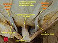
The spermatic cord is the cord-like structure in males formed by the vas deferens and surrounding tissue that runs from the deep inguinal ring down to each testicle. Its serosal covering, the tunica vaginalis, is an extension of the peritoneum that passes through the transversalis fascia. Each testicle develops in the lower thoracic and upper lumbar region and migrates into the scrotum. During its descent it carries along with it the vas deferens, its vessels, nerves etc. There is one on each side.

<span class="mw-page-title-main">Cremaster muscle</span> Muscle covering the testicles and spermatic cords

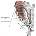
The cremaster muscle is a paired structure made of thin layers of striated and smooth muscle that covers the testicles and the spermatic cords in human males. It consists of the lateral and medial parts. Cremaster is an involuntary muscle, responsible for the cremasteric reflex; a protective and physiologic superficial reflex of the testicles. The reflex raises and lowers the testicles in order to keep them protected. Along with the dartos muscle of the scrotum, it regulates testicular temperature, thus aiding the process of spermatogenesis.

<span class="mw-page-title-main">Genitofemoral nerve</span>

The genitofemoral nerve is a mixed branch of the lumbar plexus derived from anterior rami of L1-L2. It splits a genital branch and a femoral branch. It provides sensory innervation to the upper anterior thigh, as well as the skin of the anterior scrotum in males and mons pubis in females. It also provides motor innervation to the cremaster muscle.

<span class="mw-page-title-main">Inguinal hernia</span> Medical condition in which contents of the abdominal cavity protrude through the inguinal canal

An inguinal hernia or groin hernia is a hernia (protrusion) of abdominal cavity contents through the inguinal canal. Symptoms, which may include pain or discomfort especially with or following coughing, exercise, or bowel movements, are absent in about a third of patients. Symptoms often get worse throughout the day and improve when lying down. A bulging area may occur that becomes larger when bearing down. Inguinal hernias occur more often on the right than left side. The main concern is strangulation, where the blood supply to part of the intestine is blocked. This usually produces severe pain and tenderness of the area.

<span class="mw-page-title-main">Inguinal ligament</span> Band running from the pubic tubercle to the anterior superior iliac spine

The inguinal ligament, also known as Poupart's ligament or groin ligament, is a band running from the pubic tubercle to the anterior superior iliac spine. It forms the base of the inguinal canal through which an indirect inguinal hernia may develop.

<span class="mw-page-title-main">External iliac artery</span> Arteries of the pelvis

The external iliac arteries are two major arteries which bifurcate off the common iliac arteries anterior to the sacroiliac joint of the pelvis.

<span class="mw-page-title-main">Inferior epigastric artery</span> Blood vessel

In human anatomy, the inferior epigastric artery is an artery that arises from the external iliac artery. It is accompanied by the inferior epigastric vein; inferiorly, these two inferior epigastric vessels together travel within the lateral umbilical fold The inferior epigastric artery then traverses the arcuate line of rectus sheath to enter the rectus sheath, then anastomoses with the superior epigastric artery within the rectus sheath.

<span class="mw-page-title-main">Lumbar plexus</span> Web of nerves in the lower spine

The lumbar plexus is a web of nerves in the lumbar region of the body which forms part of the larger lumbosacral plexus. It is formed by the divisions of the first four lumbar nerves (L1-L4) and from contributions of the subcostal nerve (T12), which is the last thoracic nerve. Additionally, the ventral rami of the fourth lumbar nerve pass communicating branches, the lumbosacral trunk, to the sacral plexus. The nerves of the lumbar plexus pass in front of the hip joint and mainly support the anterior part of the thigh.

<span class="mw-page-title-main">Ilioinguinal nerve</span> Branch of the first lumbar nerve

The ilioinguinal nerve is a branch of the first lumbar nerve (L1). It separates from the first lumbar nerve along with the larger iliohypogastric nerve. It emerges from the lateral border of the psoas major just inferior to the iliohypogastric, and passes obliquely across the quadratus lumborum and iliacus. The ilioinguinal nerve then perforates the transversus abdominis near the anterior part of the iliac crest, and communicates with the iliohypogastric nerve between the transversus and the internal oblique muscle.

<span class="mw-page-title-main">Conjoint tendon</span> Medial part of the posterior wall of the inguinal canal

The conjoint tendon is a sheath of connective tissue formed from the lower part of the common aponeurosis of the abdominal internal oblique muscle and the transversus abdominis muscle, joining the muscle to the pelvis. It forms the medial part of the posterior wall of the inguinal canal.

In human anatomy, the inguinal region refers to either the groin or the lower lateral regions of the abdomen. It may also refer to:

<span class="mw-page-title-main">Transversalis fascia</span> Aponeurosis between the transverse abdominal muscle and the extraperitoneal fat

The transversalis fascia is the fascial lining of the anterolateral abdominal wall situated between the inner surface of the transverse abdominal muscle, and the preperitoneal fascia. It is directly continuous with the iliac fascia, the internal spermatic fascia, and pelvic fascia.

<span class="mw-page-title-main">Fascia of Scarpa</span> Deep membranous layer of the superficial fascia of the abdomen

The fascia of Scarpa is the deep membranous layer (stratum membranosum) of the superficial fascia of the abdomen. It is a layer of the anterior abdominal wall. It is found deep to the fascia of Camper and superficial to the external oblique muscle.

<span class="mw-page-title-main">Fascia of Camper</span> Layer of the anterior abdominal wall

The fascia of Camper is a thick superficial layer of the anterior abdominal wall.

<span class="mw-page-title-main">Lateral umbilical fold</span>

The lateral umbilical fold is an elevation of the peritoneum lining the inner/posterior surface of the lower anterior abdominal wall formed by the underlying inferior epigastric artery and inferior epigastric vein which the peritoneum covers. Superiorly, the lateral umbilical fold ends where the vessels reach and enter the rectus sheath at the arcuate line of rectus sheath; in spite of the name, the lateral umbilical folds do not extend as far superiorly as the umbilicus. Inferiorly, it extends to just medial to the deep inguinal ring.

<span class="mw-page-title-main">Cremasteric artery</span>

The cremasteric artery is a branch of the inferior epigastric artery which accompanies the spermatic cord to supply the cremaster muscle as well as other coverings of the spermatic cord in the male.

<span class="mw-page-title-main">Medial umbilical ligament</span>

The medial umbilical ligament, cord of umbilical artery, or obliterated umbilical artery is a paired structure found in human anatomy. It is on the deep surface of the anterior abdominal wall, and is covered by the medial umbilical folds. It is different from the median umbilical ligament, a structure that represents the remnant of the embryonic urachus.

The cremasteric fascia is a fascia in the scrotum. As the cremaster descends, it forms a series of loops which differ in thickness and length in different subjects. At the upper part of the cord the loops are short, but they become in succession longer and longer, the longest reaching down as low as the testis, where a few are inserted into the tunica vaginalis. These loops are united together by areolar tissue, and form a thin covering over the cord and testis, the cremasteric fascia.

<span class="mw-page-title-main">Crura of superficial inguinal ring</span>

The superficial inguinal ring is bounded below by the crest of the pubis; on either side by the margins of the opening in the aponeurosis, which are called the crura of the ring; and above, by a series of curved intercrural fibers.

<span class="mw-page-title-main">Genital branch of genitofemoral nerve</span>

The genital branch of the genitofemoral nerve, also known as the external spermatic nerve in males, is a nerve in the abdomen that arises from the genitofemoral nerve. The genital branch supplies the cremaster muscle and anterior scrotal skin in males, and the skin of the mons pubis and labia majora in females.

References

  1. Tuma, Faiz; Lopez, Richard A.; Varacallo, Matthew (2023). "Anatomy, Abdomen and Pelvis: Inguinal Region (Inguinal Canal)". StatPearls. StatPearls Publishing. PMID   29261933 . Retrieved 18 June 2023.
  2. Mitura, Kryspin; Kozieł, Sławomir; Pasierbek, Michał (2018). "Ethnicity-related differences in inguinal canal dimensions between African and Caucasian populations and their potential impact on the mesh size for open and laparoscopic groin hernia repair in low-resource countries in Africa". Videosurgery and Other Miniinvasive Techniques. 13 (1): 74–81. doi:10.5114/wiitm.2018.72579. ISSN   1895-4588. PMC   5890843 . PMID   29643962.
  3. "Gross Anatomy Image". Archived from the original on 2007-11-11. Retrieved 2007-11-20.
  4. Adam Mitchell; Drake, Richard; Gray, Henry David; Wayne Vogl (2005). Gray's anatomy for students. Elsevier/Churchill Livingstone. p. 260. ISBN   0-443-06612-4.
  5. 1 2 Dalley, Arthur F.; Moore, Keith L. (2006). Clinically oriented anatomy . Hagerstown, MD: Lippincott Williams & Wilkins. pp.  217. ISBN   0-7817-3639-0.
  6. 1 2 3 4 Arthur F., II Dalley; Anne M. R. Agur (2005). Grant's Atlas of Anatomy. Hagerstown, MD: Lippincott Williams & Wilkins. p. 102. ISBN   0-7817-4255-2.
  7. Susan Standring (2004). Gray's Anatomy: The Anatomical Basis of Medicine and Surgery. Churchill-Livingstone. p.  1098. ISBN   0-443-07168-3.
  8. Koliyadan S, Narayan G, Balasekran P (2004). "Surface marking of the deep inguinal ring". Clin Anat. 17 (7): 554–7. doi:10.1002/ca.10257. PMID   15376291. S2CID   30726776.
  9. 1 2 Kyung Won, PhD. Chung (2005). Gross Anatomy (Board Review). Hagerstown, MD: Lippincott Williams & Wilkins. p. 198. ISBN   0-7817-5309-0.
  10. Sinnatamby, Chummy S. (2011). Last's Anatomy (12th ed.). Elsevier Australia. pp. 226–227. ISBN   978-0-7295-3752-0.
  11. James Harmon, M.D., Ph.D., Lecture 13. Human Gross Anatomy. University of Minnesota. September 4, 2008.
  12. "Anatomy Tables - Inguinal Region". Archived from the original on 2007-11-21. Retrieved 2007-11-20.
  13. Mayo Clinic Staff. "Retractile testicle". Mayo Clinic. Mayo Foundation for Medical Education and Research. Retrieved 10 February 2018.
  14. Moore & Agur, Essential Clinical Anatomy (2007)