Insulin-degrading enzyme

Last updated
IDE
Available structures
PDB Ortholog search: PDBe RCSB
Identifiers
Aliases IDE , INSULYSIN, insulin degrading enzyme
External IDs OMIM: 146680; MGI: 96412; HomoloGene: 3645; GeneCards: IDE; OMA:IDE - orthologs
Orthologs
SpeciesHumanMouse
Entrez
Ensembl
UniProt
RefSeq (mRNA)

NM_031156

RefSeq (protein)

NP_112419

Location (UCSC) Chr 10: 92.45 – 92.57 Mb n/a
PubMed search [2] [3]
Wikidata
View/Edit Human View/Edit Mouse

Insulin-degrading enzyme, also known as IDE, is an enzyme. [4]

Known alternatively as insulysin or insulin protease, IDE is a large zinc-binding protease of the M16 metalloprotease family known to cleave multiple short polypeptides that vary considerably in sequence. Other members of this family include the mitochondrial processing peptidase [5] and presequence protease. [6]

Structure

Gene

The gene IDE encodes protein Insulin-degrading enzyme. The human gene IDE has 28 exons and is located at chromosome band 10q23-q25. [4]

Protein

Due to alternative splicing, The human protein Insulin-degrading Enzyme has two isoforms. Isoform1 is ~118 kDa in size and composed of 1019 amino acids while the isoform 2 is ~54.2 kDa [7] size and composed of 464 amino acids (missing 1-555 amino acids). The calculated theoretical pI of this protein isoform is 6.26. [8] Structural studies of IDE by Shen et al. [9] have provided insight into the functional mechanisms of the protease. Reminiscent of the previously determined structure of the bacterial protease pitrilysin, the IDE crystal structure reveals defined N and C terminal units that form a proteolytic chamber containing the zinc-binding active site. In addition, it appears that IDE can exist in two conformations: an open conformation, in which substrates can access the active site, and a closed state, in which the active site is contained within the chamber formed by the two concave domains. Targeted mutations that favor the open conformation result in a 40-fold increase in catalytic activity. Based upon this observation, it has been proposed that a possible therapeutic approach to Alzheimer's might involve shifting the conformational preference of IDE to the open state, and thus increasing Aβ degradation, preventing aggregation, and, ideally, preventing the neuronal loss that leads to disease symptoms. [9]

Function

IDE was first identified by its ability to degrade the B chain of the hormone insulin. This activity was observed over sixty years ago, [10] though the enzyme specifically responsible for B chain cleavage was identified more recently. [11] This discovery revealed considerable amino acid sequence similarity between IDE and the previously characterized bacterial protease pitrilysin, suggesting a common proteolytic mechanism. IDE, which migrates at 110 kDa during gel electrophoresis under denaturing conditions, has since been shown to have additional substrates, including the signaling peptides glucagon, TGF alpha, and β-endorphin. [12]

Clinical Significance

Alzheimer's disease

Considerable interest in IDE has been stimulated due to the discovery that IDE can degrade amyloid beta (Aβ), a peptide implicated in the pathogenesis of Alzheimer's disease. [13] The underlying cause or causes of the disease are unclear, though the primary neuropathology observed is the formation of amyloid plaques and neurofibrillary tangles. One hypothesized mechanism of disease, called the amyloid hypothesis, suggests that the causative agent is the hydrophobic peptide Aβ, which forms quaternary structures that, by an unclear mechanism, cause neuronal death. Aβ is a byproduct generated as the result of proteolytic processing of the amyloid precursor protein (APP) by proteases referred to as the β and γ secretases. The physiological role of this processing is unclear, though it may play a role in nervous system development. [14]

Numerous in vitro and in vivo studies have shown correlations between IDE, Aβ degradation, and Alzheimer's disease. Mice engineered to lack both alleles of the IDE gene exhibit a 50% decrease in Aβ degradation, resulting in cerebral accumulation of Aβ. [15] Studies of genetically inherited forms of Alzheimer's show reduction in both IDE expression [16] and catalytic activity [17] among affected individuals. Despite the evident role of IDE in disease, relatively little is known about its physiological functions. These may be diverse, as IDE has been localized to several locations, including the cytosol, peroxisomes, endosomes, proteasome complexes, [18] and the surface of cerebrovascular endothelial cells. [19] Based upon the aforementioned observation in protein structure, it has been proposed that a possible therapeutic approach to Alzheimer's might involve shifting the conformational preference of IDE to the open state, and thus increasing Aβ degradation, preventing aggregation, and, ideally, preventing the neuronal loss that leads to disease symptoms.

Regulation of extracellular amyloid β-protein

Reports of IDE localized to the cytosol and peroxisomes [20] have raised concerns regarding how the protease could degrade endogenous Aβ. Several studies have detected insulin-degrading activity in the conditioned media of cultured cells, [21] [22] suggesting the permeability of the cell membrane and thus possible release of IDE from leaky cells. Qiu and colleagues revealed the presence of IDE in the extracellular media using antibodies to the enzyme. They also quantified levels of Aβ-degrading activity [23] using elution from column chromatography. Correlating the presence of IDE and Aβ-degrading activity in the conditioning medium confirmed that leaky membranes are responsible for extracellular IDE activity. However, other reports have indicated that it is released via exosomes. [24]

Potential role in the oligomerization of Aβ

Recent studies have observed that the oligomerization of synthetic Aβ was completely inhibited by the competitive IDE substrate, insulin. [23] These findings suggest that IDE activity is capable of joining of several Aβ fragments together. Qui et al. hypothesized that the Aβ fragments generated by IDE can either enhance oligomerization of the Aβ peptide or can oligomerize themselves. It is also entirely possible that IDE could mediate the degradation and oligomerization of Aβ by independent actions that have yet to be investigated.

Mechanism

The mechanism of the IDE enzyme remains poorly understood. The first step of one proposed mechanism [25] includes a zinc-bound hydroxide group performing a nucleophilic attack on a carbon substrate that materializes into the intermediate INT1. In this species, we can note that the zinc-bound hydroxide is completely transferred on the carbonyl carbon of substrate as a consequence of the Zn2+−OH bond breaking. In TS2, the Glu111 residue rotates to assume the right disposition to form two hydrogen bonds with the amide nitrogen and the −OH group linked to the carbon atom of substrate, thus behaving as hydrogen donor and acceptor, simultaneously. The formation of the second cited bond favors the re-establishment of the Zn2+−OH bond broken previously at the INT1 level. The nucleophilic addition and the protonation of peptide amide nitrogen is a very fast process that is believed to occur as a single step in the catalytic process. The final species on the path is the product PROD. [25] As a consequence of transfer of the proton of Glu111 onto the amide nitrogen of substrate that occurred in TS3, the peptide N—C bond is broken.

A look at the whole reaction path indicates that the rate-determining step in this process is the nucleophilic addition. After this point, the catalytic event should proceed without particular obstacles. [26] [27]

Related Research Articles

<span class="mw-page-title-main">Proteasome</span> Protein complexes which degrade unnecessary or damaged proteins by proteolysis

Proteasomes are protein complexes which degrade unneeded or damaged proteins by proteolysis, a chemical reaction that breaks peptide bonds. Enzymes that help such reactions are called proteases.

<span class="mw-page-title-main">Protease</span> Enzyme that cleaves other proteins into smaller peptides

A protease is an enzyme that catalyzes proteolysis, breaking down proteins into smaller polypeptides or single amino acids, and spurring the formation of new protein products. They do this by cleaving the peptide bonds within proteins by hydrolysis, a reaction where water breaks bonds. Proteases are involved in numerous biological pathways, including digestion of ingested proteins, protein catabolism, and cell signaling.

<span class="mw-page-title-main">Serine protease</span> Class of enzymes

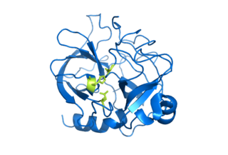
Serine proteases are enzymes that cleave peptide bonds in proteins. Serine serves as the nucleophilic amino acid at the (enzyme's) active site. They are found ubiquitously in both eukaryotes and prokaryotes. Serine proteases fall into two broad categories based on their structure: chymotrypsin-like (trypsin-like) or subtilisin-like.

<span class="mw-page-title-main">Angiotensin-converting enzyme</span> Mammalian protein found in humans

Angiotensin-converting enzyme, or ACE, is a central component of the renin–angiotensin system (RAS), which controls blood pressure by regulating the volume of fluids in the body. It converts the hormone angiotensin I to the active vasoconstrictor angiotensin II. Therefore, ACE indirectly increases blood pressure by causing blood vessels to constrict. ACE inhibitors are widely used as pharmaceutical drugs for treatment of cardiovascular diseases.

<span class="mw-page-title-main">Amylin</span> Peptide hormone that plays a role in glycemic regulation

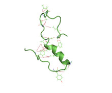
Amylin, or islet amyloid polypeptide (IAPP), is a 37-residue peptide hormone. It is co-secreted with insulin from the pancreatic β-cells in the ratio of approximately 100:1 (insulin:amylin). Amylin plays a role in glycemic regulation by slowing gastric emptying and promoting satiety, thereby preventing post-prandial spikes in blood glucose levels.

<span class="mw-page-title-main">Cathepsin</span> Family of proteases

Cathepsins are proteases found in all animals as well as other organisms. There are approximately a dozen members of this family, which are distinguished by their structure, catalytic mechanism, and which proteins they cleave. Most of the members become activated at the low pH found in lysosomes. Thus, the activity of this family lies almost entirely within those organelles. There are, however, exceptions such as cathepsin K, which works extracellularly after secretion by osteoclasts in bone resorption. Cathepsins have a vital role in mammalian cellular turnover.

<span class="mw-page-title-main">Amyloid beta</span> Group of peptides

Amyloid beta denotes peptides of 36–43 amino acids that are the main component of the amyloid plaques found in the brains of people with Alzheimer's disease. The peptides derive from the amyloid-beta precursor protein (APP), which is cleaved by beta secretase and gamma secretase to yield Aβ in a cholesterol-dependent process and substrate presentation. Aβ molecules can aggregate to form flexible soluble oligomers which may exist in several forms. It is now believed that certain misfolded oligomers can induce other Aβ molecules to also take the misfolded oligomeric form, leading to a chain reaction akin to a prion infection. The oligomers are toxic to nerve cells. The other protein implicated in Alzheimer's disease, tau protein, also forms such prion-like misfolded oligomers, and there is some evidence that misfolded Aβ can induce tau to misfold.

<span class="mw-page-title-main">Amyloid-beta precursor protein</span> Mammalian protein found in humans

Amyloid-beta precursor protein (APP) is an integral membrane protein expressed in many tissues and concentrated in the synapses of neurons. It functions as a cell surface receptor and has been implicated as a regulator of synapse formation, neural plasticity, antimicrobial activity, and iron export. It is coded for by the gene APP and regulated by substrate presentation. APP is best known as the precursor molecule whose proteolysis generates amyloid beta (Aβ), a polypeptide containing 37 to 49 amino acid residues, whose amyloid fibrillar form is the primary component of amyloid plaques found in the brains of Alzheimer's disease patients.

<span class="mw-page-title-main">Neurodegenerative disease</span> Central nervous system disease

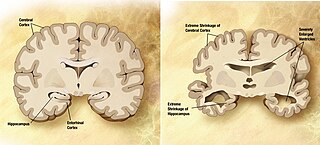
A neurodegenerative disease is caused by the progressive loss of structure or function of neurons, in the process known as neurodegeneration. Such neuronal damage may ultimately involve cell death. Neurodegenerative diseases include amyotrophic lateral sclerosis, multiple sclerosis, Parkinson's disease, Alzheimer's disease, Huntington's disease, multiple system atrophy, tauopathies, and prion diseases. Neurodegeneration can be found in the brain at many different levels of neuronal circuitry, ranging from molecular to systemic. Because there is no known way to reverse the progressive degeneration of neurons, these diseases are considered to be incurable; however research has shown that the two major contributing factors to neurodegeneration are oxidative stress and inflammation. Biomedical research has revealed many similarities between these diseases at the subcellular level, including atypical protein assemblies and induced cell death. These similarities suggest that therapeutic advances against one neurodegenerative disease might ameliorate other diseases as well.

<span class="mw-page-title-main">Beta-secretase 1</span> Enzyme

Beta-secretase 1, also known as beta-site amyloid precursor protein cleaving enzyme 1, beta-site APP cleaving enzyme 1 (BACE1), membrane-associated aspartic protease 2, memapsin-2, aspartyl protease 2, and ASP2, is an enzyme that in humans is encoded by the BACE1 gene. Expression of BACE1 is observed mainly in neurons.

The biochemistry of Alzheimer's disease, the most common cause of dementia, is not yet very well understood. Alzheimer's disease (AD) has been identified as a proteopathy: a protein misfolding disease due to the accumulation of abnormally folded amyloid beta (Aβ) protein in the brain. Amyloid beta is a short peptide that is an abnormal proteolytic byproduct of the transmembrane protein amyloid-beta precursor protein (APP), whose function is unclear but thought to be involved in neuronal development. The presenilins are components of proteolytic complex involved in APP processing and degradation.

<span class="mw-page-title-main">Gamma secretase</span> Type of protein

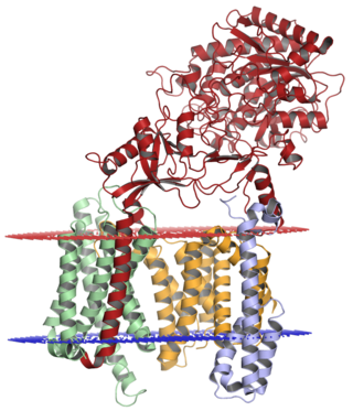
Gamma secretase is a multi-subunit protease complex, itself an integral membrane protein, that cleaves single-pass transmembrane proteins at residues within the transmembrane domain. Proteases of this type are known as intramembrane proteases. The most well-known substrate of gamma secretase is amyloid precursor protein, a large integral membrane protein that, when cleaved by both gamma and beta secretase, produces a short 37-43 amino acid peptide called amyloid beta whose abnormally folded fibrillar form is the primary component of amyloid plaques found in the brains of Alzheimer's disease patients. Gamma secretase is also critical in the related processing of several other type I integral membrane proteins, such as Notch, ErbB4, E-cadherin, N-cadherin, ephrin-B2, or CD44.

<span class="mw-page-title-main">Presenilin</span>

Presenilins are a family of related multi-pass transmembrane proteins which constitute the catalytic subunits of the gamma-secretase intramembrane protease protein complex. They were first identified in screens for mutations causing early onset forms of familial Alzheimer's disease by Peter St George-Hyslop. Vertebrates have two presenilin genes, called PSEN1 that codes for presenilin 1 (PS-1) and PSEN2 that codes for presenilin 2 (PS-2). Both genes show conservation between species, with little difference between rat and human presenilins. The nematode worm C. elegans has two genes that resemble the presenilins and appear to be functionally similar, sel-12 and hop-1.

<span class="mw-page-title-main">ADAM10</span> Protein-coding gene in the species Homo sapiens

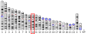
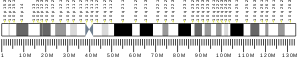
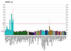
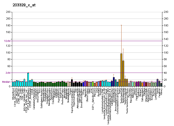
A Disintegrin and metalloproteinase domain-containing protein 10, also known as ADAM10 or CDw156 or CD156c is a protein that in humans is encoded by the ADAM10 gene.

<span class="mw-page-title-main">KLK6</span> Protein-coding gene in the species Homo sapiens

Kallikrein-6 is a protein that in humans is encoded by the KLK6 gene. Kallikrein-6 is also referred to as neurosin, protease M, hK6, or zyme. It is a 223 amino acid sequence, derived from its 244 original form, which contains a 16 residue presignal and 5 residue activation peptide.

<span class="mw-page-title-main">PITRM1</span> Protein-coding gene in humans

Pitrilysin metallopeptidase 1 also known as presequence protease, mitochondrial (PreP) and metalloprotease 1 (MTP-1) is an enzyme that in humans is encoded by the PITRM1 gene. It is also sometimes called metalloprotease 1 (MP1).PreP facilitates proteostasis by utilizing an ~13300-A(3) catalytic chamber to degrade toxic peptides, including mitochondrial presequences and β-amyloid. Deficiency of PreP is found associated with Alzheimer's disease. Reduced levels of PreP via RNAi mediated knockdown have been shown to lead to defective maturation of the protein Frataxin.

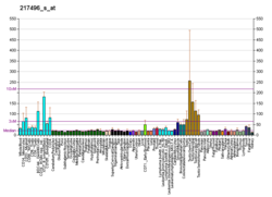
The biomarkers of Alzheimer's disease are neurochemical indicators used to assess the risk or presence of the disease. The biomarkers can be used to diagnose Alzheimer's disease (AD) in a very early stage, but they also provide objective and reliable measures of disease progress. It is imperative to diagnose AD disease as soon as possible, because neuropathologic changes of AD precede the symptoms by years. It is well known that amyloid beta (Aβ) is a good indicator of AD disease, which has facilitated doctors to accurately pre-diagnose cases of AD. When Aβ peptide is released by proteolytic cleavage of amyloid-beta precursor protein, some Aβ peptides that are solubilized are detected in CSF and blood plasma which makes AB peptides a promising candidate for biological markers. It has been shown that the amyloid beta biomarker shows 80% or above sensitivity and specificity, in distinguishing AD from dementia. It is believed that amyloid beta as a biomarker will provide a future for diagnosis of AD and eventually treatment of AD.

Intramembrane proteases (IMPs), also known as intramembrane-cleaving proteases (I-CLiPs), are enzymes that have the property of cleaving transmembrane domains of integral membrane proteins. All known intramembrane proteases are themselves integral membrane proteins with multiple transmembrane domains, and they have their active sites buried within the lipid bilayer of cellular membranes. Intramembrane proteases are responsible for proteolytic cleavage in the cell signaling process known as regulated intramembrane proteolysis (RIP).

<span class="mw-page-title-main">PLD3</span> Protein-coding gene in the species Homo sapiens

Phospholipase D3, also known as PLD3, is a protein that in humans is encoded by the PLD3 gene. PLD3 belongs to the phospholipase D superfamily because it contains the two HKD motifs common to members of the phospholipase D family, however, it has no known catalytic function similar to PLD1 or PLD2. PLD3 serves as a ssDNA 5' exonuclease in antigen presenting cells. PLD3 is highly expressed in the brain in both humans and mice, and is mainly localized in the endoplasmic reticulum (ER) and the lysosome.

The ion channel hypothesis of Alzheimer's disease (AD), also known as the channel hypothesis or the amyloid beta ion channel hypothesis, is a more recent variant of the amyloid hypothesis of AD, which identifies amyloid beta (Aβ) as the underlying cause of neurotoxicity seen in AD. While the traditional formulation of the amyloid hypothesis pinpoints insoluble, fibrillar aggregates of Aβ as the basis of disruption of calcium ion homeostasis and subsequent apoptosis in AD, the ion channel hypothesis in 1993 introduced the possibility of an ion-channel-forming oligomer of soluble, non-fibrillar Aβ as the cytotoxic species allowing unregulated calcium influx into neurons in AD.

References

  1. 1 2 3 GRCh38: Ensembl release 89: ENSG00000119912 Ensembl, May 2017
  2. "Human PubMed Reference:". National Center for Biotechnology Information, U.S. National Library of Medicine.
  3. "Mouse PubMed Reference:". National Center for Biotechnology Information, U.S. National Library of Medicine.
  4. 1 2 "Entrez Gene: IDE insulin-degrading enzyme".
  5. Aleshin AE, Gramatikova S, Hura GL, Bobkov A, Strongin AY, Stec B, et al. (November 2009). "Crystal and solution structures of a prokaryotic M16B peptidase: an open and shut case". Structure. 17 (11): 1465–75. doi:10.1016/j.str.2009.09.009. PMC   3615642 . PMID   19913481.
  6. King JV, Liang WG, Scherpelz KP, Schilling AB, Meredith SC, Tang WJ (July 2014). "Molecular basis of substrate recognition and degradation by human presequence protease". Structure. 22 (7): 996–1007. doi:10.1016/j.str.2014.05.003. PMC   4128088 . PMID   24931469.
  7. "IDE - Insulin-degrading enzyme - Homo sapiens (Human) - IDE gene & protein".
  8. "Uniprot: P14735 - IDE_HUMAN".
  9. 1 2 Shen Y, Joachimiak A, Rosner MR, Tang WJ (October 2006). "Structures of human insulin-degrading enzyme reveal a new substrate recognition mechanism". Nature. 443 (7113): 870–4. Bibcode:2006Natur.443..870S. doi:10.1038/nature05143. PMC   3366509 . PMID   17051221.
  10. Mirsky IA, Broh-Kahn RH (January 1949). "The inactivation of insulin by tissue extracts; the distribution and properties of insulin inactivating extracts". Archives of Biochemistry. 20 (1): 1–9. PMID   18104389.
  11. Affholter JA, Fried VA, Roth RA (December 1988). "Human insulin-degrading enzyme shares structural and functional homologies with E. coli protease III". Science. 242 (4884): 1415–8. Bibcode:1988Sci...242.1415A. doi:10.1126/science.3059494. PMID   3059494.
  12. Wang DS, Dickson DW, Malter JS (2006). "beta-Amyloid degradation and Alzheimer's disease". Journal of Biomedicine & Biotechnology. 2006 (3): 58406. doi: 10.1155/JBB/2006/58406 . PMC   1559921 . PMID   17047308.
  13. Kurochkin IV, Goto S (May 1994). "Alzheimer's beta-amyloid peptide specifically interacts with and is degraded by insulin degrading enzyme". FEBS Letters. 345 (1): 33–7. doi: 10.1016/0014-5793(94)00387-4 . PMID   8194595. S2CID   43917847.
  14. Kerr ML, Small DH (April 2005). "Cytoplasmic domain of the beta-amyloid protein precursor of Alzheimer's disease: function, regulation of proteolysis, and implications for drug development". Journal of Neuroscience Research. 80 (2): 151–9. doi:10.1002/jnr.20408. PMID   15672415. S2CID   31985212.
  15. Farris W, Mansourian S, Chang Y, Lindsley L, Eckman EA, Frosch MP, et al. (April 2003). "Insulin-degrading enzyme regulates the levels of insulin, amyloid beta-protein, and the beta-amyloid precursor protein intracellular domain in vivo". Proceedings of the National Academy of Sciences of the United States of America. 100 (7): 4162–7. Bibcode:2003PNAS..100.4162F. doi: 10.1073/pnas.0230450100 . PMC   153065 . PMID   12634421.
  16. Cook DG, Leverenz JB, McMillan PJ, Kulstad JJ, Ericksen S, Roth RA, et al. (January 2003). "Reduced hippocampal insulin-degrading enzyme in late-onset Alzheimer's disease is associated with the apolipoprotein E-epsilon4 allele". The American Journal of Pathology. 162 (1): 313–9. doi:10.1016/s0002-9440(10)63822-9. PMC   1851126 . PMID   12507914. Archived from the original on 2003-08-30. Retrieved 2008-02-10.
  17. Kim M, Hersh LB, Leissring MA, Ingelsson M, Matsui T, Farris W, et al. (March 2007). "Decreased catalytic activity of the insulin-degrading enzyme in chromosome 10-linked Alzheimer disease families". The Journal of Biological Chemistry. 282 (11): 7825–32. doi: 10.1074/jbc.M609168200 . PMID   17244626.
  18. Duckworth WC, Bennett RG, Hamel FG (October 1998). "Insulin degradation: progress and potential". Endocrine Reviews. 19 (5): 608–24. doi: 10.1210/edrv.19.5.0349 . PMID   9793760.
  19. Lynch JA, George AM, Eisenhauer PB, Conn K, Gao W, Carreras I, et al. (May 2006). "Insulin degrading enzyme is localized predominantly at the cell surface of polarized and unpolarized human cerebrovascular endothelial cell cultures". Journal of Neuroscience Research. 83 (7): 1262–70. doi:10.1002/jnr.20809. PMID   16511862. S2CID   23670388.
  20. Authier F, Bergeron JJ, Ou WJ, Rachubinski RA, Posner BI, Walton PA (April 1995). "Degradation of the cleaved leader peptide of thiolase by a peroxisomal proteinase". Proceedings of the National Academy of Sciences of the United States of America. 92 (9): 3859–63. Bibcode:1995PNAS...92.3859A. doi: 10.1073/pnas.92.9.3859 . PMC   42061 . PMID   7731996.
  21. Roth RA, Mesirow ML, Cassell DJ, Yokono K, Baba S (March 1985). "Characterization of an insulin degrading enzyme from cultured human lymphocytes". Diabetes Research and Clinical Practice. 1 (1): 31–9. doi:10.1016/S0168-8227(85)80026-7. PMID   3915257.
  22. Semple JW, Lang Y, Speck ER, Delovitch TL (October 1992). "Processing and presentation of insulin. III. Insulin degrading enzyme: a neutral metalloendoproteinase that is non-homologous to classical endoproteinases mediates the processing of insulin epitopes for helper T cells". International Immunology. 4 (10): 1161–7. doi:10.1093/intimm/4.10.1161. PMID   1283335.
  23. 1 2 Qiu WQ, Walsh DM, Ye Z, Vekrellis K, Zhang J, Podlisny MB, et al. (December 1998). "Insulin-degrading enzyme regulates extracellular levels of amyloid beta-protein by degradation". The Journal of Biological Chemistry. 273 (49): 32730–8. doi: 10.1074/jbc.273.49.32730 . PMID   9830016.
  24. Tamboli IY, Barth E, Christian L, Siepmann M, Kumar S, Singh S, et al. (November 2010). "Statins promote the degradation of extracellular amyloid {beta}-peptide by microglia via stimulation of exosome-associated insulin-degrading enzyme (IDE) secretion". The Journal of Biological Chemistry. 285 (48): 37405–14. doi: 10.1074/jbc.M110.149468 . PMC   2988346 . PMID   20876579.
  25. 1 2 Amata O, Marino T, Russo N, Toscano M (October 2009). "Human insulin-degrading enzyme working mechanism". Journal of the American Chemical Society. 131 (41): 14804–11. doi:10.1021/ja9037142. PMID   19785409.
  26. Leopoldini M, Russo N, Toscano M (August 2009). "Determination of the catalytic pathway of a manganese arginase enzyme through density functional investigation". Chemistry: A European Journal. 15 (32): 8026–36. doi:10.1002/chem.200802252. PMID   19288480.
  27. Hersh LB (November 2006). "The insulysin (insulin degrading enzyme) enigma". Cellular and Molecular Life Sciences. 63 (21): 2432–4. doi:10.1007/s00018-006-6238-9. PMID   16952049. S2CID   12536419.