Intraepithelial lymphocyte

Last updated

Intraepithelial lymphocytes (IEL) are lymphocytes found in the epithelial layer of mammalian mucosal linings, such as the gastrointestinal (GI) tract and reproductive tract. [1] However, unlike other T cells, IELs do not need priming. Upon encountering antigens, they immediately release cytokines and cause killing of infected target cells. In the GI tract, they are components of gut-associated lymphoid tissue (GALT). [2]

Contents

Intestinal IELs are long-lived resistant effector cells spread along the entire length of intestine, where they patrol the space between intestinal epithelial cells (IEC) and the basement membrane (the intraepithelial space). Epithelium of small intestine contains approximately 1 IEL per 10 enterocytes. [3] Due to their constant exposure to of antigens at mucosal barrier, they have unique antigen-experienced activated phenotypes and they constantly express CD103 (αE integrin), that is distinct from the conventional T cells in the intestine. [3] IELs are mainly T cells with mixture of subsets. They are divided into two groups – conventional and unconventional IELs. [4]

In mice both groups are retained in almost equal proportions. [5] In humans, the majority of IELs are alpha beta T cells. 15% of IELs are gamma delta T cells and thus represent a minor component of human IELs. However, IELs significantly increase under certain conditions, such as celiac disease. [1]

primary biliary cirrhosis. Bile duct intraepithelial lymphocytes Primary biliary cirrhosis low mag.jpg
primary biliary cirrhosis. Bile duct intraepithelial lymphocytes

Phenotype

The majority of IELs (80%) are CD3+, and over 75% of these also express CD8. IELs can be divided into two major subsets based on their CD8 coreceptor expression. [5] One subset of IELs typically express activation marker CD8αα and some IELs express CD8αβ+ marker (CD8αβ promotes TCR activation, whereas CD8αα suppresses TCR signals).

In both humans and mice IELs express higher levels of CD103, activation marker CD69, granzyme B and perforin cytolytic granules. CD25 expression is lower in comparison with effector memory T cells. [6] [7]

CD8αα

Expression of CD8αα is an important phenotypic marker of IELs, but not all IELs subpopulations express this molecule. CD8αα homodimer is an alternative isoform to classical CD8αβ heterodimer, which is expressed on conventional CD8 T-cells. CD8αα is mainly expressed by effector or mature antigen-experienced cells in the gut. This molecule can bind MHC I, but, opposed to the function of CD8αβ, CD8αα reduces sensitivity of TCR towards antigens. Thus, when recognizing MHC I, CD8αα functions as a repressor of activation. [8]

CD8αα can also recognize thymus leukemia (TL) antigen, which is a non-classical MHC I molecule that is expressed in thymus and in intestinal epithelium. Interaction between TL and CD8αα does not serve for migration of IELs into the epithelium, but it is important for modulating immune response of IELs. [9] It has been suggested that cross-talk between TL and CD8αα might regulate IELs survival and proliferation. [8] More accurately, TL prevents proliferation of IELs, when there is co-occurrence of weak TCR stimulation. [9]

Development

Induced IELs (TCRαβ+ CD8αβ+) are generated from naive T cells during an immune response. TCRαβ+ CD8αα (natural IELs) cells differentiate in the thymus. [6] [10]

Development and cytolytic activation are independent of live micro-organisms but they become cytolytic in response to the exogenous antigenic substances other than live micro-organisms in the gut. IEL T cells acquire their activated memory phenotype post-thymically, in response to antigens encountered in the periphery. [11]

Function

Their role in immune system is crucial because IELs provide a first line of defense at this extensive barrier with the outside world. All IEL T cells are antigen-experienced T cells, which typically display a cytotoxic functional phenotype. IELs mediate antigen-specific delayed-type hypersensitivity (DTH) responses, exhibit virus-specific CTL function, to express natural killer (NK)-like activity and produce a local graft-versus-host reaction (GVHR) when transferred to semiallogeneic hosts. IELs are also able to produce a variety of cytokines which are characteristically produced by Th1- and Th2-type cells and can also provide help for B cell responses. [6] [10] [11]

Pathology

An elevated IEL population, as determined by biopsy, typically indicates ongoing inflammation within the mucosa. In diseases such as celiac sprue, IEL elevation throughout the small intestine is one of many specific markers. [1] IELs have heightened activated status that can lead to inflammatory disease such as IBD, promote cancer development and progression, [12] or become the malignant cells in enteropathy-associated T-cell lymphoma, a lymphoma that is a complication of celiac sprue. [13] [14]

Alternatively, elevated IEL populations can be a marker for developing neoplasia in the tissue such as found in cervical and prostate cancers, as well as some colorectal cancers, particularly those associated with Lynch syndrome (hereditary non-polyposis colon cancer <HNPCC>). [15] IELs themselves can, when chronically activated, undergo mutation that can lead to lymphoma. [16]

Classification

IELs can be divided into different subpopulations based on molecular markers expression, mainly by expression of TCR and CD8αα, and by origin.

Induced TCR+ IELs

Also termed conventional IELs, express TCRαβ together with CD4 or CD8αβ and are derived from antigen-experienced T cells that home to intraepithelial space. Contrary to natural IELs, induced IELs are the progeny of MHCI-restricted CD8αβ or MHCII-restricted CD4 naïve T cells that further undergo a post-thymic differentiation. These cells express activation markers (CD44, CD69) and unlike natural TCR+IELs express CD2, CD5, CD28, LFA-1, and Thy1. Upon the entry into the intestinal epithelium, these cells can start express also CD8αα. [4] [3]

TCRαβ+CD4+ IELs

TCRαβ+CD4+ IELs arise from conventional peripheral CD4+ T-cells. These cells migrate into the intestinal epithelium as effector or tissue-resident memory T cells.

In mice, up to 50% of these IELs can express CD8αα homodimer, which they acquire in the intestinal epithelium after external stimuli such as TGF-β, IFN-γ, IL-27 and retinoic acid. Function of TCRαβ+ CD4+ CD8αα+ IELs is unclear. Even though they express granzymes and have cytolytic properties, it has been suggested that they can also have regulatory properties in the context of chronic intestinal inflammation. [3] [17]

TCRαβ+CD8αβ+ IELs

These IELs emerge from peripherally activated conventional CD8+ T-cells and home to the intestinal epithelium, where they function as effector or memory cells. They continuously express integrin β7, granzyme B, CD103 and CD69 and produce lower amounts of TNF-α and IFN-γ as opposed to the conventional CD8+ T-cells. [4]

Some of these cells also express CD8αα homodimer and can be pathogenic during coeliac disease in humans. [3]

Double positive (DP) TCRαβ+CD4+CD8αα+ IELs

These DP IELs are subset of induced IELs, which are CD4+ IELs with some functions of CD8+ IELs and under physiological condition their number in the intestine is very small. During the intestinal inflammation, levels of DP IELs significantly increase. [18]

DP IELs develop independently of the thymus and contrary to natural IELs, these cells increase with age, especially when they are exposed to exogenous antigens. Their migration into the intestinal epithelium depends mainly on the luminal bacteria and the dietary antigens. [18]

DP IELs induction is directed by the transcriptional regulation. During the development of IELs, CD4+ T cells downregulate ThPOK and instead start to express Runx3 transcription factor, because CD4+CD8aa+ IELs have low levels of ThPOK expression while the expression of Runx3 is very high. T-bet inducing environment is also required for the Runx3 upregulation, most likely containing IFN-y, IL-27, IL-15 and Retionic acid (RA). [19] RA have the ability to induce an expression of the intetsine-homing receptors, such as α4β7-integrin and CC-chemokine receptor 9 (CCR9). [20] Another transcription factor responsible for DP IELs induction is the Aryl hydrocarbon receptor (AhR). AhR is ligand-dependent transcription factor, and its activation is responsible for ThPOK downregulation. AhR is activated by indole metabolites of tryptophan induced by microbiota, such as Lactobacillus reuteri . [21] Therefore, the DP IELs induction is dependent on the microbiota composition and the diet.

The function of CD4CD8aa IELs is due to their CD8 phenotype and granzyme B expression to prevent pathogens from invading and to maintain integrity of the intestinal epithelial barrier. Their CD4 phenotype is responsible for IL-10 and TGF-β secretion that prevents Th1-induced inflammation in the intestine, therefore their role can be complementary to T regulatory cells. [18] [22]

DP IELs probably play role in intestinal homeostasis because of their immunosuppressive function. But for their cytotoxic responses they may play an important role in the pathological process of IBD. [18]

Natural TCR+ IELs

Also termed unconventional IELs, express either TCRαβ or TCRγδ and do not express either CD4 or CD8αβ, but express CD8αα homodimers. In contrast to induced TCR+ IELs lack expression of CD2, CD5, CD28, LFA-1, and Thy1. [4]

TCRαβ+ IELs

In mice, these IELs are the most abundant at birth and with age their numbers decrease. In humans, these cells are present during gestation but are very rare in adulthood. TCRαβ+ IELs develop in thymus where they undergo agonist positive selection and thereby are self-reactive. Nevertheless, they have regulatory properties and protect against colitis in animal experiments. These cells are influenced by normal intestinal microbiota and vitamin D. NOD2 receptor expressed by antigen presenting cells and epithelial cells in the intestine recognizes microbes and triggers the production of IL-15 cytokine, which promotes TCRαβ+CD8αα+ IELs. [3]

TCRγδ+ IELs

TCRγδ+ IELs develop outside of thymus and their maintenance and function in the intestinal epithelium is influenced by a cross-talk with enterocytes. Moreover, they can migrate through the epithelium with the help of interactions with epithelial cells. [23] Most of these cells express Vγ7 in mice and Vγ4 in humans. Their function resides in the protection of the intestinal barrier against pathogens early in the infection and later they quench the inflammation and protect the barrier from tissue damage. The mechanism is not clear, but TCRγδ+ IELs have cytotoxic properties and can produce cytokines TGF-β, TNF-α, IFN-γ, IL-13 and IL-10 and antimicrobial peptides, all of which can contribute to the diverse functions. [3]

Similar functions have been found in the context of colitis, where these cells seem to have pathogenic role at the beginning, whereas later they protect the epithelium against tissue damage. [17]

TCR IELs

IELs that do not express TCR.

ILC-like IELs

These cells show properties of NK cells. In humans, they are elevated during Crohn´s disease and in mice, they are pathogenic during colitis. [4]

iCD8α

These innate lymphocytes express homodimer CD8αα and CD3 and develop outside of thymus. They have cytotoxic and phagocytic properties, express MHC II and thereby can present antigens to conventional CD4+ T-cells. iCD8α protect against bacterial infections and promotes experimental colitis. [3]

TCRiCD3+CD8αα IELs

These cells are very similar to iCD8α population and it is unclear if this is a different subset of cells or only precursors of iCD8α. [3]

See also

IEL of the GI tract

Related Research Articles

<span class="mw-page-title-main">T cell</span> White blood cells of the immune system

T cells are one of the important types of white blood cells of the immune system and play a central role in the adaptive immune response. T cells can be distinguished from other lymphocytes by the presence of a T-cell receptor (TCR) on their cell surface.

<span class="mw-page-title-main">Cytotoxic T cell</span> T cell that kills infected, damaged or cancerous cells

A cytotoxic T cell (also known as TC, cytotoxic T lymphocyte, CTL, T-killer cell, cytolytic T cell, CD8+ T-cell or killer T cell) is a T lymphocyte (a type of white blood cell) that kills cancer cells, cells that are infected by intracellular pathogens (such as viruses or bacteria), or cells that are damaged in other ways.

<span class="mw-page-title-main">T helper cell</span> Type of immune cell

The T helper cells (Th cells), also known as CD4+ cells or CD4-positive cells, are a type of T cell that play an important role in the adaptive immune system. They aid the activity of other immune cells by releasing cytokines. They are considered essential in B cell antibody class switching, breaking cross-tolerance in dendritic cells, in the activation and growth of cytotoxic T cells, and in maximizing bactericidal activity of phagocytes such as macrophages and neutrophils. CD4+ cells are mature Th cells that express the surface protein CD4. Genetic variation in regulatory elements expressed by CD4+ cells determines susceptibility to a broad class of autoimmune diseases.

The regulatory T cells (Tregs or Treg cells), formerly known as suppressor T cells, are a subpopulation of T cells that modulate the immune system, maintain tolerance to self-antigens, and prevent autoimmune disease. Treg cells are immunosuppressive and generally suppress or downregulate induction and proliferation of effector T cells. Treg cells express the biomarkers CD4, FOXP3, and CD25 and are thought to be derived from the same lineage as naïve CD4+ cells. Because effector T cells also express CD4 and CD25, Treg cells are very difficult to effectively discern from effector CD4+, making them difficult to study. Research has found that the cytokine transforming growth factor beta (TGF-β) is essential for Treg cells to differentiate from naïve CD4+ cells and is important in maintaining Treg cell homeostasis.

In immunology, central tolerance is the process of eliminating any developing T or B lymphocytes that are autoreactive, i.e. reactive to the body itself. Through elimination of autoreactive lymphocytes, tolerance ensures that the immune system does not attack self peptides. Lymphocyte maturation occurs in primary lymphoid organs such as the bone marrow and the thymus. In mammals, B cells mature in the bone marrow and T cells mature in the thymus.

Gut-associated lymphoid tissue (GALT) is a component of the mucosa-associated lymphoid tissue (MALT) which works in the immune system to protect the body from invasion in the gut.

Memory T cells are a subset of T lymphocytes that might have some of the same functions as memory B cells. Their lineage is unclear.

Microfold cells are found in the gut-associated lymphoid tissue (GALT) of the Peyer's patches in the small intestine, and in the mucosa-associated lymphoid tissue (MALT) of other parts of the gastrointestinal tract. These cells are known to initiate mucosal immunity responses on the apical membrane of the M cells and allow for transport of microbes and particles across the epithelial cell layer from the gut lumen to the lamina propria where interactions with immune cells can take place.

MHC-restricted antigen recognition, or MHC restriction, refers to the fact that a T cell can interact with a self-major histocompatibility complex molecule and a foreign peptide bound to it, but will only respond to the antigen when it is bound to a particular MHC molecule.

In immunology, a naive T cell (Th0 cell) is a T cell that has differentiated in the thymus, and successfully undergone the positive and negative processes of central selection in the thymus. Among these are the naive forms of helper T cells (CD4+) and cytotoxic T cells (CD8+). Any naive T cell is considered immature and, unlike activated or memory T cells, has not encountered its cognate antigen within the periphery. After this encounter, the naive T cell is considered a mature T cell.

In immunology, peripheral tolerance is the second branch of immunological tolerance, after central tolerance. It takes place in the immune periphery. Its main purpose is to ensure that self-reactive T and B cells which escaped central tolerance do not cause autoimmune disease. Peripheral tolerance prevents immune response to harmless food antigens and allergens, too.

Gamma delta T cells are T cells that have a γδ T-cell receptor (TCR) on their surface. Most T cells are αβ T cells with TCR composed of two glycoprotein chains called α (alpha) and β (beta) TCR chains. In contrast, γδ T cells have a TCR that is made up of one γ (gamma) chain and one δ (delta) chain. This group of T cells is usually less common than αβ T cells. Their highest abundance is in the gut mucosa, within a population of lymphocytes known as intraepithelial lymphocytes (IELs).

<span class="mw-page-title-main">BTLA</span> Protein-coding gene in the species Homo sapiens

B- and T-lymphocyte attenuator or BTLA is a protein that belongs to the CD28 immunoglobulin superfamily (IgSF) which is encoded by the BTLA gene located on the 3rd human chromosome. BTLA was first discovered in 2003 as an inhibitor of Th1 expansion and it became the 3rd member of the CD28 IgSF. However, its discovered ligand herpes virus entry mediator or HVEM belongs to the tumor necrosis factor receptor superfamily (TNFRSF). This finding was surprising because until the discovery of HVEM it was believed that receptors and ligands always belong to the same family.

<span class="mw-page-title-main">Enteropathy-associated T-cell lymphoma</span> Complication of coeliac disease

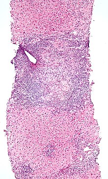
Enteropathy-associated T-cell lymphoma (EATL), previously termed enteropathy-associated T-cell lymphoma, type I and at one time termed enteropathy-type T-cell lymphoma (ETTL), is a complication of coeliac disease in which a malignant T-cell lymphoma develops in areas of the small intestine affected by the disease's intense inflammation. While a relatively rare disease, it is the most common type of primary gastrointestinal T-cell lymphoma.

T helper 3 cells (Th3) are a subset of T lymphocytes with immunoregulary and immunosuppressive functions, that can be induced by administration of foreign oral antigen. Th3 cells act mainly through the secretion of anti-inflammatory cytokine transforming growth factor beta (TGF-β). Th3 have been described both in mice and human as CD4+FOXP3 regulatory T cells. Th3 cells were first described in research focusing on oral tolerance in the experimental autoimmune encephalitis (EAE) mouse model and later described as CD4+CD25FOXP3LAP+ cells, that can be induced in the gut by oral antigen through T cell receptor (TCR) signalling.

<span class="mw-page-title-main">Mucosal immunology</span> Field of study

Mucosal immunology is the study of immune system responses that occur at mucosal membranes of the intestines, the urogenital tract, and the respiratory system. The mucous membranes are in constant contact with microorganisms, food, and inhaled antigens. In healthy states, the mucosal immune system protects the organism against infectious pathogens and maintains a tolerance towards non-harmful commensal microbes and benign environmental substances. Disruption of this balance between tolerance and deprivation of pathogens can lead to pathological conditions such as food allergies, irritable bowel syndrome, susceptibility to infections, and more.

Mucosal-associated invariant T cells make up a subset of T cells in the immune system that display innate, effector-like qualities. In humans, MAIT cells are found in the blood, liver, lungs, and mucosa, defending against microbial activity and infection. The MHC class I-like protein, MR1, is responsible for presenting bacterially-produced vitamin B2 and B9 metabolites to MAIT cells. After the presentation of foreign antigen by MR1, MAIT cells secrete pro-inflammatory cytokines and are capable of lysing bacterially-infected cells. MAIT cells can also be activated through MR1-independent signaling. In addition to possessing innate-like functions, this T cell subset supports the adaptive immune response and has a memory-like phenotype. Furthermore, MAIT cells are thought to play a role in autoimmune diseases, such as multiple sclerosis, arthritis and inflammatory bowel disease, although definitive evidence is yet to be published.

<span class="mw-page-title-main">Intestinal mucosal barrier</span>

The intestinal mucosal barrier, also referred to as intestinal barrier, refers to the property of the intestinal mucosa that ensures adequate containment of undesirable luminal contents within the intestine while preserving the ability to absorb nutrients. The separation it provides between the body and the gut prevents the uncontrolled translocation of luminal contents into the body proper. Its role in protecting the mucosal tissues and circulatory system from exposure to pro-inflammatory molecules, such as microorganisms, toxins, and antigens is vital for the maintenance of health and well-being. Intestinal mucosal barrier dysfunction has been implicated in numerous health conditions such as: food allergies, microbial infections, irritable bowel syndrome, inflammatory bowel disease, celiac disease, metabolic syndrome, non-alcoholic fatty liver disease, diabetes, and septic shock.

Nasal- or nasopharynx- associated lymphoid tissue (NALT) represents immune system of nasal mucosa and is a part of mucosa-associated lymphoid tissue (MALT) in mammals. It protects body from airborne viruses and other infectious agents. In humans, NALT is considered analogous to Waldeyer's ring.

Thymic epithelial cells (TECs) are specialized cells with high degree of anatomic, phenotypic and functional heterogeneity that are located in the outer layer (epithelium) of the thymic stroma. The thymus, as a primary lymphoid organ, mediates T cell development and maturation. The thymic microenvironment is established by TEC network filled with thymocytes in different developing stages. TECs and thymocytes are the most important components in the thymus, that are necessary for production of functionally competent T lymphocytes and self tolerance. Dysfunction of TECs causes several immunodeficiencies and autoimmune diseases.

References

  1. 1 2 3 Hopper AD, Hurlstone DP, Leeds JS, McAlindon ME, Dube AK, Stephenson TJ, Sanders DS (November 2006). "The occurrence of terminal ileal histological abnormalities in patients with coeliac disease". Digestive and Liver Disease. 38 (11): 815–819. doi:10.1016/j.dld.2006.04.003. PMID   16787773.
  2. DeFranco AL, Locksley RM, Robertson M (2007). The Immune Response in Infection and Inflammatory Disease. London: New Science Press. pp. 218–219. ISBN   978-0-19-920614-8.
  3. 1 2 3 4 5 6 7 8 9 Olivares-Villagómez D, Van Kaer L (April 2018). "Intestinal Intraepithelial Lymphocytes: Sentinels of the Mucosal Barrier". Trends in Immunology. 39 (4): 264–275. doi:10.1016/j.it.2017.11.003. PMC   8056148 . PMID   29221933.
  4. 1 2 3 4 5 McDonald BD, Jabri B, Bendelac A (August 2018). "Diverse developmental pathways of intestinal intraepithelial lymphocytes". Nature Reviews. Immunology. 18 (8): 514–525. doi:10.1038/s41577-018-0013-7. PMC   6063796 . PMID   29717233.
  5. 1 2 Sheridan BS, Lefrançois L (December 2010). "Intraepithelial lymphocytes: to serve and protect". Current Gastroenterology Reports. 12 (6): 513–521. doi:10.1007/s11894-010-0148-6. PMC   3224371 . PMID   20890736.
  6. 1 2 3 Mayassi T, Jabri B (September 2018). "Human intraepithelial lymphocytes". Mucosal Immunology. 11 (5): 1281–1289. doi:10.1038/s41385-018-0016-5. PMC   6178824 . PMID   29674648.
  7. Lambolez F, Mayans S, Cheroutre H (2013). "Lymphocytes: Intraepithelial". eLS. American Cancer Society. doi:10.1002/9780470015902.a0001197.pub3. ISBN   9780470015902.
  8. 1 2 Cheroutre H, Lambolez F (February 2008). "Doubting the TCR coreceptor function of CD8alphaalpha". Immunity. 28 (2): 149–159. doi: 10.1016/j.immuni.2008.01.005 . PMID   18275828.
  9. 1 2 Olivares-Villagómez D, Van Kaer L (November 2010). "TL and CD8αα: Enigmatic partners in mucosal immunity". Immunology Letters. 134 (1): 1–6. doi:10.1016/j.imlet.2010.09.004. PMC   2967663 . PMID   20850477.
  10. 1 2 Sim GK (1995-01-01). Intraepithelial lymphocytes and the immune system. Advances in Immunology. Vol. 58. pp. 297–343. doi:10.1016/s0065-2776(08)60622-7. ISBN   9780120224586. PMID   7741030.
  11. 1 2 McGhee JR (1998-01-01). "Mucosa-Associated Lymphoid Tissue (MALT)" . In Delves PJ (ed.). Encyclopedia of Immunology (Second ed.). Elsevier. pp.  1774–1780. doi:10.1006/rwei.1999.0448. ISBN   9780122267659.
  12. Cheroutre H (2015-01-01). "Chapter 35 - Intraepithelial TCRαβ T Cells in Health and Disease". In Mestecky J, Lefrancois L, Strober W, Russell MW, Kelsall BL (eds.). Mucosal Immunology (Fourth ed.). Academic Press. pp. 733–748. doi:10.1016/b978-0-12-415847-4.00035-5. ISBN   9780124158474.
  13. Ondrejka S, Jagadeesh D (December 2016). "Enteropathy-Associated T-Cell Lymphoma". Current Hematologic Malignancy Reports. 11 (6): 504–513. doi:10.1007/s11899-016-0357-7. PMID   27900603. S2CID   13329863.
  14. Chander U, Leeman-Neill RJ, Bhagat G (August 2018). "Pathogenesis of Enteropathy-Associated T Cell Lymphoma". Current Hematologic Malignancy Reports. 13 (4): 308–317. doi:10.1007/s11899-018-0459-5. PMID   29943210. S2CID   49430640.
  15. Bellizzi AM, Frankel WL (November 2009). "Colorectal cancer due to deficiency in DNA mismatch repair function: a review". Advances in Anatomic Pathology. 16 (6): 405–417. doi:10.1097/PAP.0b013e3181bb6bdc. PMID   19851131. S2CID   25600795.
  16. Meresse B, Malamut G, Cerf-Bensussan N (June 2012). "Celiac disease: an immunological jigsaw". Immunity. 36 (6): 907–919. doi: 10.1016/j.immuni.2012.06.006 . PMID   22749351.
  17. 1 2 Ma H, Qiu Y, Yang H (February 2021). "Intestinal intraepithelial lymphocytes: Maintainers of intestinal immune tolerance and regulators of intestinal immunity". Journal of Leukocyte Biology. 109 (2): 339–347. doi:10.1002/JLB.3RU0220-111. PMC   7891415 . PMID   32678936.
  18. 1 2 3 4 Park Y, Moon SJ, Lee SW (January 2016). "Lineage re-commitment of CD4CD8αα intraepithelial lymphocytes in the gut". BMB Reports. 49 (1): 11–17. doi:10.5483/BMBRep.2016.49.1.242. PMC   4914207 . PMID   26592937.
  19. Reis BS, Hoytema van Konijnenburg DP, Grivennikov SI, Mucida D (August 2014). "Transcription factor T-bet regulates intraepithelial lymphocyte functional maturation". Immunity. 41 (2): 244–256. doi:10.1016/j.immuni.2014.06.017. PMC   4287410 . PMID   25148025.
  20. Iwata M, Hirakiyama A, Eshima Y, Kagechika H, Kato C, Song SY (October 2004). "Retinoic acid imprints gut-homing specificity on T cells". Immunity. 21 (4): 527–538. doi: 10.1016/j.immuni.2004.08.011 . PMID   15485630.
  21. Cervantes-Barragan L, Chai JN, Tianero MD, Di Luccia B, Ahern PP, Merriman J, et al. (August 2017). "Lactobacillus reuteri induces gut intraepithelial CD4+CD8αα+ T cells". Science. 357 (6353): 806–810. doi:10.1126/science.aah5825. PMC   5687812 . PMID   28775213.
  22. Sujino T, London M, Hoytema van Konijnenburg DP, Rendon T, Buch T, Silva HM, et al. (June 2016). "Tissue adaptation of regulatory and intraepithelial CD4⁺ T cells controls gut inflammation". Science. 352 (6293): 1581–1586. Bibcode:2016Sci...352.1581S. doi:10.1126/science.aaf3892. PMC   4968079 . PMID   27256884.
  23. Hoytema van Konijnenburg DP, Reis BS, Pedicord VA, Farache J, Victora GD, Mucida D (November 2017). "Intestinal Epithelial and Intraepithelial T Cell Crosstalk Mediates a Dynamic Response to Infection". Cell. 171 (4): 783–794.e13. doi:10.1016/j.cell.2017.08.046. PMC   5670000 . PMID   28942917.