Enteropathy-associated T-cell lymphoma

Last updated
Enteropathy-associated T-cell lymphoma (formerly termed enteropathy-associated T-cell lymphoma, type 1)
Other namesEnteropathy-associated T-cell lymphoma, type I
Enteropathy-associated T cell lymphoma - low mag.jpg
Micrograph of enteropathy-associated T cell lymphoma (upper right of image). H&E stain.
Specialty Oncology, hematology, gastroenterology
Complications Bowel obstructions, bowel perforations
CausesComplication of celiac disease
Risk factors Genetic predisposition
Prevention Gluten-free diet
Prognosis guarded

Enteropathy-associated T-cell lymphoma (EATL), previously termed enteropathy-associated T-cell lymphoma, type I and at one time termed enteropathy-type T-cell lymphoma (ETTL), is a complication of coeliac disease in which a malignant T-cell lymphoma develops in areas of the small intestine affected by the disease's intense inflammation. [1] While a relatively rare disease, it is the most common type of primary gastrointestinal T-cell lymphoma. [2]

Contents

EATL had been defined as a single type of small intestine lymphoma, but in 2008, the World Health Organization (WHO) divided the disease into two subtypes: 1) EATL type I, which occurs in individuals with coeliac disease, a chronic immune disorder causing inflammatory responses to dietary gluten primarily in the upper reaches (i.e. jejunum and duodenum) of the small intestine; and 2) EATL type II, a disorder similar to EATL type I that occurs without coeliac disease. While type I and II EATL share many similar features, post-2008 studies found some significant differences between the two types. In 2016, the WHO redefined the two diseases as separate entities, keeping the term enteropathy-associated T-cell lymphoma for the coeliac disease-associated lymphoma and terming type 2 disease as monomorphic epitheliotropic intestinal T cell lymphoma (MEITL). [3] EATL is five to ten times more common than is MEITL. [4] The WHO also defined a third type of intestinal T-cell lymphoma that cannot not be classified as EATL or MEITL as peripheral T-cell lymphoma not otherwise specified (ITCL-NOS). [5]

EATL arises from the malignant transformation of small-intestinal intraepithelial lymphocytes (IEL). IEL are a heterogeneous group of principally T-cell lymphocytes residing in epithelial tissues that interface the environment, such as the mucosa of the bronchi, reproductive tract and gastrointestinal tract. [6] At these sites, IEL are exposed and regulate immune responses to non-dietary and dietary antigens, pathogenic and non-pathogenic organisms and injured self tissues. [7] Gastrointestinal tract IEL appear in the epithelium of the small intestine, colon, stomach and esophagus, residing between the epithelial cells that line these organs' lumens. [7] These IEL often exhibit natural killer and cytotoxic T-cell cell activation markers, [6] contain various toxic agents (e.g. perforin, granzyme) and therefore are capable, if activated, of causing severe tissue injuries. [4] With coeliac disease, the IEL react to the glutelins in dietary gluten by increasing their numbers, becoming pathologically active, producing chronic inflammation that injures intestinal cells, interfering with nutrient absorption and creating an environment conducive to their malignant transformation into EATL. [1]

Optimal treatment of EATL has used regimens consisting of intensive chemotherapy, hematopoietic stem cell transplantation and, in cases with bulky, obstructive and/or perforated bowel disease, surgical intervention. [3] The disease has a five-year overall survival rate of only ~20%. [8] However, recent studies focusing on the malignant IEL in EATL have increased understanding of the disease and suggested newer chemotherapy-based strategies and novel molecular targets that might be attacked therapeutically to improve the disease's prognosis. [7]

Presentation

EATL typically occurs in patients with a history of coeliac disease and who may have been previously diagnosed with Type I or II refractory disease, [9] but in any case, affected individuals present with worsening coeliac disease symptoms of abdominal pain, malabsorption, diarrhea, weight loss, fever or night sweats. [10] [3] EATL diagnosis is more likely if symptoms develop suddenly, [4] or if the serious symptoms of bowel obstruction and/or bowel perforation caused by bulky EATL masses develop. [10] Patients with ulcerative jejunitis usually present with more severe symptoms, including more frequent bowel perforations and obstructions. [10] Some patients with no history of coeliac disease present with symptoms or signs of a small-intestinal lymphoma but on diagnostic workup are found to have coeliac disease.[ citation needed ]

Pathophysiology

Genetics

The cause of EATL, while not fully understood, is by definition related to celiac disease. Individuals are genetically predisposed to develop celiac disease because of the specific types of HLA-DQ proteins expressed by their antigen-presenting cells (APC). HLA-DQ proteins are on the surface of APC and function to present foreign or self antigens to the T cell receptors (TCR) expressed on the surface of T-cells and thereby to stimulate these cells either to initiate or suppress immune responses to the presented antigens. HLA-DQ proteins are composed of α and β polypeptide chains encoded by the HLA-DQA1 and HLA-DQB1 genes, respectively. Since there are several different alleles (i.e. gene variants) at these two genetic loci, individuals are usually heterozygous, i.e. have inherited different alleles from each parent at each locus; uncommonly, however, individuals are homozygous at one or both loci because their parents have the same alleles at one or both loci. The HLA-DQ proteins that predispose individuals to coeliac disease bind and respond specifically to gluten-related antigens presented to them by APC. [11] The genetic predisposition to develop coeliac disease is clinically determined by identifying the serotypes of an individual's APC's HLA-DQ proteins using serotype-specific antibody preparations and/or by identifying the alleles at an individual's HLA-DQA1 and HLA-DQB1 genetic loci. Studies show that:

Intraepithelial lymphocytes

IEL are a diverse population of lymphocytes, which unlike most peripheral lymphocytes, do not recirculate through the blood and/or lymphatic system but rather reside permanently in the epithelium of various organs. [11] In the GI tract, IEL localize between the epithelial cells lining the colon, small intestine, stomach, and esophagus where they serve to maintain the mucosal barrier, combat infection by pathogens, and regulate immune responses to antigens originating from the diet, pathogens, and damaged tissues. [6] Human IEL are classified into those which express the TCR (i.e. TCL+IEL) and those which do not (i.e. TCR-IEL). TCL+IEL are further divided into 4 subtypes, TCRαβ+CD4+ IEL, TCRαβ+CD4+CD8αα+ IEL, TCRαβ+CD8αβ+ IEL, and TCRλδ+CD8αα+. These subtypes are based on the expression of alpha (α) and beta (β) chain-containing TCR (i.e. αβTCR); gamma (γ) and delta (δ) chain-containing TCR (i.e. γδTCR); CD4; CD8αβ; and/or CD8αα by individual IEL.. A fifth TCL+IEL subtype, TCRαβ+CD8αα+, occurs in mice but its presence in the human intestine is disputed. [11] Human TCR-IEL are also divided into 4 sub-types: their: ILC1-like IEL have morphological and functional similarities to normal intestinal epithelial cells and express NKp46; ICL3-like IEL have morphological similarities to normal epithelial cells and, similar to Th1 cells, make Interleukin 17 (IL-17) and Interleukin 22 (IL-22) cytokines and express the RORγt transcription factor and NKp44; iCD3-IEL express iCD3; and iCD8α-IEL express iCD3 and CD8α. [7] CD3 designates a protein complex that is attached to the cell surface membrane whereas iCD3 refers to a CD3 protein complex in which one or more of its proteins resides abnormally in the cell's cytosol [11] Studies suggest that iCD3+IEL are the principal cell type that becomes malignant in EATL cases that are not classified as de novo (see next section). [7] These cells also express CD103 and, frequently, CD30. [4]

Acquisition of malignancy

Coeliac disease patients may be asymptomatic, minimally symptomatic, and/or well-controlled on a gluten-free diet (i.e. a diet free of cereal, rye, wheat, and barley [16] ) but nonetheless develop EATL. About 46% of all AETL cases occur in this setting and have had their malignancy described as de novo EATL. The remaining ~54% of EATL cases develop in coeliac disease patients whose disease becomes refractory to dietary control, exhibits increasing symptoms, and progress over ~4–10 years through Type I refractor coeliac disease (Type 1 RCD) and Type II refractory coeliac disease (Type II RCD) to become EATL. [3] The rates at which non-refractory celiac disease, Type I RCD, and Type II RCD progress to de novo or EATL are <1%, 3-14%, and 33-52%, respectively. [9]

De novo EATL

De novo EATL can occur in individuals whose coeliac disease was undiagnosed until EATL was found or who have mild/well-controlled coeliac disease. The findings in these patients usually differ little from those found in mild/well-controlled cases that do not progress to EATL; their small intestinal mucosa is populated by increased number of IEL and exhibits tissue destruction (e.g. small intestinal villus atrophy), Nonetheless, their IEL are normal-appearing, small cells that on examination are polyclonal (i.e. genetically diverse), express CD3 and CD8, and do not have genetic abnormalities. The mechanism behind the development of EATL in these individuals is not understood. [3]

Type I refractory coeliac disease

Type I RCD patients, who constitute 15–23% of all patients with RCD, [17] are refractory to the gluten diet as evidenced by their worsening symptoms, increased tissue destruction, [10] and rising numbers of TCRαβ+CD*αβ+IEL in tissue lesions. [3] Some Type I RCD patients may have failed to respond to the diet from the onset of their disease. If either cases, these patients show no change in the normal appearance and polyclonal nature of their small intestinal IEL and these IEL show no genetic abnormalities. [10] The cause for these coeliac disease patients progressing to Type I RCD, after excluding the very common problem of failure to fully exclude gluten from their diets, is either due to their genetic makeup (see above section on genetics) or is unknown. [3]

Type II refractory coeliac disease

Type I RCD patients may progress to Type II RCD as evidenced by their more severe symptoms, increased intestinal tissue destruction, and expanding numbers of intestinal IEL, particularly iCD3+IEL. [3] Their IEL typically consist of genetically different subpopulations of cells that have a monoclonal rearrangement of their TCR and therefore are descendant from a single ancestral cell. [10] Subpopulations of these IEL also have one or more of the following genetic abnormalities: trisomy of chromosome 1's long (or "q") arm at position 22–44 (abbreviated 1-q22-24); genomic alterations around the TP53 tumor suppressor gene at position 13.1 on the short or "p" arm of chromosome 17; genomic alterations around the CDKN2A tumor suppressor and CDKN2B cell proliferation regulator at position p21.3 on chromosome 9 that result in loss of heterozygosity for both genes; and/or activating mutations in JAK1 (75% of cases) and STAT3 (25% of cases). In Type II RCD, the same types of abnormal ILE found in the small intestine may be detected in the colon, stomach, [9] mesenteric lymph nodes, blood, bone marrow, and epithelium of the airways and skin. [9] Finally, the small intestinal lesions in Type II RCD contain IL-2 and IL-21 [18] as well as increased levels of IL-15. [3] Laboratory studies indicate that these 3 cytokines acting individually or in synergy are potent stimulators of the JAK1/STAT3 signaling pathway in iCD3+IEL and thereby promote these cells survival (by blocking apoptosis) and proliferation. [18] [19] The small intestinal lesions also contain a tumor necrosis factor which stimulates iCD3+IEL survival and proliferation but does so by activating NF-κB, MAPK, and/or c-Jun N-terminal kinases rather than JAK1/STAT3 signaling pathway. [18] These data suggest that: a) Type II RCD is a low grade lymphoma; [9] 2) the intense inflammation in Type II RCD, perhaps amplified by the cited cytokines, promotes the proliferation, survival, genome instability, and consequential genetic abnormalities in IEL; and 3) one or more of these factors cause the transformation of Type II RD to EATL. [3] [9] [19] As currently understood, the release of IL-15 by mucosal epithelial cells, the binding of IL-15 to the IL-15Rβ cell surface receptor on iCD3+IEL, and the stimulation thereby of these cells appears particularly important in driving Type II RCD to EATL in a significant number of cases. [3]

Ulcerative jejunitis

Ulcerative jejunitis [16] (also termed chronic ulcerative jejunitis, multifocal ulcerated microlymphomas, [8] ulcerative jejunoilitis, [10] and chronic ulcerative jejunoilitis [3] ) is regarded as a rare complications or severe form of Type II RCD in which the jejunum or jejunum plus ileum portions of the small intestine contain multifocal ulcers. Patients with this disorder have a higher risk of developing EATL than other Type II RCD patients. [10] [16]

EATL

Besides the genetic gene abnormalities found in Type II RCD, the malignant IEL in EATL consist of one or more subpopulations that have mutations: in other JAK-STAT pathway genes viz., STAT5B, JAK3 , and SOCS1 (SOCS1 inhibits STAT signaling); tumor suppressor genes BCL11B and SETD2 (SETD2 is also involved in regulating lymphocyte development); another gene involved in lymphocyte development, PRDM1 ; a gene promoting activation of the tumor suppressor p53, IRF1 ; DNA repair genes BRIP1 and TERT (TERT is also involved in maintaining DNA telomeres and thereby chromosome stability); the NRAS and KRAS oncogenes; a gene involved in progression of the cell cycle and thereby cellular proliferation, STK10 ; a gene involved in promoting cell death by apoptosis, DAPK3 ; a gene involved in regulating Interferon gamma actions, inhibiting toll-like receptor signaling, and regulating activation of innate and adaptive immune systems, IRF4 ; a gene involved in cell signaling through various cell receptors, GNAS ; a gene involved in production of the immunoglobulin, IgA, BBX ; and two chromatin remodeling genes, TET2 and YLPM1. These cells also overexpress or under express various genes that impact cell survival, growth, and malignancy. It is likely that one or more of these genetic and gene expression abnormalities contribute to the malignant behavior of EATL. [3] [20]

Diagnosis

The diagnosis of EATL is based on endoscopic findings of: 1) flattened duodenal folds and small intestinal fissures and ulcers; 2) biopsy findings of small intestinal inflammation, increased IEL, villous atrophy, and crypt hyperplasia; 3) HLA-DG serology typing and/or gene allele analyses showing results compatible with coeliac disease (see above section on genetics); and 4) positive serology tests for IgA antitissue translutamase antibodies, IgA antibodies to deamidated gliadin peptides, IgG antibodies to deamidated gliadin peptides, and/or IgA antibodies to antitissue translutamase. [10] About 35% of EATL cases will be found to have spread of the disease to extra-intestinal sites [3] with lesions in the mesenteric lymph nodes (~35% of cases), bone marrow (<10% of cases), and, uncommonly, blood that contain IEL with the same genetic abnormalities and cell markers as those found in the IEL of their intestinal lesions. [4] Intestinal biopsy specimens of EATL lesions also commonly show the presence of mucosal inflammatory cells (particularly eosinophils and histiocytes); a greatly expanded population of medium- to large-sized or anaplastic IEL expressing iCD3 as well as cytotoxic and cell activation markers (e.g. granzyme B and usually TIA1 and perforin); and, frequently CD30. [8] In most cases these IEL also show genetic abnormalities, particularly activating mutations in JAK1 and/or STAT3 and to lesser extents those cited in the above section on EATL. The malignant IEL in EATL do not express CD56. [3] [20] Rarely, patients present with EATL who have no gastrointestinal symptoms of celiac disease but rather with extra-intestinal manifestations that are associated with the disease such as dermatitis herpetiformis, [3] psoriasis, other chronic skin conditions, dental enamel defects, gluten-induced cerebellar ataxia, arthritis, and arthralgias. [10]

Differential diagnosis

Other gastrointestinal T-cell lymphomas can resemble, and therefore need to be differentiated from, EATL. These include:

Prevention

Strict adherence to a gluten-free diet has been shown in some but not all studies to prevent in a significant number of cases the progression of coeliac disease to Type I RCD, Type II RCD, and EATL. [9] For example, an Italian study of 1757 patients found that the morbidity of EATL over 3 years fell from 6.42 to 0.22 in coeliac disease patients kept on a strict gluten-free diet. While two other studies found that the risk of malignancy in the diseases did not fall on this diet, current opinion strongly favors using it in all stages of coeliac disease. [4] [16]

Management

Treatment of refractory celiac disease

Efforts have also been made to treat refractory coeliac disease in order to prevent EATL. Treatment with corticosteroids, particularly budesonide gives temporary improvement in symptoms and histological responses in 30–40% of patients [9] but few have attained a good overall response. [16] The addition of azathioprine, cyclosporin, or a monoclonal antibody directed against tumor necrosis factor-α to the corticosteroid regimen, the use, as single agents, of purine analogs (i.e. pentostatin, cladribine) or monoclonal antibody directed against CD52 as well as the use of intensive chemotherapy regimens have shown little therapeutic effects. Furthermore, azathioprine, anti-CD52 antibody, and cladribine have been reported to increase the disease's progression to EATL. [9] In summary, the role these drugs, intensive chemotherapy regimens, and hematopoietic stem cell transplantation in the treatment of refractory coeliac disease is unclear and has not been shown to improve, and in some cases may worsen, the chances that Type I and Type II RCD, will progress to EATL. [3] [10] [16]

Patients with refractory coeliac disease, especially those with Type II RCD, should be examined at regular intervals for the development of EATL using magnetic resonance imaging, capsule endoscopy, CT scan, and Positron emission tomography;. [9] These examinations should also be used whenever patients with refractory disease experience worsening symptoms. [16]

Treatment of EATL

In eligible patients, surgery where necessary (required in >80% of patients) to repair obstructed or perforated bowel or remove bulky disease followed by a conditioning regimen of high-dose chemotherapy (usually the CHOP regimen) and autologous stem cell transplantion has been the mainstay of treating EATL. [4] [32] Previous chemotherapy treatment regimens that did not use autologous stem cell transplantation reported poor prognoses with overal survival, progression free, and mortality rates over a 5-year period of 22%, 3%, and 81%. respectively whereas a regimen that included intensive chemotherapy, conditioning, and autologous stem cell transplantation had rates of 60%, 52%, and 39%, respectively. [4]

Research

Clinical trials

A phase 2 study sponsored by the Imagine Institute [33] and being conducted in Paris, France is recruiting patients to examine the efficacy and side effects of a new treatment regimen on EATL. The regimen consists of treatment with brentuximab vedotin plus CHP (i.e. cyclophosphamide, Adriamycin, prednisone) followed by consolidation chemotherapy and autologous hematopoietic stem cell transplantation. Brentuximab vedotin is a chimeric monoclonal antibody that is complexed to the antimitotic agent, monomethyl auristatin E; the drug binds to the cell-membrane protein CD30 to deliver thereby the antimitotic aged into CD30-bearing target cells. This study is based on a phase 1 study finding that a regimen consisting of brentuximab vedotin plus CHP achieved objective responses in all 26 patients tested, with complete remissions obtained in the only patient with EATL, all 6 patients with ALCL, and 16 of 19 patients with other types of T-cell/NK-cell lymphomas. [34]

A phase 1 study sponsored by the National Institutes of Health Clinical Center is recruiting patients that have CD30-expressing lymphomas such as EATL to examine the effects of a conditioning drug regimen (i.e. cyclophosphamide and fludarabine) followed by infusions of the patients' chimeric antigen receptor T cells that have been modified to target and destroy cells bearing CD30. [35]

A phase 1 study sponsored by the NIH and Mayo Clinic USA is recruiting patients with peripheral T-cell lymphomas, including EATL, to study the efficacy and toxicity of nivolumab. Nivolumab is monoclonal antibody checkpoint inhibitor that binds to the programmed cell death protein 1 (PD-1) thereby blocking this protein from being activated by programmed death-ligand 1 (PD-L1). Many types of cancer cells increase their expression of PD-LI in order to inhibit immune cells that express PD-1 from killing them. Nivolumab blocks this inhibition and has been found effective in suppressing the growth of certain cancers. [36]

Clinical trials

A recently completed Phase 2, randomized, double-blinded, placebo-controlled, parallel group study evaluated the efficacy and safety of a monoclonal antibody (termed AMG 714) directed against IL-15 in adult patients with Type II RCD. [37] The study found potential therapeutic effects of the treatment in that it halted the malignant progression of IEL in these patients. [17] Expanded access or compassionate use requests for AMG 714 may be considered for adult patients with biopsy-proven Type II RCD who have failed all available treatment options and do not have EATL. To request access, use Responsible Party contact information found by hitting the "More info..." linkage on the following clinical trials page. [38]

See also

Related Research Articles

<span class="mw-page-title-main">Coeliac disease</span> Autoimmune disorder that results in a reaction to gluten

Coeliac disease or celiac disease is a long-term autoimmune disorder, primarily affecting the small intestine, where individuals develop intolerance to gluten, present in foods such as wheat, rye and barley. Classic symptoms include gastrointestinal problems such as chronic diarrhoea, abdominal distention, malabsorption, loss of appetite, and among children failure to grow normally. This often begins between six months and two years of age. Non-classic symptoms are more common, especially in people older than two years. There may be mild or absent gastrointestinal symptoms, a wide number of symptoms involving any part of the body, or no obvious symptoms. Coeliac disease was first described in childhood; however, it may develop at any age. It is associated with other autoimmune diseases, such as Type 1 diabetes mellitus and Hashimoto's thyroiditis, among others.

<span class="mw-page-title-main">Human leukocyte antigen</span> Genes on human chromosome 6

The human leukocyte antigen (HLA) system or complex is a complex of genes on chromosome 6 in humans which encode cell-surface proteins responsible for regulation of the immune system. The HLA system is also known as the human version of the major histocompatibility complex (MHC) found in many animals.

<span class="mw-page-title-main">Anaplastic large-cell lymphoma</span> Medical condition

Anaplastic large-cell lymphoma (ALCL) refers to a group of non-Hodgkin lymphomas in which aberrant T cells proliferate uncontrollably. Considered as a single entity, ALCL is the most common type of peripheral lymphoma and represents ~10% of all peripheral lymphomas in children. The incidence of ALCL is estimated to be 0.25 cases per 100,000 people in the United States of America. There are four distinct types of anaplastic large-cell lymphomas that on microscopic examination share certain key histopathological features and tumor marker proteins. However, the four types have very different clinical presentations, gene abnormalities, prognoses, and/or treatments.

<span class="mw-page-title-main">Intravascular lymphomas</span> Medical condition

Intravascular lymphomas (IVL) are rare cancers in which malignant lymphocytes proliferate and accumulate within blood vessels. Almost all other types of lymphoma involve the proliferation and accumulation of malignant lymphocytes in lymph nodes, other parts of the lymphatic system, and various non-lymphatic organs but not in blood vessels.

<span class="mw-page-title-main">HLA-DQ</span> Cell surface receptor protein found on antigen-presenting cells.

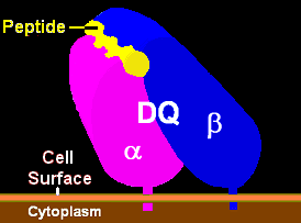
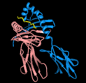
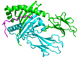
HLA-DQ (DQ) is a cell surface receptor protein found on antigen-presenting cells. It is an αβ heterodimer of type MHC class II. The α and β chains are encoded by two loci, HLA-DQA1 and HLA-DQB1, that are adjacent to each other on chromosome band 6p21.3. Both α-chain and β-chain vary greatly. A person often produces two α-chain and two β-chain variants and thus 4 isoforms of DQ. The DQ loci are in close genetic linkage to HLA-DR, and less closely linked to HLA-DP, HLA-A, HLA-B and HLA-C.

HLA DR3-DQ2 is double serotype that specifically recognizes cells from individuals who carry a multigene HLA DR, DQ haplotype. Certain HLA DR and DQ genes have known involvement in autoimmune diseases. DR3-DQ2, a multigene haplotype, stands out in prominence because it is a factor in several prominent diseases, namely coeliac disease and juvenile diabetes. In coeliac disease, the DR3-DQ2 haplotype is associated with highest risk for disease in first degree relatives, highest risk is conferred by DQA1*0501:DQB1*0201 homozygotes and semihomozygotes of DQ2, and represents the overwhelming majority of risk. HLA DR3-DQ2 encodes DQ2.5cis isoform of HLA-DQ, this isoform is described frequently as 'the DQ2 isoform', but in actuality there are two major DQ2 isoform. The DQ2.5 isoform, however, is many times more frequently associated with autoimmune disease, and as a result to contribution of DQ2.2 is often ignored.

<span class="mw-page-title-main">Intraepithelial lymphocyte</span>

Intraepithelial lymphocytes (IEL) are lymphocytes found in the epithelial layer of mammalian mucosal linings, such as the gastrointestinal (GI) tract and reproductive tract. However, unlike other T cells, IELs do not need priming. Upon encountering antigens, they immediately release cytokines and cause killing of infected target cells. In the GI tract, they are components of gut-associated lymphoid tissue (GALT).

<span class="mw-page-title-main">Gluten-related disorders</span> Set of diseases caused by gluten exposure

Gluten-related disorders is the term for the diseases triggered by gluten, including celiac disease (CD), non-celiac gluten sensitivity (NCGS), gluten ataxia, dermatitis herpetiformis (DH) and wheat allergy. The umbrella category has also been referred to as gluten intolerance, though a multi-disciplinary physician-led study, based in part on the 2011 International Coeliac Disease Symposium, concluded that the use of this term should be avoided due to a lack of specificity.

Gluten-sensitive enteropathy–associated conditions are comorbidities or complications of gluten-related gastrointestinal distress. GSE has key symptoms typically restricted to the bowel and associated tissues; however, there are a wide variety of associated conditions. These include bowel disorders, eosinophilic gastroenteritis and increase with coeliac disease (CD) severity. With some early onset and a large percentage of late onset disease, other disorders appear prior to the coeliac diagnosis or allergic-like responses markedly increased in GSE. Many of these disorders persist on a strict gluten-free diet, and are thus independent of coeliac disease after triggering. For example, autoimmune thyroiditis is a common finding with GSE.

<span class="mw-page-title-main">HLA-DQ2</span>

HLA-DQ2 (DQ2) is a serotype group within HLA-DQ (DQ) serotyping system. The serotype is determined by the antibody recognition of β2 subset of DQ β-chains. The β-chain of DQ is encoded by HLA-DQB1 locus and DQ2 are encoded by the HLA-DQB1*02 allele group. This group currently contains two common alleles, DQB1*0201 and DQB1*0202. HLA-DQ2 and HLA-DQB1*02 are almost synonymous in meaning. DQ2 β-chains combine with α-chains, encoded by genetically linked HLA-DQA1 alleles, to form the cis-haplotype isoforms. These isoforms, nicknamed DQ2.2 and DQ2.5, are also encoded by the DQA1*0201 and DQA1*0501 genes, respectively.

<span class="mw-page-title-main">HLA-DQ7</span>

HLA-DQ7 (DQ7) is an HLA-DQ serotype that recognizes the common HLA DQB1*0301 and the less common HLA DQB1*0304 gene products. DQ7 is a form of 'split antigen' of the broad antigen group DQ3 which also contains DQ8 and DQ9.

<span class="mw-page-title-main">HLA-DQB1</span> Protein-coding gene in the species Homo sapiens

Major histocompatibility complex, class II, DQ beta 1, also known as HLA-DQB1, is a human gene and also denotes the genetic locus that contains this gene. The protein encoded by this gene is one of two proteins that are required to form the DQ heterodimer, a cell surface receptor essential to the function of the immune system.

Oat sensitivity represents a sensitivity to the proteins found in oats, Avena sativa. Sensitivity to oats can manifest as a result of allergy to oat seed storage proteins either inhaled or ingested. A more complex condition affects individuals who have gluten-sensitive enteropathy in which there is an autoimmune response to avenin, the glutinous protein in oats similar to the gluten within wheat. Sensitivity to oat foods can also result from their frequent contamination by wheat, barley, or rye particles.

The immunochemistry of Triticeae glutens is important in several inflammatory diseases. It can be subdivided into innate responses, class II mediated presentation, class I mediated stimulation of killer cells, and antibody recognition. The responses to gluten proteins and polypeptide regions differs according to the type of gluten sensitivity. The response is also dependent on the genetic makeup of the human leukocyte antigen genes. In gluten sensitive enteropathy, there are four types of recognition, innate immunity, HLA-DQ, and antibody recognition of gliadin and transglutaminase. With idiopathic gluten sensitivity only antibody recognition to gliadin has been resolved. In wheat allergy, the response pathways are mediated through IgE against other wheat proteins and other forms of gliadin.

HLA A1-B8-DR3-DQ2 haplotype is a multigene haplotype that covers a majority of the human major histocompatibility complex on chromosome 6. A multigene haplotype is set of inherited alleles covering several genes, or gene-alleles; common multigene haplotypes are generally the result of descent by common ancestry. Chromosomal recombination fragments multigene haplotypes as the distance to that ancestor increases in number of generations.

<span class="mw-page-title-main">Extranodal NK/T-cell lymphoma, nasal type</span> Medical condition

Extranodal NK/T-cell lymphoma, nasal type (ENKTCL-NT) is a rare type of lymphoma that commonly involves midline areas of the nasal cavity, oral cavity, and/or pharynx At these sites, the disease often takes the form of massive, necrotic, and extremely disfiguring lesions. However, ENKTCL-NT can also involve the eye, larynx, lung, gastrointestinal tract, skin, and various other tissues. ENKTCL-NT mainly affects adults; it is relatively common in Asia and to lesser extents Mexico, Central America, and South America but is rare in Europe and North America. In Korea, ENKTCL-NT often involves the skin and is reported to be the most common form of cutaneous lymphoma after mycosis fungoides.

Duodenal lymphocytosis, sometimes called lymphocytic duodenitis, lymphocytic duodenosis, or duodenal intraepithelial lymphocytosis, is a condition where an increased number of intra-epithelial lymphocytes is seen in biopsies of the duodenal mucosa when these are examined microscopically. This form of lymphocytosis is often a feature of coeliac disease but may be found in other disorders.

Natural killer cell enteropathy, also termed NK cell enteropathy (NKCE), and a closely related disorder, lymphomatoid gastropathy (LG), are non-malignant diseases in which one type of lymphocyte, the natural killer cell, proliferates excessively in the gastrointestinal tract. This proliferation causes red, sore-like spots, raised lesions, erosions, and ulcers in the mucosal layer surrounding the GI tract lumen. Both disorders cause either no or only vague symptoms of GI tract disturbances such as nausea, vomiting, and bleeding.

Indolent T cell lymphoproliferative disorder of the gastrointestinal tract or Indolent T cell lymphoproliferative disorder of the GI tract (ITCLD-GT) is a rare and recently recognized disorder in which mature T cell lymphocytes accumulation abnormally in the gastrointestinal tract. This accumulation causes various lesions in the mucosal layer lining the GI tract. Individuals with ITCLD-GT commonly complain of chronic GI tract symptoms such as nausea, vomiting, diarrhea, abdominal pain, and rectal bleeding.

Monomorphic epitheliotropic intestinal T cell lymphoma (MEITL) is an extremely rare peripheral T-cell lymphoma that involves the malignant proliferation of a type of lymphocyte, the T cell, in the gastrointestinal tract. Over time, these T cells commonly spread throughout the mucosal lining of a portion of the GI tract, lead to GI tract nodules and ulcerations, and cause symptoms such as abdominal pain, weight loss, diarrhea, obstruction, bleeding, and/or perforation.

References

  1. 1 2 Nijeboer P, Malamut G, Mulder CJ, Cerf-Bensussan N, Sibon D, Bouma G, Cellier C, Hermine O, Visser O (2015). "Enteropathy-associated T-cell lymphoma: improving treatment strategies". Digestive Diseases. 33 (2): 231–5. doi:10.1159/000369542. PMID   25925928. S2CID   25886266.
  2. Isaacson PG (October 1994). "Gastrointestinal lymphoma". Hum. Pathol. 25 (10): 1020–9. doi:10.1016/0046-8177(94)90060-4. PMID   7927306.
  3. 1 2 3 4 5 6 7 8 9 10 11 12 13 14 15 16 17 18 19 20 Chander U, Leeman-Neill RJ, Bhagat G (August 2018). "Pathogenesis of Enteropathy-Associated T Cell Lymphoma". Current Hematologic Malignancy Reports. 13 (4): 308–317. doi:10.1007/s11899-018-0459-5. PMID   29943210. S2CID   49430640.
  4. 1 2 3 4 5 6 7 8 9 Ondrejka S, Jagadeesh D (December 2016). "Enteropathy-Associated T-Cell Lymphoma". Current Hematologic Malignancy Reports. 11 (6): 504–513. doi:10.1007/s11899-016-0357-7. PMID   27900603. S2CID   13329863.
  5. Matutes E (May 2018). "The 2017 WHO update on mature T- and natural killer (NK) cell neoplasms". International Journal of Laboratory Hematology. 40 (Suppl 1): 97–103. doi: 10.1111/ijlh.12817 . PMID   29741263.
  6. 1 2 3 Hoytema van Konijnenburg DP, Mucida D (August 2017). "Intraepithelial lymphocytes". Current Biology. 27 (15): R737–R739. doi: 10.1016/j.cub.2017.05.073 . PMID   28787597.
  7. 1 2 3 4 5 Olivares-Villagómez D, Van Kaer L (April 2018). "Intestinal Intraepithelial Lymphocytes: Sentinels of the Mucosal Barrier". Trends in Immunology. 39 (4): 264–275. doi:10.1016/j.it.2017.11.003. PMC   8056148 . PMID   29221933.
  8. 1 2 3 4 5 6 7 Foukas PG, de Leval L (January 2015). "Recent advances in intestinal lymphomas". Histopathology. 66 (1): 112–36. doi:10.1111/his.12596. PMID   25639480. S2CID   20669863.
  9. 1 2 3 4 5 6 7 8 9 10 11 12 Malamut G, Cellier C (June 2015). "Complications of coeliac disease". Best Practice & Research. Clinical Gastroenterology. 29 (3): 451–8. doi:10.1016/j.bpg.2015.05.005. PMID   26060109.
  10. 1 2 3 4 5 6 7 8 9 10 11 12 13 Al-Bawardy B, Codipilly DC, Rubio-Tapia A, Bruining DH, Hansel SL, Murray JA (February 2017). "Celiac disease: a clinical review". Abdominal Radiology. 42 (2): 351–360. doi:10.1007/s00261-016-1034-y. PMID   28078381. S2CID   3974481.
  11. 1 2 3 4 Mayassi T, Jabri B (September 2018). "Human intraepithelial lymphocytes". Mucosal Immunology. 11 (5): 1281–1289. doi:10.1038/s41385-018-0016-5. PMC   6178824 . PMID   29674648.
  12. Paul SP, Hoghton M, Sandhu B (February 2017). "Limited role of HLA DQ2/8 genotyping in diagnosing coeliac disease". Scottish Medical Journal. 62 (1): 25–27. doi:10.1177/0036933016689008. PMID   28084899. S2CID   206428837.
  13. "Alleles Report < IMGT/HLA < IPD < EMBL-EBI". Archived from the original on 2016-03-06. Retrieved 2019-04-14.
  14. "Alleles Report < IMGT/HLA < IPD < EMBL-EBI". Archived from the original on 2016-04-15. Retrieved 2019-04-14.
  15. Jores RD, Frau F, Cucca F, et al. (2007). "HLA-DQB1*0201 homozygosis predisposes to severe intestinal damage in celiac disease". Scand. J. Gastroenterol. 42 (1): 48–53. doi:10.1080/00365520600789859. PMID   17190762. S2CID   7675714.
  16. 1 2 3 4 5 6 7 Nasr I, Nasr I, Campling H, Ciclitira PJ (2016). "Approach to patients with refractory coeliac disease". F1000Research. 5: 2544. doi: 10.12688/f1000research.9051.1 . PMC   5074352 . PMID   27803799.
  17. 1 2 Cichewicz AB, Mearns ES, Taylor A, Boulanger T, Gerber M, Leffler DA, Drahos J, Sanders DS, Thomas Craig KJ, Lebwohl B (March 2019). "Diagnosis and Treatment Patterns in Celiac Disease". Digestive Diseases and Sciences. 64 (8): 2095–2106. doi:10.1007/s10620-019-05528-3. PMID   30820708. S2CID   71143826.
  18. 1 2 3 Kooy-Winkelaar YM, Bouwer D, Janssen GM, Thompson A, Brugman MH, Schmitz F, de Ru AH, van Gils T, Bouma G, van Rood JJ, van Veelen PA, Mearin ML, Mulder CJ, Koning F, van Bergen J (February 2017). "CD4 T-cell cytokines synergize to induce proliferation of malignant and nonmalignant innate intraepithelial lymphocytes". Proceedings of the National Academy of Sciences of the United States of America. 114 (6): E980–E989. Bibcode:2017PNAS..114E.980K. doi: 10.1073/pnas.1620036114 . PMC   5307453 . PMID   28049849.
  19. 1 2 Chan JY, Lim ST (April 2018). "Novel findings from the Asian Lymphoma Study Group: focus on T and NK-cell lymphomas". International Journal of Hematology. 107 (4): 413–419. doi: 10.1007/s12185-018-2406-6 . PMID   29380182.
  20. 1 2 3 Moffitt AB, Ondrejka SL, McKinney M, Rempel RE, Goodlad JR, Teh CH, Leppa S, Mannisto S, Kovanen PE, Tse E, Au-Yeung RK, Kwong YL, Srivastava G, Iqbal J, Yu J, Naresh K, Villa D, Gascoyne RD, Said J, Czader MB, Chadburn A, Richards KL, Rajagopalan D, Davis NS, Smith EC, Palus BC, Tzeng TJ, Healy JA, Lugar PL, Datta J, Love C, Levy S, Dunson DB, Zhuang Y, Hsi ED, Dave SS (May 2017). "Enteropathy-associated T cell lymphoma subtypes are characterized by loss of function of SETD2". The Journal of Experimental Medicine. 214 (5): 1371–1386. doi:10.1084/jem.20160894. PMC   5413324 . PMID   28424246.
  21. 1 2 Richir M, Songun I, Wientjes C, Snel P, Dwars B (October 2010). "Small Bowel Adenocarcinoma in a Patient with Coeliac Disease: Case Report and Review of the Literature". Case Reports in Gastroenterology. 4 (3): 416–420. doi:10.1159/000313547. PMC   2975010 . PMID   21060711.
  22. 1 2 Caio G, Volta U, Ursini F, Manfredini R, De Giorgio R (March 2019). "Small bowel adenocarcinoma as a complication of celiac disease: clinical and diagnostic features". BMC Gastroenterology. 19 (1): 45. doi: 10.1186/s12876-019-0964-6 . PMC   6437995 . PMID   30917787.
  23. Zullo A, De Francesco V, Manta R, Ridola L, Lorenzetti R (December 2017). "A Challenging Diagnosis of Jejunal Adenocarcinoma in a Celiac Patient: Case Report and Systematic Review of the Literature". Journal of Gastrointestinal and Liver Diseases. 26 (4): 411–415. doi: 10.15403/jgld.2014.1121.264.zet . PMID   29253057.
  24. Green PH, Cellier C (October 2007). "Celiac disease". The New England Journal of Medicine. 357 (17): 1731–43. doi:10.1056/NEJMra071600. PMID   17960014.
  25. Benhammane H, El M'rabet FZ, Idrissi Serhouchni K, El Yousfi M, Charif I, Toughray I, Mellas N, Riffi Amarti A, Maazaz K, Ibrahimi SA, El Mesbahi O (2012). "Small bowel adenocarcinoma complicating coeliac disease: a report of three cases and the literature review". Case Reports in Oncological Medicine. 2012: 935183. doi: 10.1155/2012/935183 . PMC   3517832 . PMID   23243535.
  26. 1 2 Broccoli A, Zinzani PL (March 2017). "Peripheral T-cell lymphoma, not otherwise specified". Blood. 129 (9): 1103–1112. doi: 10.1182/blood-2016-08-692566 . PMID   28115372.
  27. Hapgood G, Savage KJ (July 2015). "The biology and management of systemic anaplastic large cell lymphoma". Blood. 126 (1): 17–25. doi: 10.1182/blood-2014-10-567461 . PMID   25869285.
  28. Yamaguchi M, Oguchi M, Suzuki R (September 2018). "Extranodal NK/T-cell lymphoma: Updates in biology and management strategies". Best Practice & Research. Clinical Haematology. 31 (3): 315–321. doi:10.1016/j.beha.2018.07.002. PMID   30213402. S2CID   52272644.
  29. Farrell PJ (2019). "Epstein–Barr Virus and Cancer". Annual Review of Pathology. 14: 29–53. doi:10.1146/annurev-pathmechdis-012418-013023. PMID   30125149. S2CID   52051261.
  30. 1 2 Matnani R, Ganapathi KA, Lewis SK, Green PH, Alobeid B, Bhagat G (March 2017). "Indolent T- and NK-cell lymphoproliferative disorders of the gastrointestinal tract: a review and update". Hematological Oncology. 35 (1): 3–16. doi: 10.1002/hon.2317 . PMID   27353398. S2CID   21364706.
  31. Lemonnier F, Gaulard P, de Leval L (September 2018). "New insights in the pathogenesis of T-cell lymphomas" (PDF). Current Opinion in Oncology. 30 (5): 277–284. doi:10.1097/CCO.0000000000000474. PMID   30028743. S2CID   51704108.
  32. Jantunen, E; Boumendil, A; Finel, H; Luan, J. J.; Johnson, P; Rambaldi, A; Haynes, A; Duchosal, M. A.; Bethge, W; Biron, P; Carlson, K; Craddock, C; Rudin, C; Finke, J; Salles, G; Kroschinsky, F; Sureda, A; Dreger, P; Lymphoma Working Party of the EBMT (2013). "Autologous stem cell transplantation for enteropathy-associated T-cell lymphoma: A retrospective study by the EBMT". Blood. 121 (13): 2529–32. doi: 10.1182/blood-2012-11-466839 . PMID   23361910.
  33. "Home Page". Archived from the original on 2019-11-15. Retrieved 2019-04-14.
  34. "Phase 2 Study of Brentuximab Vedotin Associated with CHP Followed by Consolidation with High-dose Therapy / Autologous Stem-cell Transplantation as Frontline Treatment of Patients with Enteropathy-associated T-cell Lymphoma Type 1". 9 February 2021.
  35. "T Cells Expressing a Fully-Human Anti-CD30 Chimeric Antigen Receptor for Treating CD30-Expressing Lymphomas - Tabular View - ClinicalTrials.gov". clinicaltrials.gov.
  36. "Nivolumab in Treating Patients With Relapsed or Refractory Peripheral T-cell Lymphoma - Tabular View - ClinicalTrials.gov". clinicaltrials.gov.
  37. "A Phase 2a, Randomized, Double-Blind, Placebo-Controlled, Parallel-Group Study to Evaluate the Efficacy and Safety of AMG 714 in Adult Patients with Type II Refractory Celiac Disease, an in Situ Small Bowel T Cell Lymphoma". 11 December 2019.
  38. "A Phase 2a, Randomized, Double-Blind, Placebo-Controlled, Parallel-Group Study to Evaluate the Efficacy and Safety of AMG 714 in Adult Patients with Type II Refractory Celiac Disease, an in Situ Small Bowel T Cell Lymphoma". 11 December 2019.