Kernohan's notch

Last updated

Kernohan's notch is a cerebral peduncle indentation associated with some forms of transtentorial herniation (uncal herniation). [1] [2] It is a secondary condition caused by a primary injury on the opposite hemisphere of the brain. [3] Kernohan's notch is an ipsilateral condition, in that a left-sided primary lesion (in which Kernohan's notch would be on the right side) evokes motor impairment in the left side of the body and a right-sided primary injury evokes motor impairment in the right side of the body. [4] The seriousness of Kernohan's notch varies depending on the primary problem causing it, which may range from benign brain tumors to advanced subdural hematoma.

Contents

Mechanism

Kernohan's notch phenomenon is a result of the compression of the cerebral peduncle, which is part of the mesencephalon, against the tentorium cerebelli due to transtentorial herniation. This produces ipsilateral hemiparesis or hemiplegia [5]

The skull is an incompressible closed space with a limited volume (Monro-Kellie Doctrine). When there is increased cranial pressure in the brain, a shift in the brain forms towards the only opening of the skull, the foramen magnum. Thus, when an increase of pressure in a hemisphere of the brain exists, the cerebral peduncle on the opposite hemisphere is pushed up against the tentorium, which separates the posterior fossa from the middle fossa. This produces a visible "notch" in the cerebral peduncle. [4] Because a Kernohan's notch is caused by an injury creating pressure on the opposite hemisphere of the brain, it is characterized as a false localizing sign. [3]

The Kernohan's notch phenomenon is unique in that it is not only a false localizing sign, but is also ipsilateral or same-sided. The left cerebral peduncle contains motor fibers that cross over to the right side of the body. Thus, if you have a right hemisphere trans-tentorial herniation, it causes a Kernohan's notch in the left cerebral peduncle which results in right-sided motor impairment. Therefore, you get, paradoxically, impairment of motor function on the same side of the body as the herniation which caused the Kernohan's notch on the controlateral side. [3]

Causes

The Kernohan's notch is a secondary phenomenon that results from a major primary injury. Non-tumoral, non-traumatic, intracranial haemorrhage rarely causes this phenomenon.

A wide range of serious injuries can cause an increase in intracranial pressure to trigger the formation of a Kernohan's notch. In general, this phenomenon occurs in patients with advanced brain tumor or severe head injury. [6] In the case of severe head injury, a clot can occur over the surface of the brain and can often cause shift of the middle part of the brain against the tentorium, which creates the Kernohan's notch. Chronic subdural hematomas have been known to be a familiar cause of Kernohan's notch. [7]

MRIs have shown evidence of Kernohan's notch from patients with traumatic head injury that are related to acute space-occupying lesions such as subdural hematoma, epidural hematoma, depressed skull fracture, or spontaneous intracerebral hematoma. [8] [9]

Also, it is important to note that the anatomical size of tentorial notches vary considerably between individuals; however, very little evidence supports that a more narrow notch creates a predisposition towards Kernohan's notch. [8]

Signs and symptoms

Symptoms directly related to the Kernohan's notch is most commonly paralysis or weakness on one side of the body (ipsilateral paralysis / paresis), [8] the so-called Kernohan's sign. Paralysis and weakness is known as hemiplegia and hemiparesis, respectively. This is due to destruction or pressure applied to the motor fibers located in the cerebral peduncle. A more rare sign of Kernohan's notch is ipsilateral oculomotor nerve palsy. [10]

However, most patients come into the clinic citing symptoms associated with the primary injury causing the Kernohan's notch. Since so many types of head injuries exist, virtually any symptom of brain trauma can be seen accompanying Kernohan's notch. These symptoms may range from total paralysis to simple headache, nausea, and vomiting. [3]

Diagnosis

Because of the ipsilateral characteristic of Kernohan's notch, diagnosis is unique. Many clinicians assume a right sided paralysis corresponds with an injury in the left hemisphere of the brain and misdiagnose Kernohan's notch. [3] Despite this complication that ipsilateral paralysis causes, it makes it very easy to diagnose when coupled with imaging techniques. For example, when seeing an MRI of a blood clot on the left side of the brain coupled with left-sided paralysis, it immediately points to Kernohan's notch.

In most head trauma cases, CT scans are the standard diagnostic method; however it is not ideal for imaging small lesions, so MRI is used to identify Kernohan's notch. It is important to distinguish Kernohan's notch from direct brain stem injuries. Case studies have shown that in patients with chronic subdural hematoma, a compressive deformity of the crus cerebri without an abnormal MRI signal may predict a better recovery in patients with Kernohan's notch. [8]

Treatments

There is no special treatment for Kernohan's notch since treatment is completely dependent on the injury causing it. The pressure against the tentorium should be relieved and the notch should go away. However, this does not mean that damage to the motor fibers disappears. Depending on the severity of the injury, there may or may not be persistent damage after pressure relief. Neurological deficits may resolve after surgery, but some degree of deficit, especially motor weakness, generally remains. [8]

Pressure relief to "un-notch" the cerebral peduncle may include the removal of brain tumors and blood clots or the suction of blood from a drilled hole in the skull. [3]

Case studies

Kernohan's notch and misdiagnosis

The ipsilateral and false localizing signs characteristic of Kernohan's notch sometimes cause confusion and result in misdiagnosis. Such a case is described in an account by Wolf at the University Hospital in Groningen, Netherlands: [3]

"A 39 year-old man sustained a minor head injury when he was struck on the head by a golf club. 5 hours later, he had sudden onset of headache with nausea and vomiting, Simultaneously he developed muscle weakness on his left side. He then became somnolent. A CT scan showed a subdural blood collection on the left side...these findings puzzled the attending neurologist as well as the radiologist who both expected the abnormalities to be on the opposite (right) side. The radiological technician remarked that the left-right marks were not in the usual place on the monitor screen and argued that the latest scan had been a coronal infundibulum scan in which a top-view is used instead of a bottom view...It was then decided that the patient had a right-sided acute subdural haematoma. In the operating room a right-sided drilling hole was made but no blood was aspirated, nor was it from a second right-sided drilling hole. A right-sided craniotomy was then done, which did not show any signs of a subdural blood collection and the operation was ended...a postoperative CT scan the next day showed a subdural haematoma on the left side and the signs of craniotomy on the right...Reconstruction of the events led to the conclusions that the false localizing signs were caused by a Kernohan notch...the left-right marks on the screen had initially been correct but were wrongly switched to fit the patients' clinical symptoms. This sad, but unique, example of misdiagnosis has prompted us to re-evaluate the index settings before examination of each new patient."

Ruptured occipital arteriovenous malformation

Kernohan's notch phenomenon associated with a ruptured arteriovenous malformation (AVM) is rare, but not unknown. This was accounted for in a case study described by Fujimoto at the Tsukuba Memorial Hospital in Ibaraki, Japan: [10]

"The patient, a 23 year-old woman, visited our outpatient clinic with a chief complaint of severe headache, nausea and vomiting. Her pupils did not react to light and her left pupil was mydriasic...The patient was immediately operated on to decompress the brain and remove the subdural hematoma. The onset-operation interval was 46 min...Intraoperatively, a parenchymal AVM was found in the occipital area, which was removed. On day four part onset, we noticed left hemiparesis with a partial left oculomotor nerve palsy, the so called Kernohan's phenomenon...One month after onset, the patient had no significant neurological deficit...We believe that her good outcome with little neurological deficit was due to the short interval from onset to the first operation."

Another case-report involving AVM:

"Right AVM produced a Kernohan's notch phenomenon in a 33 year-old male who was admitted to the Inland Empire Medical Center with full seizures and a GCS of 6. CT evaluation revealed a subdural hematoma that was evacuated with an emergency craniotomy about 45 minutes after initial onset. Patient experienced transient right-sided paralysis of limbs that resolved after 48 hours. The volume of blood flow across the dura created when the AVM burst caused compression of the brainstem on the level of the tentorium on the opposite side. Subsequently, the patient was transferred to Cedars-Sinai under the surgical care of Wouter Schievink, MD and has made a complete recovery after one-year."

History

Early findings

One of the first references to casual brain herniation was that of James Colier, who clearly described cerebellar tonsilar herniation in 1904. [11] He observed accompanying false localizing signs and commented:

"In many cases of intracranial tumour of long duration, it was found postmortem that the posterior inferior part of the cerebellum had been pushed down and backwards into the foramen magnum and the medulla itself somewhat caudally displaced, the two structures together forming a cone-shaped plug tightly filling up the foramen magnum."

In 1920, Adolf Meyer confirmed the pathologies of brain herniation. [12] He commented:

"The falx and tentorium constitute an important protection against any sudden impacts of pressure by keeping apart heavy portions of the brain, but they also provide an opportunity for trouble in case of swelling or need of displacement."

The work of Collier and Meyer described ipsilateral hemiparesis, a false localizing sign. However, it became known as the Kernohan-Woltman phenomenon. [13]

Kernohan-Woltman

In 1929, Kernohan and Woltman published their work on brain lesions that showed ipsilateral hemiplegia. [14] In their paper, they state:

"The tumour was often large enough to displace the brain toward the opposite side and also to cause herniation through the tentorium. Such herniation and displacement may be evidenced by a groove sweeping over the uncinate gyrus on the side of the tumour. On the opposite side the groove may be absent...".(p. 282)

Kernohan fully described the Kernohan's notch in 1929 and is given credit for its discovery.

See also

This is an interesting story in that Kernohan and Woltman's article never did support their own contention that the ipsilateral finding they reported were due to the notching from the displacement of the hemisphere containing the tumor (as follows): As seen in their Fig. 7, There were a total of 35 supratentorial cases all of them with midbrain notching opposite the hemisphere bearing the lesion. However, only 17 of these 35 had "false" localizing signs (ipsilateral to the tumor; hyperactive reflexes, upgoing toes, etc.).The rest did not have such signs. Therefore, logically speaking, there were no cause and effect relationship between notching and the "herniation" signs (again, since exactly half of those with the notches were "asymptomatic." The reason for occurrence of these signs in half of the population affected must have been the laterality of the hemisphere harboring the tumor, not the presence or absence of a notch resulting from the tumor (which was a fact present in all cases). We now know that influences of the hemisphere of speech (i.e. the command center, major hemisphere) on the other hemisphere (the minor) is excitatory and via the corpus callosum. Therefore, only those cases with tumors in the major hemisphere suffered deafferentation of the contralateral (minor) hemisphere; rendering it nonfunctional, causing appearance of signs on the same side as the tumor. Those with tumors in the nondominant hemisphere (minor) did not have ipsilateral signs as there is no motor communication from the minor to the major hemisphere to cause deafferentation of the major hemisphere (diaschisis) (see www.mimickingman.com, for details).

Notes

  1. "Definition: Kernohan's notch from Online Medical Dictionary".
  2. "Medcyclopaedia - Kernohan's notch".
  3. 1 2 3 4 5 6 7 Wolf et al, 1995
  4. 1 2 Pearce, 2006
  5. Moon, 2006
  6. Jones et al, 1991
  7. Yamasaki et al, 1997
  8. 1 2 3 4 5 Moon et al, 2006
  9. Kunii et al, 2005
  10. 1 2 Fujimoto et al, 1995
  11. Collier J, 1904
  12. Meyer A, 1920
  13. Pearce J, 2006
  14. Kernohan et al, 1929

Carrasco et al., 2017; Zhang et al. 2017 review

Related Research Articles

<span class="mw-page-title-main">Brain abscess</span> Medical condition

Brain abscess is an abscess within the brain tissue caused by inflammation and collection of infected material coming from local or remote infectious sources. The infection may also be introduced through a skull fracture following a head trauma or surgical procedures. Brain abscess is usually associated with congenital heart disease in young children. It may occur at any age but is most frequent in the third decade of life.

Hemiparesis, or unilateral paresis, is weakness of one entire side of the body. Hemiplegia is, in its most severe form, complete paralysis of half of the body. Hemiparesis and hemiplegia can be caused by different medical conditions, including congenital causes, trauma, tumors, or stroke.

<span class="mw-page-title-main">Head injury</span> Serious trauma to the cranium

A head injury is any injury that results in trauma to the skull or brain. The terms traumatic brain injury and head injury are often used interchangeably in the medical literature. Because head injuries cover such a broad scope of injuries, there are many causes—including accidents, falls, physical assault, or traffic accidents—that can cause head injuries.

<span class="mw-page-title-main">Intracranial pressure</span> Pressure exerted by fluids inside the skull and on the brain

Intracranial pressure (ICP) is the pressure exerted by fluids such as cerebrospinal fluid (CSF) inside the skull and on the brain tissue. ICP is measured in millimeters of mercury (mmHg) and at rest, is normally 7–15 mmHg for a supine adult. The body has various mechanisms by which it keeps the ICP stable, with CSF pressures varying by about 1 mmHg in normal adults through shifts in production and absorption of CSF.

<span class="mw-page-title-main">Subdural hematoma</span> Hematoma usually associated with traumatic brain injury

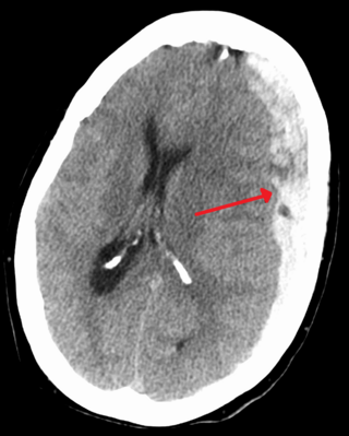
A subdural hematoma (SDH) is a type of bleeding in which a collection of blood—usually but not always associated with a traumatic brain injury—gathers between the inner layer of the dura mater and the arachnoid mater of the meninges surrounding the brain. It usually results from tears in bridging veins that cross the subdural space.

<span class="mw-page-title-main">Intracranial hemorrhage</span> Hemorrhage, or bleeding, within the skull

Intracranial hemorrhage (ICH), also known as intracranial bleed, is bleeding within the skull. Subtypes are intracerebral bleeds, subarachnoid bleeds, epidural bleeds, and subdural bleeds.

<span class="mw-page-title-main">Epidural hematoma</span> Build-up of blood between the dura mater and skull

Epidural hematoma is when bleeding occurs between the tough outer membrane covering the brain and the skull. Often there is loss of consciousness following a head injury, a brief regaining of consciousness, and then loss of consciousness again. Other symptoms may include headache, confusion, vomiting, and an inability to move parts of the body. Complications may include seizures.

Monoplegia is paralysis of a single limb, usually an arm. Common symptoms associated with monoplegic patients are weakness, numbness, and pain in the affected limb. Monoplegia is a type of paralysis that falls under hemiplegia. While hemiplegia is paralysis of half of the body, monoplegia is localized to a single limb or to a specific region of the body. Monoplegia of the upper limb is sometimes referred to as brachial monoplegia, and that of the lower limb is called crural monoplegia. Monoplegia in the lower extremities is not as common of an occurrence as in the upper extremities. Monoparesis is a similar, but less severe, condition because one limb is very weak, not paralyzed. For more information, see paresis.

<span class="mw-page-title-main">Intracerebral hemorrhage</span> Type of intracranial bleeding that occurs within the brain tissue itself

Intracerebral hemorrhage (ICH), also known as hemorrhagic stroke, is a sudden bleeding into the tissues of the brain, into its ventricles, or into both. An ICH is a type of bleeding within the skull and one kind of stroke. Symptoms can vary dramatically depending on the severity, acuity, and location (anatomically) but can include headache, one-sided weakness, numbness, tingling, or paralysis, speech problems, vision or hearing problems, memory loss, attention problems, coordination problems, balance problems, dizziness or lightheadedness or vertigo, nausea/vomiting, seizures, decreased level of consciousness or total loss of consciousness, neck stiffness, and fever.

<span class="mw-page-title-main">Intraparenchymal hemorrhage</span> Medical condition

Intraparenchymal hemorrhage (IPH) is one form of intracerebral bleeding in which there is bleeding within brain parenchyma. The other form is intraventricular hemorrhage (IVH).

<span class="mw-page-title-main">Brain herniation</span> Potentially deadly side effect of very high pressure within the skull

Brain herniation is a potentially deadly side effect of very high pressure within the skull that occurs when a part of the brain is squeezed across structures within the skull. The brain can shift across such structures as the falx cerebri, the tentorium cerebelli, and even through the foramen magnum. Herniation can be caused by a number of factors that cause a mass effect and increase intracranial pressure (ICP): these include traumatic brain injury, intracranial hemorrhage, or brain tumor.

<span class="mw-page-title-main">Duret haemorrhages</span> Medical condition

Duret haemorrhages are small linear areas of bleeding in the midbrain and upper pons of the brainstem. They are caused by a traumatic downward displacement of the brainstem.

<span class="mw-page-title-main">Uncus</span> Structure in the brain

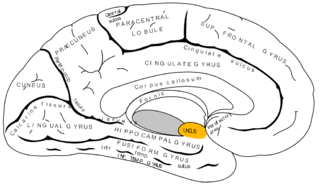
The uncus is an anterior extremity of the parahippocampal gyrus. It is separated from the apex of the temporal lobe by a slight fissure called the incisura temporalis.

In medicine, a mass effect is the effect of a growing mass that results in secondary pathological effects by pushing on or displacing surrounding tissue.

Central facial palsy is a symptom or finding characterized by paralysis or paresis of the lower half of one side of the face. It usually results from damage to upper motor neurons of the facial nerve.

<span class="mw-page-title-main">Brown-Séquard syndrome</span> Human spinal cord disorder

Brown-Séquard syndrome is caused by damage to one half of the spinal cord, i.e. hemisection of the spinal cord resulting in paralysis and loss of proprioception on the same side as the injury or lesion, and loss of pain and temperature sensation on the opposite side as the lesion. It is named after physiologist Charles-Édouard Brown-Séquard, who first described the condition in 1850.

James Watson Kernohan, M.D. (1896–1981) was an Irish-American pathologist born October 1, 1896, in County Antrim, Ireland. He studied medicine at Queen's University Belfast, and in 1922 he emigrated to the United States and subsequently worked as a pathologist at the Mayo Clinic in Rochester, Minnesota. Kernohan retired from active medical practice in 1962 and died May 5, 1981.

Amorphosynthesis, also called a hemi-sensory deficit, is a neuropsychological condition in which a patient experiences unilateral inattention to sensory input. This phenomenon is frequently associated with damage to the right cerebral hemisphere resulting in severe sensory deficits that are observed on the contralesional (left) side of the body. A right-sided deficit is less commonly observed and the effects are reported to be temporary and minor. Evidence suggests that the right cerebral hemisphere has a dominant role in attention and awareness to somatic sensations through ipsilateral and contralateral stimulation. In contrast, the left cerebral hemisphere is activated only by contralateral stimuli. Thus, the left and right cerebral hemispheres exhibit redundant processing to the right-side of the body and a lesion to the left cerebral hemisphere can be compensated by the ipsiversive processes of the right cerebral hemisphere. For this reason, right-sided amorphosynthesis is less often observed and is generally associated with bilateral lesions.

A sports-related traumatic brain injury is a serious accident which may lead to significant morbidity or mortality. Traumatic brain injury (TBI) in sports are usually a result of physical contact with another person or stationary object, These sports may include boxing, gridiron football, field/ice hockey, lacrosse, martial arts, rugby, soccer, wrestling, auto racing, cycling, equestrian, rollerblading, skateboarding, skiing or snowboarding.

<span class="mw-page-title-main">Raymond–Céstan syndrome</span> Medical condition

Raymond–Céstan syndrome is caused by blockage of the long circumferential branches of the basilar artery. It was described by Fulgence Raymond and Étienne Jacques Marie Raymond Céstan. Along with other related syndromes such as Millard–Gubler syndrome, Foville's syndrome, and Weber's syndrome, the description was instrumental in establishing important principles in brain-stem localization.

References