Foramen magnum

Last updated
Foramen magnum
Crane4 Foramen magnum.png
Upper surface of base of the skull. The hole indicated by an arrow is the foramen magnum
Gray130.png
Occipital bone. Inner surface.
Details
Part of Occipital bone
System Skeletal
Identifiers
Latin foramen magnum
MeSH D005539
TA98 A02.1.04.002
TA2 553
FMA 75306
Anatomical terms of bone

The foramen magnum (Latin : great hole) is a large, oval-shaped opening in the occipital bone of the skull. It is one of the several oval or circular openings (foramina) in the base of the skull. The spinal cord, an extension of the medulla oblongata, passes through the foramen magnum as it exits the cranial cavity. Apart from the transmission of the medulla oblongata and its membranes, the foramen magnum transmits the vertebral arteries, the anterior and posterior spinal arteries, the tectorial membranes and alar ligaments. It also transmits the accessory nerve into the skull.

Contents

The foramen magnum is a very important feature in bipedal mammals. One of the attributes of a biped's foramen magnum is a forward shift of the anterior border of the cerebellar tentorium; this is caused by the shortening of the cranial base. Studies on the foramen magnum position have shown a connection to the functional influences of both posture and locomotion. The forward shift of the foramen magnum is apparent in bipedal hominins, including modern humans, Australopithecus africanus, and Paranthropus boisei. This common feature of bipedal hominins is the driving argument used by Michel Brunet that Sahelanthropus tchadensis was also bipedal, and may be the earliest known bipedal ape. The discovery of this feature has given scientists another form of identifying bipedal mammals. [1]

Structure

The foramen magnum is a large, oval-shaped opening (foramen) in the occipital bone of the skull. [2] It is present in humans, and in many other animals. Anteriorly, it is bounded by the basiocciput. [2] Posteriorly, it is bounded by the supraocciput. [2] Laterally, it is bounded by the occipital condyles. [2]

On the occipital bone, the foramen magnum presents two midline cephalometric landmarks. The opisthion is the midpoint on the posterior margin of the foramen magnum. The basion is located at the midpoint on the anterior margin of the foramen magnum.

The alar ligament, which is attached on each side to the tubercle of occipital condyle, divides the foramen magnum into an anterior smaller compartment and a posterior larger compartment. [3]

Variation

From the size, position and where it is located the foramen magnum can help determine many factors. In humans, the foramen magnum is found to be located and positioned anteriorly. [4] In some rodents and mammals, the foramen magnum can be found anteriorly in the cranium. [4]

The foramen magnum varies in size between individuals. Earlier ossification of the occipital bone leads to a smaller foramen. [5]

The foramen magnum varies in size and shape when comparing different populations to each other. In humans, men tend to have a larger sized foramen magnum than women, but the overall shape is consistent. [6]

In humans, the foramen magnum is farther underneath the head than in the other great apes. Thus, in humans, the neck muscles (including the occipitofrontalis muscle) do not need to be as robust in order to hold the head upright. Comparisons of the position of the foramen magnum in early hominid species are useful to determine how comfortable a particular species was when walking on two limbs (bipedalism) rather than four (quadrupedalism).

Heart-shaped foramen magnum

In an early hominin, Paranthropus boisei, also known as Australopithecus boisei, has a foramen magnum that is similar to a shape of a heart or a cardioid. [7] When KNM-ER 406 was examined among other species, the foramen magnum was different. Compared to the usual oval-shaped foramen magnum, the shape of the Australopithecus boisei foramen magnum appears to be shortened. In the foramen magnum of Australopithecus boisei, both the anterior and posterior are different in size and diameter. The anterior of the foramen magnum had a greater lateral diameter and was more broad compared to other early hominins. While the lateral diameter of the posterior foramen magnum was shortened to create more of a cardioid shape. The anterior edges of the foramen magnum in Australopithecus boisei lacks the oval curve that would normally appear in other species that have an oval to circular-shaped foramen magnum. The unique shape of the foramen magnum was also seen in some casts of Hadar species. [7] The Hadar shows traits and characteristics of having a similar cardioid shape. [7] The change in the shape of the foramen magnum has been studied to identify the possibility of different functions. There is a potential correlation between the change of shape of the foramen magnum to how it functions in the body. Although not proven, compared to other hominins, the route that blood travels through the foramen magnum in Australopithecus boisei are more straight and direct. [7] With the enlargement and broadening of the anterior foramen magnum, allow for more venous drainage from the occipital and marginal sinuses. [7]

Function

The foramen magnum transmits a number of important structures between the neck and the neurocranium.

Structures passing through anterior compartment (osseoligamentous compartment) include:

Structures passing through posterior compartment (neurovascular compartment) include:

Clinical significance

The foramen magnum may be too large, too small, or the wrong shape. [2] A small foramen magnum can cause neurological problems, and the reduced circulation of cerebrospinal fluid can cause hydrocephalus. [2] This may be treated with suboccipital craniectomy. [2]

Other animals

An anterior foramen magnum can be found in other bipedal mammals besides humans. [8] The jerboa, a bipedal rodent, also has a foramen magnum. [8]

Evolution of foramen magnum in fossils

Although not fully proven, there are many studies that show the possibility [9] that where the foramen magnum is positioned in the cranium is significant in fossils. By being able to locate where the foramen magnum is positioned, anthropologists and other researchers are able to determine whether or not the species were bipedal (among other factors). The positioning of the foramen magnum changing over time can be seen in different fossils. [10] Analyzing the foramen magnum in fossils like Ardipithecus ramidus, links the fossil to other earlier and later hominid who share similar anterior foramen magnum and other bipedalism features. Although not proven, there are correlations with the foramen magnum being more anterior positioned to the ability of mammals to walk on two limbs. [9] Comparing this to other animals, such as some primates, the foramen magnum are located more of a posterior position in the cranium. [10] With the foramen magnum being position anterior in the cranium, the body of bipedal mammals is given a different center of gravity compared to quadrupedal mammals. The anterior foramen magnum shifts the weight of the body more to the mammals' pelvis and femur, present in some primates, like great apes. With a posterior foramen magnum, the alignment and weight of the body falls more lateral under the head This allows for humans and other bipedal mammals to be able to walk on two limbs. Even with a posterior foramen magnum, some primates are still able to use their lower two limbs to walk.

With a common anterior foramen magnum in fossils, the fossils can be studied and researched to link mammals to others who are potentially bipedal. [10] With more correlations and similarities of fossils who have an anterior foramen magnum; it links to the idea that these species might be bipedal. The shift in the foramen magnum moving towards a more anterior position in the cranium, gives rise to the idea of possible bipedalism.


Additional images

See also

Related Research Articles

<i>Homo habilis</i> Archaic human species from 2.8 to 1.65 mya

Homo habilis is an extinct species of archaic human from the Early Pleistocene of East and South Africa about 2.8 million years ago to 1.65 million years ago (mya). Upon species description in 1964, H. habilis was highly contested, with many researchers recommending it be synonymised with Australopithecus africanus, the only other early hominin known at the time, but H. habilis received more recognition as time went on and more relevant discoveries were made. By the 1980s, H. habilis was proposed to have been a human ancestor, directly evolving into Homo erectus which directly led to modern humans. This viewpoint is now debated. Several specimens with insecure species identification were assigned to H. habilis, leading to arguments for splitting, namely into "H. rudolfensis" and "H. gautengensis" of which only the former has received wide support.

<i>Paranthropus</i> Contested extinct genus of hominins

Paranthropus is a genus of extinct hominin which contains two widely accepted species: P. robustus and P. boisei. However, the validity of Paranthropus is contested, and it is sometimes considered to be synonymous with Australopithecus. They are also referred to as the robust australopithecines. They lived between approximately 2.9 and 1.2 million years ago (mya) from the end of the Pliocene to the Middle Pleistocene.

<span class="mw-page-title-main">Atlas (anatomy)</span> First cervical vertebra of the spine which supports the skull

In anatomy, the atlas (C1) is the most superior (first) cervical vertebra of the spine and is located in the neck.

<i>Australopithecus</i> Genus of hominin ancestral to modern humans

Australopithecus is a genus of early hominins that existed in Africa during the Pliocene and Early Pleistocene. The genera Homo, Paranthropus, and Kenyanthropus evolved from some Australopithecus species. Australopithecus is a member of the subtribe Australopithecina, which sometimes also includes Ardipithecus, though the term "australopithecine" is sometimes used to refer only to members of Australopithecus. Species include A. garhi, A. africanus, A. sediba, A. afarensis, A. anamensis, A. bahrelghazali and A. deyiremeda. Debate exists as to whether some Australopithecus species should be reclassified into new genera, or if Paranthropus and Kenyanthropus are synonymous with Australopithecus, in part because of the taxonomic inconsistency.

<i>Sahelanthropus</i> Extinct hominid from Miocene Africa

Sahelanthropus tchadensis is an extinct species of hominid dated to about 7 million years ago, during the Miocene epoch. The species, and its genus Sahelanthropus, was announced in 2002, based mainly on a partial cranium, nicknamed Toumaï, discovered in northern Chad.

<span class="mw-page-title-main">Occipital bone</span> Bone of the neurocranium

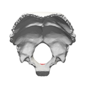
The occipital bone is a cranial dermal bone and the main bone of the occiput. It is trapezoidal in shape and curved on itself like a shallow dish. The occipital bone overlies the occipital lobes of the cerebrum. At the base of the skull in the occipital bone, there is a large oval opening called the foramen magnum, which allows the passage of the spinal cord.

<i>Meganthropus</i> Hominin fossil

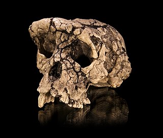
Meganthropus is an extinct genus of non-hominin hominid ape, known from the Pleistocene of Indonesia. It is known from a series of large jaw and skull fragments found at the Sangiran site near Surakarta in Central Java, Indonesia, alongside several isolated teeth. The genus has a long and convoluted taxonomic history. The original fossils were ascribed to a new species, Meganthropus palaeojavanicus, and for a long time was considered invalid, with the genus name being used as an informal name for the fossils.

Paleoanthropology or paleo-anthropology is a branch of paleontology and anthropology which seeks to understand the early development of anatomically modern humans, a process known as hominization, through the reconstruction of evolutionary kinship lines within the family Hominidae, working from biological evidence and cultural evidence.

<span class="mw-page-title-main">Mrs. Ples</span> Hominin fossil

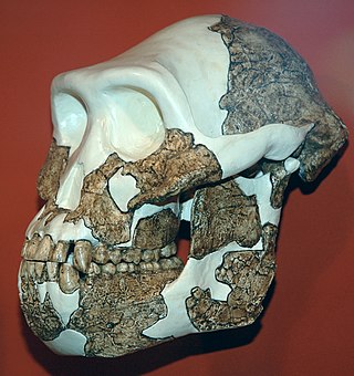
Mrs. Ples is the popular nickname for the most complete skull of an Australopithecus africanus ever found in South Africa. Many Australopithecus fossils have been found near Sterkfontein, about 40 kilometres (25 mi) northwest of Johannesburg, in a region of Gauteng now designated as the Cradle of Humankind World Heritage Site. Mrs. Ples was discovered by Robert Broom and John T. Robinson on April 18, 1947. Because of Broom's use of dynamite and pickaxe while excavating, Mrs. Ples's skull was blown into pieces and some fragments are missing. Nonetheless, Mrs./Mr. Ples is one of the most "perfect" pre-human skulls ever found. The skull is currently held at the Ditsong National Museum of Natural History in Pretoria.

<i>Australopithecus africanus</i> Extinct hominid from South Africa

Australopithecus africanus is an extinct species of australopithecine which lived between about 3.3 and 2.1 million years ago in the Late Pliocene to Early Pleistocene of South Africa. The species has been recovered from Taung, Sterkfontein, Makapansgat, and Gladysvale. The first specimen, the Taung child, was described by anatomist Raymond Dart in 1924, and was the first early hominin found. However, its closer relations to humans than to other apes would not become widely accepted until the middle of the century because most had believed humans evolved outside of Africa. It is unclear how A. africanus relates to other hominins, being variously placed as ancestral to Homo and Paranthropus, to just Paranthropus, or to just P. robustus. The specimen "Little Foot" is the most completely preserved early hominin, with 90% of the skeleton intact, and the oldest South African australopith. However, it is controversially suggested that it and similar specimens be split off into "A. prometheus".

<i>Homo rhodesiensis</i> Species of primate (fossil)

Homo rhodesiensis is the species name proposed by Arthur Smith Woodward (1921) to classify Kabwe 1, a Middle Stone Age fossil recovered from Broken Hill mine in Kabwe, Northern Rhodesia. In 2020, the skull was dated to 324,000 to 274,000 years ago. Other similar older specimens also exist.

<i>Australopithecus anamensis</i> Extinct hominin from Pliocene east Africa

Australopithecus anamensis is a hominin species that lived approximately between 4.2 and 3.8 million years ago and is the oldest known Australopithecus species, living during the Plio-Pleistocene era.

<i>Paranthropus aethiopicus</i> Extinct species of hominin of East Africa

Paranthropus aethiopicus is an extinct species of robust australopithecine from the Late Pliocene to Early Pleistocene of East Africa about 2.7–2.3 million years ago. However, it is much debated whether or not Paranthropus is an invalid grouping and is synonymous with Australopithecus, so the species is also often classified as Australopithecus aethiopicus. Whatever the case, it is considered to have been the ancestor of the much more robust P. boisei. It is debated if P. aethiopicus should be subsumed under P. boisei, and the terms P. boisei sensu lato and P. boisei sensu stricto can be used to respectively include and exclude P. aethiopicus from P. boisei.

<i>Paranthropus robustus</i> Extinct species of hominin of South Africa

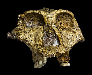
Paranthropus robustus is a species of robust australopithecine from the Early and possibly Middle Pleistocene of the Cradle of Humankind, South Africa, about 2.27 to 0.87 million years ago. It has been identified in Kromdraai, Swartkrans, Sterkfontein, Gondolin, Cooper's, and Drimolen Caves. Discovered in 1938, it was among the first early hominins described, and became the type species for the genus Paranthropus. However, it has been argued by some that Paranthropus is an invalid grouping and synonymous with Australopithecus, so the species is also often classified as Australopithecus robustus.

<i>Paranthropus boisei</i> Extinct species of hominin of East Africa

Paranthropus boisei is a species of australopithecine from the Early Pleistocene of East Africa about 2.5 to 1.15 million years ago. The holotype specimen, OH 5, was discovered by palaeoanthropologist Mary Leakey in 1959 at Olduvai Gorge, Tanzania and described by her husband Louis a month later. It was originally placed into its own genus as "Zinjanthropus boisei", but is now relegated to Paranthropus along with other robust australopithecines. However, it is also argued that Paranthropus is an invalid grouping and synonymous with Australopithecus, so the species is also often classified as Australopithecus boisei.

Orthograde is a term derived from Greek ὀρθός, orthos + Latin gradi that describes a manner of walking which is upright, with the independent motion of limbs. Both New and Old World monkeys are primarily arboreal, and they have a tendency to walk with their limbs swinging in parallel to one another. This differs from the manner of walking demonstrated by the apes.

<span class="mw-page-title-main">Lateral parts of occipital bone</span> Parts of bone in base of skull

The lateral parts of the occipital bone are situated at the sides of the foramen magnum; on their under surfaces are the condyles for articulation with the superior facets of the atlas.

<span class="mw-page-title-main">Human skeletal changes due to bipedalism</span> Evoltionary changes to the human skeleton as a consequence of bipedalism

The evolution of human bipedalism, which began in primates approximately four million years ago, or as early as seven million years ago with Sahelanthropus, or approximately twelve million years ago with Danuvius guggenmosi, has led to morphological alterations to the human skeleton including changes to the arrangement, shape, and size of the bones of the foot, hip, knee, leg, and the vertebral column. These changes allowed for the upright gait to be overall more energy efficient in comparison to quadrupeds. The evolutionary factors that produced these changes have been the subject of several theories that correspond with environmental changes on a global scale.

<span class="mw-page-title-main">Endocranium</span> The lower and inner parts of the skull

The endocranium in comparative anatomy is a part of the skull base in vertebrates and it represents the basal, inner part of the cranium. The term is also applied to the outer layer of the dura mater in human anatomy.

<span class="mw-page-title-main">Lunate sulcus</span>

In brain anatomy, the lunate sulcus or simian sulcus, also known as the sulcus lunatus, is a fissure in the occipital lobe variably found in humans and more often larger when present in apes and monkeys. The lunate sulcus marks the transition between V1 and V2.

References

PD-icon.svgThis article incorporates text in the public domain from page 129 of the 20th edition of Gray's Anatomy (1918)

  1. Russo, Gabrielle A.; Kirk, Christopher E. (November 2013). "Foramen magnum position in bipedal mammals". Journal of Human Evolution. 65 (5): 656–670. CiteSeerX   10.1.1.591.2458 . doi:10.1016/j.jhevol.2013.07.007. PMID   24055116.
  2. 1 2 3 4 5 6 7 Sanchez, Pedro; Graham, John M. (2017). "31 - Congenital Anomalies of the Skull". Swaiman's Pediatric Neurology (6th ed.). Elsevier. pp. 233–241. doi:10.1016/B978-0-323-37101-8.00031-X. ISBN   978-0-323-37101-8.
  3. Dutta, Asim Kumar (2013). Essentials of Human Anatomy Head & Neck. kolkata: Current books international. pp. 56–57. ISBN   978-81-86793-79-4.
  4. 1 2 Russo, Gabrielle A.; Kirk, E. Christopher (2017-04-01). "Another look at the foramen magnum in bipedal mammals". Journal of Human Evolution. 105: 24–40. doi:10.1016/j.jhevol.2017.01.018. ISSN   0047-2484. PMID   28366198.
  5. Sanchez, Pedro; Graham, John M. (2017). "31 - Congenital Anomalies of the Skull". Swaiman's Pediatric Neurology (6th ed.). Elsevier. pp. 233–241. doi:10.1016/B978-0-323-37101-8.00031-X. ISBN   978-0-323-37101-8.
  6. Zdilla, Matthew J; Russell, Michelle L; Bliss, Kaitlyn N; Mangus, Kelsey R; Koons, Aaron W (2017). "The size and shape of the foramen magnum in man". Journal of Craniovertebral Junction & Spine. 8 (3): 205–221. doi: 10.4103/jcvjs.JCVJS_62_17 . ISSN   0974-8237. PMC   5634107 . PMID   29021672.
  7. 1 2 3 4 5 Tobias, Phillip V.; Symons, Joel A. (1992). "Functional, Morphogenetic and Phylogenetic Significance of Conjunction between Cardioid Foramen Magnum and Enlarged Occipital and Marginal Venous Sinuses". Archaeology in Oceania. 27 (3): 120–127. doi:10.1002/j.1834-4453.1992.tb00295.x. ISSN   0728-4896. JSTOR   40386950.
  8. 1 2 Russo, Gabrielle A.; Kirk, E. Christopher (2013-11-01). "Foramen magnum position in bipedal mammals". Journal of Human Evolution. 65 (5): 656–670. doi:10.1016/j.jhevol.2013.07.007. ISSN   0047-2484. PMID   24055116.
  9. 1 2 Russo, Gabrielle A.; Kirk, E. Christopher (2013-11-01). "Foramen magnum position in bipedal mammals". Journal of Human Evolution. 65 (5): 656–670. doi:10.1016/j.jhevol.2013.07.007. ISSN   0047-2484. PMID   24055116.
  10. 1 2 3 Russo, Gabrielle A.; Kirk, E. Christopher (2017-04-01). "Another look at the foramen magnum in bipedal mammals". Journal of Human Evolution. 105: 24–40. doi:10.1016/j.jhevol.2017.01.018. ISSN   0047-2484. PMID   28366198.