Related Research Articles
Spasticity is a feature of altered skeletal muscle performance with a combination of paralysis, increased tendon reflex activity, and hypertonia. It is also colloquially referred to as an unusual "tightness", stiffness, or "pull" of muscles.

Tetraplegia, also known as quadriplegia, is defined as the dysfunction or loss of motor and/or sensory function in the cervical area of the spinal cord. A loss of motor function can present as either weakness or paralysis leading to partial or total loss of function in the arms, legs, trunk, and pelvis; paraplegia is similar but affects the thoracic, lumbar, and sacral segments of the spinal cord and arm function is retained. The paralysis may be flaccid or spastic. A loss of sensory function can present as an impairment or complete inability to sense light touch, pressure, heat, pinprick/pain, and proprioception. In these types of spinal cord injury, it is common to have a loss of both sensation and motor control.
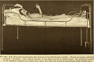
Paraplegia, or paraparesis, is an impairment in motor or sensory function of the lower extremities. The word comes from Ionic Greek (παραπληγίη) "half-stricken". It is usually caused by spinal cord injury or a congenital condition that affects the neural (brain) elements of the spinal canal. The area of the spinal canal that is affected in paraplegia is either the thoracic, lumbar, or sacral regions. If four limbs are affected by paralysis, tetraplegia or quadriplegia is the correct term. If only one limb is affected, the correct term is monoplegia. Spastic paraplegia is a form of paraplegia defined by spasticity of the affected muscles, rather than flaccid paralysis.
Anosognosia is a condition in which a person with a disability is cognitively unaware of having it due to an underlying physical condition. Anosognosia results from physiological damage to brain structures, typically to the parietal lobe or a diffuse lesion on the fronto-temporal-parietal area in the right hemisphere, and is thus a neuropsychiatric disorder. A deficit of self-awareness, it was first named by the neurologist Joseph Babinski in 1914.
The primary goals of stroke management are to reduce brain injury and promote maximum patient recovery. Rapid detection and appropriate emergency medical care are essential for optimizing health outcomes. When available, patients are admitted to an acute stroke unit for treatment. These units specialize in providing medical and surgical care aimed at stabilizing the patient's medical status. Standardized assessments are also performed to aid in the development of an appropriate care plan. Current research suggests that stroke units may be effective in reducing in-hospital fatality rates and the length of hospital stays.
The Bobath concept is an approach to neurological rehabilitation that is applied in patient assessment and treatment. The goal of applying the Bobath concept is to promote motor learning for efficient motor control in various environments, thereby improving participation and function. This is done through specific patient handling skills to guide patients through the initiation and completing of intended tasks. This approach to neurological rehabilitation is multidisciplinary, primarily involving physiotherapists, occupational therapists, and speech and language therapists. In the United States, the Bobath concept is also known as 'neuro-developmental treatment' (NDT).

Mirror therapy (MT) or mirror visual feedback (MVF) is a therapy for pain or disability that affects one side of the patient more than the other side. It was invented by Vilayanur S. Ramachandran to treat post-amputation patients who had phantom limb pain (PLP). Ramachandran created a visual illusion of two intact limbs by putting the patient's affected limb into a "mirror box," with a mirror down the center.
Monoplegia is paralysis of a single limb, usually an arm. Common symptoms associated with monoplegic patients are weakness, numbness, and pain in the affected limb. Monoplegia is a type of paralysis that falls under hemiplegia. While hemiplegia is paralysis of half of the body, monoplegia is localized to a single limb or to a specific region of the body. Monoplegia of the upper limb is sometimes referred to as brachial monoplegia, and that of the lower limb is called crural monoplegia. Monoplegia in the lower extremities is not as common of an occurrence as in the upper extremities. Monoparesis is a similar, but less severe, condition because one limb is very weak, not paralyzed. For more information, see paresis.
Hypertonia is a term sometimes used synonymously with spasticity and rigidity in the literature surrounding damage to the central nervous system, namely upper motor neuron lesions. Impaired ability of damaged motor neurons to regulate descending pathways gives rise to disordered spinal reflexes, increased excitability of muscle spindles, and decreased synaptic inhibition. These consequences result in abnormally increased muscle tone of symptomatic muscles. Some authors suggest that the current definition for spasticity, the velocity-dependent over-activity of the stretch reflex, is not sufficient as it fails to take into account patients exhibiting increased muscle tone in the absence of stretch reflex over-activity. They instead suggest that "reversible hypertonia" is more appropriate and represents a treatable condition that is responsive to various therapy modalities like drug or physical therapy.
Diplegia, when used singularly, refers to paralysis affecting symmetrical parts of the body. This is different from hemiplegia which refers to spasticity restricted to one side of the body, paraplegia which refers to paralysis restricted to the legs and hip, and quadriplegia which requires the involvement of all four limbs but not necessarily symmetrical. Diplegia is the most common cause of crippling in children, specifically in children with cerebral palsy. Other causes may be due to injury of the spinal cord. There is no set course of progression for people with diplegia. Symptoms may get worse but the neurological part does not change. The primary parts of the brain that are affected by diplegia are the ventricles, fluid filled compartments in the brain, and the wiring from the center of the brain to the cerebral cortex. There is also usually some degeneration of the cerebral neurons, as well as problems in the upper motor neuron system. The term diplegia can refer to any bodily area, such as the face, arms, or legs.
Central facial palsy is a symptom or finding characterized by paralysis or paresis of the lower half of one side of the face. It usually results from damage to upper motor neurons of the facial nerve.

Weber's syndrome, also known as midbrain stroke syndrome or superior alternating hemiplegia, is a form of stroke that affects the medial portion of the midbrain. It involves oculomotor fascicles in the interpeduncular cisterns and cerebral peduncle so it characterizes the presence of an ipsilateral lower motor neuron type oculomotor nerve palsy and contralateral hemiparesis or hemiplegia.
Constraint-induced movement therapy is a form of rehabilitation therapy that improves upper extremity function in stroke and other central nervous system damage patients by increasing the use of their affected upper limb. Due to its high duration of treatment, the therapy has been found to frequently be infeasible when attempts have been made to apply it to clinical situations, and both patients and treating clinicians have reported poor compliance and concerns with patient safety. In the United States, the high duration of the therapy has also made the therapy not able to get reimbursed in most clinical environments.
Kernohan's notch is a cerebral peduncle indentation associated with some forms of transtentorial herniation. It is a secondary condition caused by a primary injury on the opposite hemisphere of the brain. Kernohan's notch is an ipsilateral condition, in that a left-sided primary lesion evokes motor impairment in the left side of the body and a right-sided primary injury evokes motor impairment in the right side of the body. The seriousness of Kernohan's notch varies depending on the primary problem causing it, which may range from benign brain tumors to advanced subdural hematoma.

Over time, the approach to cerebral palsy management has shifted away from narrow attempts to fix individual physical problems – such as spasticity in a particular limb – to making such treatments part of a larger goal of maximizing the person's independence and community engagement. Much of childhood therapy is aimed at improving gait and walking. Approximately 60% of people with CP are able to walk independently or with aids at adulthood. However, the evidence base for the effectiveness of intervention programs reflecting the philosophy of independence has not yet caught up: effective interventions for body structures and functions have a strong evidence base, but evidence is lacking for effective interventions targeted toward participation, environment, or personal factors. There is also no good evidence to show that an intervention that is effective at the body-specific level will result in an improvement at the activity level, or vice versa. Although such cross-over benefit might happen, not enough high-quality studies have been done to demonstrate it.
Upper motor neuron syndrome (UMNS) is the motor control changes that can occur in skeletal muscle after an upper motor neuron lesion.

Spastic cerebral palsy is the type of cerebral palsy characterized by spasticity or high muscle tone often resulting in stiff, jerky movements. Cases of spastic CP are further classified according to the part or parts of the body that are most affected. Such classifications include spastic diplegia, spastic hemiplegia, spastic quadriplegia, and in cases of single limb involvement, spastic monoplegia.

Spastic hemiplegia is a neuromuscular condition of spasticity that results in the muscles on one side of the body being in a constant state of contraction. It is the "one-sided version" of spastic diplegia. It falls under the mobility impairment umbrella of cerebral palsy. About 20–30% of people with cerebral palsy have spastic hemiplegia. Due to brain or nerve damage, the brain is constantly sending action potentials to the neuromuscular junctions on the affected side of the body. Similar to strokes, damage on the left side of the brain affects the right side of the body and damage on the right side of the brain affects the left side of the body. Other side can be effected for lesser extent. The affected side of the body is rigid, weak and has low functional abilities. In most cases, the upper extremity is much more affected than the lower extremity. This could be due to preference of hand usage during early development. If both arms are affected, the condition is referred to as double hemiplegia. Some patients with spastic hemiplegia only experience minor impairments, where in severe cases one side of the body could be completely paralyzed. The severity of spastic hemiplegia is dependent upon the degree of the brain or nerve damage.
Alternating hemiplegia is a form of hemiplegia that has an ipsilateral cranial nerve palsies and contralateral hemiplegia or hemiparesis of extremities of the body. The disorder is characterized by recurrent episodes of paralysis on one side of the body. There are multiple forms of alternating hemiplegia, Weber's syndrome, middle alternating hemiplegia, and inferior alternating hemiplegia. This type of syndrome can result from a unilateral lesion in the brainstem affecting both upper motor neurons and lower motor neurons. The muscles that would receive signals from these damaged upper motor neurons result in spastic paralysis. With a lesion in the brainstem, this affects the majority of limb and trunk muscles on the contralateral side due to the upper motor neurons decussation after the brainstem. The cranial nerves and cranial nerve nuclei are also located in the brainstem making them susceptible to damage from a brainstem lesion. Cranial nerves III (Oculomotor), VI (Abducens), and XII (Hypoglossal) are most often associated with this syndrome given their close proximity with the pyramidal tract, the location which upper motor neurons are in on their way to the spinal cord. Damages to these structures produce the ipsilateral presentation of paralysis or palsy due to the lack of cranial nerve decussation before innervating their target muscles. The paralysis may be brief or it may last for several days, many times the episodes will resolve after sleep. Some common symptoms of alternating hemiplegia are mental impairment, gait and balance difficulties, excessive sweating and changes in body temperature.
Pusher syndrome is a condition observed in some people following a stroke which has left them with one side weakened due to hemiparesis. Sufferers exhibit a tendency to actively push away from the unweakened side, thus leading to a loss of postural balance. It can be a result of left or right brain damage. In contrast to most stroke patients, who typically prefer more weight-bearing on their non-hemiparetic side, this abnormal condition can vary in severity and leads to a loss of postural balance. The lesion involved in this syndrome is thought to be in the posterior thalamus on either side, or multiple areas of the right cerebral hemisphere.
References
- 1 2 3 4 Detailed article about hemiparesis Archived 2022-02-02 at the Wayback Machine at Disabled-World.com
- 1 2 3 4 5 6 7 Karnath HO, Broetz D (December 2003). "Understanding and treating "pusher syndrome"". Phys Ther. 83 (12): 1119–25. doi: 10.1093/ptj/83.12.1119 . PMID 14640870.
- ↑ Karnath HO, Ferber S, Dichgans J (November 2000). "The origin of contraversive pushing: evidence for a second graviceptive system in humans". Neurology. 55 (9): 1298–304. doi:10.1212/wnl.55.9.1298. PMID 11087771. S2CID 19399616.
- ↑ Karnath HO, Ferber S, Dichgans J (December 2000). "The neural representation of postural control in humans". Proceedings of the National Academy of Sciences of the United States of America. 97 (25): 13931–6. Bibcode:2000PNAS...9713931K. doi: 10.1073/pnas.240279997 . PMC 17678 . PMID 11087818.
- 1 2 Pedersen PM, Wandel A, Jørgensen HS, Nakayama H, Raaschou HO, Olsen TS (January 1996). "Ipsilateral pushing in stroke: incidence, relation to neuropsychological symptoms, and impact on rehabilitation. The Copenhagen Stroke Study". Archives of Physical Medicine and Rehabilitation. 77 (1): 25–8. doi:10.1016/s0003-9993(96)90215-4. PMID 8554469.
- ↑ Davies PM (1985). Steps to follow: A guide to the treatment of adult hemiplegia : Based on the concept of K. and B. Bobath. New York: Springer-Verlag.
- ↑ O'Sullivan S (2007). "Ch. 12: Stroke". In O'Sullivan S, Schmitz T (eds.). Physical Rehabilitation (5th ed.). Philadelphia: F.A. Davis. pp. 705–769.
- ↑ Lagerqvist, J.; Skargren, E. (2006). "Pusher syndrome: reliability, validity, and sensitivity to change of a classification instrument". Advances in Physiotherapy. 8 (4): 154–160. doi:10.1080/14038190600806596. S2CID 145015737.
- 1 2 3 4 Babyar SR, Peterson MG, Bohannon R, Pérennou D, Reding M (July 2009). "Clinical examination tools for lateropulsion or pusher syndrome following stroke: a systematic review of the literature". Clinical Rehabilitation. 23 (7): 639–50. doi:10.1177/0269215509104172. PMID 19403555. S2CID 40016612.
- ↑ "hemiplegia in children". Children's Hemiplegia and Stroke Association (CHASA). Archived from the original on February 4, 2012.
- ↑ Shapiro M, Blanco DA (February 2017). "Neurological Complications of Gastrointestinal Disease". Seminars in Pediatric Neurology (Review). 24 (1): 43–53. doi:10.1016/j.spen.2017.02.001. PMID 28779865.
- ↑ "What is hemiplegia? | HemiHelp: for children and young people with hemiplegia (hemiparesis)". HemiHelp. Archived from the original on 2013-03-05. Retrieved 2013-03-08.
- ↑ Martin L (2009). "I was awake -- and could not move!". Lakesidepress.com. Retrieved 2013-03-08.
Sleep paralysis, parasomnia, sleep apnea, sleep eat, parasomnias, paresthesias, dysesthesias, obstructive sleep apnea, REM, Stage 1, Sinemet narcolepsy, insomnia, cataplexy, benzodiazepines, opioids, sleepiness, sleep walking, daytime sleepiness, upper airway, CPAP, hypoxemia, UVVP, uvula, Somnoplasty, obesity, airway obstruction, EEG, electroencephalogram, Klonopine, night terrors, bruxism, parasomnias, EMG, Epworth Sleepiness Scale, BiPAP, sleep efficiency
- ↑ Ago T, Kitazono T, Ooboshi H, Takada J, Yoshiura T, Mihara F, et al. (August 2003). "Deterioration of pre-existing hemiparesis brought about by subsequent ipsilateral lacunar infarction". Journal of Neurology, Neurosurgery, and Psychiatry. 74 (8): 1152–3. doi:10.1136/jnnp.74.8.1152. PMC 1738578 . PMID 12876260.
- ↑ Song YM, Lee JY, Park JM, Yoon BW, Roh JK (May 2005). "Ipsilateral hemiparesis caused by a corona radiata infarct after a previous stroke on the opposite side". Archives of Neurology. 62 (5): 809–11. doi: 10.1001/archneur.62.5.809 . PMID 15883270.
- ↑ Yamamoto S, Takasawa M, Kajiyama K, Baron JC, Yamaguchi T (2007). "Deterioration of hemiparesis after recurrent stroke in the unaffected hemisphere: Three further cases with possible interpretation". Cerebrovascular Diseases. 23 (1): 35–9. doi:10.1159/000095756. PMID 16968984. S2CID 40273792.
- ↑ Terakawa H, Abe K, Nakamura M, Okazaki T, Obashi J, Yanagihara T (May 2000). "Ipsilateral hemiparesis after putaminal hemorrhage due to uncrossed pyramidal tract" (PDF). Neurology. 54 (9): 1801–5. doi:10.1212/WNL.54.9.1801. PMID 10802787. S2CID 15086685.
- ↑ "Spastic Hemiplegia : Cerebral Palsy". OriginsOfCerebralPalsy.com. Archived from the original on 2018-01-26. Retrieved 2013-03-08.
- ↑ "Gait Abnormalities". The Stanford 25. Archived from the original on October 11, 2010.
- ↑ Fugl-Meyer AR, Jääskö L, Leyman I, Olsson S, Steglind S (1975). "The post-stroke hemiplegic patient. 1. a method for evaluation of physical performance". Scandinavian Journal of Rehabilitation Medicine. 7 (1): 13–31. doi: 10.2340/1650197771331 . PMID 1135616. S2CID 19245788.
- ↑ Sullivan KJ, Tilson JK, Cen SY, Rose DK, Hershberg J, Correa A, et al. (February 2011). "Fugl-Meyer assessment of sensorimotor function after stroke: standardized training procedure for clinical practice and clinical trials". Stroke. 42 (2): 427–32. doi: 10.1161/STROKEAHA.110.592766 . PMID 21164120.
- ↑ Sullivan SB (2007). "Stroke". In O'Sullivan SB, Schmitz TJ (eds.). Physical Rehabilitation (5th ed.). Philadelphia PA: F.A. Davis.
- ↑ "Fugl-Meyer Assessment of Motor Recovery after". Rehab Measures. Archived from the original on 2016-09-24. Retrieved 2013-03-08.
- ↑ Gowland C, Stratford P, Ward M, Moreland J, Torresin W, Van Hullenaar S, et al. (January 1993). "Measuring physical impairment and disability with the Chedoke-McMaster Stroke Assessment". Stroke. 24 (1): 58–63. doi:10.1161/01.STR.24.1.58. PMID 8418551.
- ↑ Valach L, Signer S, Hartmeier A, Hofer K, Steck GC (June 2003). "Chedoke-McMaster stroke assessment and modified Barthel Index self-assessment in patients with vascular brain damage". International Journal of Rehabilitation Research. 26 (2): 93–9. doi:10.1097/00004356-200306000-00003. PMID 12799602.
- ↑ "Chedoke-McMaster Stroke Assessment Measure". Rehab Measures. Archived from the original on 2014-10-06. Retrieved 2013-03-08.
- ↑ Daley K, Mayo N, Wood-Dauphinée S (January 1999). "Reliability of scores on the Stroke Rehabilitation Assessment of Movement (STREAM) measure". Physical Therapy. 79 (1): 8–19, quiz 20–3. doi: 10.1093/ptj/79.1.8 . PMID 9920188.
- ↑ O'sullivan S, Schmitz T (2007). Physical Rehabilitation (5th ed.). Philadelphia PA: F.A. Davis. p. 736.
- ↑ Knutson, Jayme S.; Fu, Michael J.; Sheffler, Lynne R.; Chae, John (November 26, 2015). "Neuromuscular Electrical Stimulation for Motor Restoration in Hemiplegia". Physical Medicine and Rehabilitation Clinics of North America. 26 (4): 729–745. doi:10.1016/j.pmr.2015.06.002. ISSN 1558-1381. PMC 4630679 . PMID 26522909.
- ↑ Beebe, Justin A.; Lang, Catherine E. (March 5, 2009). "Active range of motion predicts upper extremity function 3 months after stroke". Stroke. 40 (5): 1772–1779. doi:10.1161/STROKEAHA.108.536763. ISSN 1524-4628. PMC 2718540 . PMID 19265051.
- ↑ Sterr A, Freivogel S (September 2003). "Motor-improvement following intensive training in low-functioning chronic hemiparesis". Neurology. 61 (6): 842–4. doi:10.1212/wnl.61.6.842. PMID 14504336. S2CID 43563527.
- ↑ Dohle C, Püllen J, Nakaten A, Küst J, Rietz C, Karbe H (2009). "Mirror therapy promotes recovery from severe hemiparesis: a randomized controlled trial". Neurorehabilitation and Neural Repair. 23 (3): 209–17. doi: 10.1177/1545968308324786 . PMID 19074686. S2CID 14252958.
- ↑ Stroke in Physical Rehabilitation 2007 , p. 746
- ↑ "Right Hemisphere Damage". American Speech-Language-Hearing Association. Retrieved 2023-10-06.
- ↑ Patten C, Lexell J, Brown HE (May 2004). "Weakness and strength training in persons with poststroke hemiplegia: rationale, method, and efficacy". Journal of Rehabilitation Research and Development. 41 (3A): 293–312. doi:10.1682/JRRD.2004.03.0293. PMID 15543447. S2CID 563507.
- ↑ "Hemiplegia/Hemiparesis". Archived from the original on 2009-04-20. Retrieved 2014-10-02.
- ↑ Farag SM, Mohammed MO, El-Sobky TA, ElKadery NA, ElZohiery AK (March 2020). "Botulinum Toxin A Injection in Treatment of Upper Limb Spasticity in Children with Cerebral Palsy: A Systematic Review of Randomized Controlled Trials". JBJS Reviews. 8 (3): e0119. doi:10.2106/JBJS.RVW.19.00119. PMC 7161716 . PMID 32224633.
- ↑ Blumetti FC, Belloti JC, Tamaoki MJ, Pinto JA (October 2019). "Botulinum toxin type A in the treatment of lower limb spasticity in children with cerebral palsy". The Cochrane Database of Systematic Reviews. 2019 (10): CD001408. doi:10.1002/14651858.CD001408.pub2. PMC 6779591 . PMID 31591703.
- ↑ Dutta, Abhijit; Singh, Subhas; Saha, Subhranil; Rath, Prasanta; Sehrawat, Nisha; Singh, Navin Kumar (2022-08-29). "Efficacy of individualized homeopathic medicines in treatment of post-stroke hemiparesis: A randomized trial". Explore. 19 (2): S1550–8307(22)00160–4. doi:10.1016/j.explore.2022.08.017. ISSN 1878-7541. PMID 36115790. S2CID 251943815.
- ↑ Barreca S, Wolf SL, Fasoli S, Bohannon R (December 2003). "Treatment interventions for the paretic upper limb of stroke survivors: a critical review". Neurorehabilitation and Neural Repair. 17 (4): 220–6. doi: 10.1177/0888439003259415 . PMID 14677218. S2CID 23055506.
- ↑ Price CI, Pandyan AD (February 2001). "Electrical stimulation for preventing and treating post-stroke shoulder pain: a systematic Cochrane review". Clinical Rehabilitation. 15 (1): 5–19. doi:10.1191/026921501670667822. PMID 11237161. S2CID 1792159.
- ↑ Ada L, Foongchomcheay A, Canning C (January 2005). Ada L (ed.). "Supportive devices for preventing and treating subluxation of the shoulder after stroke". The Cochrane Database of Systematic Reviews. 2005 (1): CD003863. doi:10.1002/14651858.CD003863.pub2. PMC 6984447 . PMID 15674917. S2CID 10451803.
- ↑ Zorowitz RD, Hughes MB, Idank D, Ikai T, Johnston MV (March 1996). "Shoulder pain and subluxation after stroke: correlation or coincidence?". The American Journal of Occupational Therapy. 50 (3): 194–201. doi:10.5014/ajot.50.3.194. PMID 8822242.
- ↑ Wittenberg GF, Schaechter JD (December 2009). "The neural basis of constraint-induced movement therapy". Current Opinion in Neurology. 22 (6): 582–8. doi:10.1097/WCO.0b013e3283320229. PMID 19741529. S2CID 16050784.
- ↑ Choo, Yoo Jin; Chang, Min Cheol (2021-08-13). "Commonly Used Types and Recent Development of Ankle-Foot Orthosis: A Narrative Review". Healthcare. 9 (8): 1046. doi: 10.3390/healthcare9081046 . ISSN 2227-9032. PMC 8392067 . PMID 34442183.
- 1 2 "Hemiplegia (Hemiparalysis)". Healthopedia.com. 2009-04-06. Retrieved 2013-03-08.
- ↑ "Kingsolver, Barbara : The Poisonwood Bible". Litmed.med.nyu.edu. 2000-05-17. Retrieved 2013-03-08.
- ↑ "The Poisonwood Bible Barbara Kingsolver Study Guide, Lesson Plan & more". eNotes.com. Retrieved 2013-03-08.
- ↑ "HAERTS Announce Debut EP Hemiplegia, Out 9/17 on Columbia Records". broadwayworld.com. 2013-08-08. Retrieved 2013-10-19.