Trismus

Last updated
Trismus
Other namesLockjaw
Lockjawsymptoms.jpg
Trismus caused due to muscle rigidity. Diagram of jaw muscles are shown here.

Trismus is a condition of restricted opening of the mouth. [1] [2] The term was initially used in the setting of tetanus. [2] Trismus may be caused by spasm of the muscles of mastication or a variety of other causes. [3] Temporary trismus occurs much more frequently than permanent trismus. [4] It is known to interfere with eating, speaking, and maintaining proper oral hygiene. This interference, specifically with an inability to swallow properly, results in an increased risk of aspiration. In some instances, trismus presents with altered facial appearance. The condition may be distressing and painful. Examination and treatments requiring access to the oral cavity can be limited, or in some cases impossible, due to the nature of the condition itself.

Contents

Definition

Trismus is defined as painful restriction in opening the mouth due to a muscle spasm, [5] however it can also refer to limited mouth opening of any cause. [6] Another definition of trismus is simply a limitation of movement. [4] Historically and commonly, the term lockjaw was sometimes used as a synonym for both trismus [2] and tetanus. [7]

Normal mouth-opening ranges from 35 to 45 mm. [6] Males usually have slightly greater mouth opening than females. (40–60 mm, average of 50 mm). The normal lateral movement is 8–12 mm, [8] and normal protrusive movement is approximately 10 mm.[ medical citation needed ] Some have distinguished mild trismus as 20–30 mm interincisal opening, moderate as 10–20 mm and severe as less than 10 mm. [9]

Trismus is derived from the Greek word trigmos/trismos meaning "a scream; a grinding, rasping or gnashing". [10]

Differential diagnosis

Traditionally causes of trismus are divided into intra-articular (factors within the temporomandibular joint [TMJ]) and extra-articular (factors outside the joint, see table). [4]

Commonly listed causes of trismus

Intra-articular:

  • Internal derangement of TMJ / meniscus displacement. [3] [6]
  • Fractured mandibular condyle or intracapsular fracture. [3]
  • TMJ dislocation. [4]
  • Traumatic synovitis. [3]
  • Septic arthritis. [6]
  • Osteoarthritis. [3]
  • Inflammatory arthritis (e.g. rheumatoid or psoriatic). [3]
  • Ankylosis. [3]
  • Osteophyte formation. [6]

Extra-articular:

  • Trauma not involving the mandibular condyle (e.g. a fracture of another part of the mandible, fractures of the middle third of the facial skeleton, fractures of the zygoma or zygomatic arch). [3]
  • Post surgical edema, e.g. removal of impacted lower wisdom teeth, [3] or other dentoalveolar surgery. [6]
  • Recent prolonged dental treatment (e.g. root canal therapy). [3]
  • Following administration of inferior alveolar nerve block with local anesthetic (medial pterygoid). [6]
  • Hematoma of medial pterygoid. [4]
  • Acute infections of the oral tissues, especially involving the buccal space or muscles of mastication. [3]
    • Odontogenic infection. [3] [6]
    • Peritonsillar abscess. [6]
    • Acute parotitis, e.g. mumps. [4]
    • Pericoronitis. [4]
    • Submasseteric abscess. [4]
  • Tetanus. [4] [6]
  • Tetany. [4]
  • Local malignancy. [6]
  • Myofascial pain / temporomandibular joint dysfunction. [6]
  • Radiation fibrosis. [6]
  • Fibrosis from burns. [4]
  • Submucous fibrosis. [6]
  • Systemic sclerosis. [11]
  • Myositis ossificans. [11]
  • Coronoid hyperplasia. [6]
  • Malignant hyperpyrexia. [6]
  • Epidermolysis bullosa. [8]
  • Drug associated dyskinesia. [4]
  • Psychotic disturbances, hysteria. [4]

Joint problems

Ankylosis

  • True bony ankylosis: can result from trauma to chin, infections and from prolonged immobilization following condylar fracture
    • Treatment – several surgical procedures are used to treat bony ankylosis, e.g.: Gap arthroplasty using interpositional materials between the cut segments.
  • Fibrous ankylosis: usually results due to trauma and infection
    • Treatment – trismus appliances in conjunction with physical therapy.

Arthritis synovitis

Meniscus pathology

Extra-articular causes

Infection

  • Odontogenic- Pulpal
    • Periodontal
    • Pericoronal
  • Non-odontogenic- Peritonsillar abscess
    • Tetanus
    • Meningitis
    • Brain abscess
    • Parotid abscess
  • The hallmark of a masticatory space infection is trismus or infection in anterior compartment of lateral pharyngeal space results in trismus. If these infections are unchecked, can spread to various facial spaces of the head and neck and lead to serious complications such as cervical cellulitis or mediastinitis.
    • Treatment: Elimination of etiologic agent along with antibiotic coverage
  • Trismus or lock jaw due to masseter muscle spasm, can be a primary presenting symptom in tetanus, Caused by Clostridium tetani, where tetanospasmin (toxin) is responsible for muscle spasms.
    • Prevention: primary immunization (DPT)

Dental treatment

  • Dental trismus is defined by difficulty in opening the jaw. It is a temporary condition that usually lasts no more than two weeks. Dental trismus is caused by an injury to the masticatory muscles, such as opening the jaw for an extended period of time or having a needle pass through a muscle. Typical dental anesthesia for the lower jaw involves inserting a needle into or through a muscle. In these cases it is usually the medial pterygoid or the buccinator muscles.
  • Oral surgery procedures, as in the extraction of lower molar teeth, may cause trismus as a result either of inflammation to the muscles of mastication or direct trauma to the TMJ.
  • Barbing of needles at the time of injection followed by tissue damage on withdrawal of the barbed needle causes post-injection persistent paresthesia, trismus and paresis.
    • Treatment: in acute phase:
      • Heat therapy
      • Analgesics
      • A soft diet
      • Muscle relaxants (if necessary)
      • Note: When acute phase is over the patient should be advised to initiate physiotherapy for opening and closing mouth.

Trauma

Fractures, particularly those of the mandible and fractures of zygomatic arch and zygomatic arch complex, accidental incorporation of foreign bodies due to external traumatic injury. Treatment: fracture reduction, removal of foreign bodies with antibiotic coverage[ citation needed ]

TMJ disorders

  • Extra-capsular disorders – Myofascial pain dysfunction syndrome
  • Intra-capsular problems – Disc displacement, arthritis, fibrosis, etc.
  • Acute closed locked conditions – displaced meniscus

Tumors and oral care

Rarely, trismus is a symptom of nasopharyngeal or infratemporal tumors/ fibrosis of temporalis tendon, when patient has limited mouth opening, always premalignant conditions like oral submucous fibrosis (OSMF) should also be considered in differential diagnosis.

Drug therapy

Succinyl choline, phenothiazines and tricyclic antidepressants causes trismus as a secondary effect. Trismus can be seen as an extra-pyramidal side-effect of metoclopromide, phenothiazines and other medications.

Radiotherapy and chemotherapy

  • Complications of radiotherapy:
    • Osteoradionecrosis may result in pain, trismus, suppuration and occasionally a foul smelling wound.
    • When muscles of mastication are within the field of radiation, it leads to fibrosis and result in decreased mouth opening.
  • Complications of Chemotherapy:
    • Oral mucosal cells have high growth rate and are susceptible to the toxic effects of chemotherapy, which lead to stomatitis.

Congenital and developmental causes

  • Hypertrophy of coronoid process causes interference of coronoid against the anteromedial margin of the zygomatic arch.
    • Treatment: Coronoidectomy
  • Trismus-pseudo-camtodactyly syndrome is a rare combination of hand, foot and mouth abnormalities and trismus.

Miscellaneous disorders

  • Hysteric patients: Through the mechanisms of conversion, the emotional conflict are converted into a physical symptom. E.g.: trismus
  • Scleroderma: A condition marked by edema and induration of the skin involving facial region can cause trismus

Common causes

Other causes

Diagnostic approach

X-ray/CT scan taken from the TMJ to see if there is any damage to the TMJ and surrounding structures.[ citation needed ]

Treatment

Treatment requires treating the underlying condition with dental treatments, speech therapy for swallowing difficulty and mouth opening restrictions, physical therapy, and passive range of motion devices. Additionally, control of symptoms with pain medications (NSAIDs), muscle relaxants, and warm compresses may be used.

Splints have been used. [13]

History

Historically, the term trismus was used to describe the early effects of tetany, also called "lockjaw".

Related Research Articles

<span class="mw-page-title-main">Temporomandibular joint dysfunction</span> Medical condition

Temporomandibular joint dysfunction is an umbrella term covering pain and dysfunction of the muscles of mastication and the temporomandibular joints. The most important feature is pain, followed by restricted mandibular movement, and noises from the temporomandibular joints (TMJ) during jaw movement. Although TMD is not life-threatening, it can be detrimental to quality of life; this is because the symptoms can become chronic and difficult to manage.

<span class="mw-page-title-main">Tetanus</span> Bacterial infection characterized by muscle spasms

Tetanus, also known as lockjaw, is a bacterial infection caused by Clostridium tetani and characterized by muscle spasms. In the most common type, the spasms begin in the jaw, and then progress to the rest of the body. Each spasm usually lasts for a few minutes. Spasms occur frequently for three to four weeks. Some spasms may be severe enough to fracture bones. Other symptoms of tetanus may include fever, sweating, headache, trouble swallowing, high blood pressure, and a fast heart rate. Onset of symptoms is typically 3 to 21 days following infection. Recovery may take months; about 10% of cases prove to be fatal.

<span class="mw-page-title-main">Temporomandibular joint</span> Joints connecting the jawbone to the skull

In anatomy, the temporomandibular joints (TMJ) are the two joints connecting the jawbone to the skull. It is a bilateral synovial articulation between the temporal bone of the skull above and the mandible below; it is from these bones that its name is derived. The joints are unique in their bilateral function, being connected via the mandible.

Lockjaw can refer to:

<span class="mw-page-title-main">Peritonsillar abscess</span> Pus behind the tonsil due to an infection

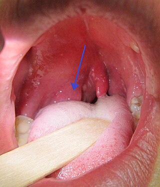
Peritonsillar abscess (PTA), also known as quinsy, is an accumulation of pus due to an infection behind the tonsil. Symptoms include fever, throat pain, trouble opening the mouth, and a change to the voice. Pain is usually worse on one side. Complications may include blockage of the airway or aspiration pneumonitis.

<span class="mw-page-title-main">Pericoronitis</span> Inflammation of the soft tissues surrounding the crown of a partially erupted tooth

Pericoronitis is inflammation of the soft tissues surrounding the crown of a partially erupted tooth, including the gingiva (gums) and the dental follicle. The soft tissue covering a partially erupted tooth is known as an operculum, an area which can be difficult to access with normal oral hygiene methods. The hyponym operculitis technically refers to inflammation of the operculum alone.

<span class="mw-page-title-main">Meige's syndrome</span> Medical condition

Meige's syndrome is a type of dystonia. It is also known as Brueghel's syndrome and oral facial dystonia. It is actually a combination of two forms of dystonia, blepharospasm and oromandibular dystonia (OMD).

<span class="mw-page-title-main">Masticatory muscle myositis</span>

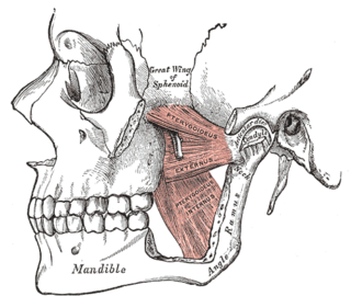
Masticatory muscle myositis (MMM) is an inflammatory disease in dogs affecting the muscles of mastication (chewing). It is also known as atrophic myositis or eosinophilic myositis. MMM is the most common inflammatory myopathy in dogs. The disease mainly affects large breed dogs. German Shepherd Dogs and Cavalier King Charles Spaniels may be predisposed. There is a similar disease of the eye muscles found in Golden Retrievers. Symptoms of acute MMM include swelling of the jaw muscles, drooling, and pain on opening the mouth. Ophthalmic signs may include third eyelid protrusion, red eyes, and exophthalmos. In chronic MMM there is atrophy of the jaw muscles, and scarring of the masticatory muscles due to fibrosis may result in inability to open the mouth (trismus). The affected muscles include the temporalis, masseter, and pterygoid muscles. The disease is usually bilateral.

A dental emergency is an issue involving the teeth and supporting tissues that are of high importance to be treated by the relevant professional. Dental emergencies do not always involve pain, although this is a common signal that something needs to be looked at. Pain can originate from the tooth, surrounding tissues or can have the sensation of originating in the teeth but be caused by an independent source. Depending on the type of pain experienced an experienced clinician can determine the likely cause and can treat the issue as each tissue type gives different messages in a dental emergency.

<span class="mw-page-title-main">Buccal space</span>

The buccal space is a fascial space of the head and neck. It is a potential space in the cheek, and is paired on each side. The buccal space is superficial to the buccinator muscle and deep to the platysma muscle and the skin. The buccal space is part of the subcutaneous space, which is continuous from head to toe.

Mouth infections, also known as oral infections, are a group of infections that occur around the oral cavity. They include dental infection, dental abscess, and Ludwig's angina. Mouth infections typically originate from dental caries at the root of molars and premolars that spread to adjacent structures. In otherwise healthy patients, removing the offending tooth to allow drainage will usually resolve the infection. In cases that spread to adjacent structures or in immunocompromised patients, surgical drainage and systemic antibiotics may be required in addition to tooth extraction. Since bacteria that normally reside in the oral cavity cause mouth infections, proper dental hygiene can prevent most cases of infection. As such, mouth infections are more common in populations with poor access to dental care or populations with health-related behaviors that damage one's teeth and oral mucosa. This is a common problem, representing nearly 36% of all encounters within the emergency department related to dental conditions.

Oral submucous fibrosis (OSF) is a chronic, complex, premalignant condition of the oral cavity, characterized by juxta-epithelial inflammatory reaction and progressive fibrosis of the submucosal tissues. As the disease progresses, the oral mucosa becomes fibrotic to the point that the person is unable to open the mouth. The condition is remotely linked to oral cancers and is associated with the chewing of areca nut and/or its byproducts, commonly practiced in South and South-East Asian countries. The incidence of OSF has also increased in western countries due to changing habits and population migration.

Fibrous ankylosis is a condition that affects fibrous connective tissue causing a limited range of movement.

Dentomandibular sensorimotor dysfunction (DMSD) is a medical condition involving the mandible, upper three cervical (neck) vertebrae, and the surrounding muscle and nerve areas.

Fascial spaces are potential spaces that exist between the fasciae and underlying organs and other tissues. In health, these spaces do not exist; they are only created by pathology, e.g. the spread of pus or cellulitis in an infection. The fascial spaces can also be opened during the dissection of a cadaver. The fascial spaces are different from the fasciae themselves, which are bands of connective tissue that surround structures, e.g. muscles. The opening of fascial spaces may be facilitated by pathogenic bacterial release of enzymes which cause tissue lysis. The spaces filled with loose areolar connective tissue may also be termed clefts. Other contents such as salivary glands, blood vessels, nerves and lymph nodes are dependent upon the location of the space. Those containing neurovascular tissue may also be termed compartments.

<span class="mw-page-title-main">Submasseteric space</span>

The submasseterric space is a fascial space of the head and neck. It is a potential space in the face over the angle of the jaw, and is paired on each side. It is located between the lateral aspect of the mandible and the medial aspect of the masseter muscle and its investing fascia. The term is derived from sub- meaning "under" in Latin and masseteric which refers to the masseter muscle. The submasseteric space is one of the four compartments of the masticator space. Sometimes the submasseteric space is described as a series of spaces, created because the masseter muscle has multiple insertions that cover most of the lateral surface of the ramus of the mandible.

<span class="mw-page-title-main">Pterygomandibular space</span>

The pterygomandibular space is a fascial space of the head and neck. It is a potential space in the head and is paired on each side. It is located between the lateral pterygoid muscle and the medial surface of the ramus of the mandible. The pterygomandibular space is one of the four compartments of the masticator space.

<span class="mw-page-title-main">Infratemporal space</span>

The infratemporal space is a fascial space of the head and neck. It is a potential space in the side of the head, and is paired on either side. It is located posterior to the maxilla, between the lateral pterygoid plate of the sphenoid bone medially and by the base of skull superiorly. The term is derived from infra- meaning below and temporal which refers to the temporalis muscle.

Osteomyelitis of the jaws is osteomyelitis which occurs in the bones of the jaws. Historically, osteomyelitis of the jaws was a common complication of odontogenic infection. Before the antibiotic era, it was frequently a fatal condition.

A parapharyngeal abscess is a deep neck space abscess of the parapharyngeal space, which is lateral to the superior pharyngeal constrictor muscle and medial to the masseter muscle. This space is divided by the styloid process into anterior and posterior compartments. The posterior compartment contains the carotid artery, internal jugular vein, and many nerves.

References

  1. "Trismus – The Oral Cancer Foundation" . Retrieved 5 April 2022.
  2. 1 2 3 Santiago-Rosado, Livia M.; Lewison, Cheryl S. (2023), "Trismus", StatPearls, Treasure Island (FL): StatPearls Publishing, PMID   29630255 , retrieved 2023-11-17
  3. 1 2 3 4 5 6 7 8 9 10 11 12 Odell, Edward W., ed. (2010). Clinical problem solving in dentistry (3rd ed.). Edinburgh: Churchill Livingstone. pp. 37–41. ISBN   9780443067846.
  4. 1 2 3 4 5 6 7 8 9 10 11 12 13 Soames, J.V.; Southam, J.C. (1998). Oral pathology (3rd ed.). Oxford: Oxford University Press. p.  330. ISBN   019262895X.
  5. "trismus". Miller-Keane Encyclopedia and Dictionary of Medicine, Nursing, and Allied Health (Seventh ed.). 2003.
  6. 1 2 3 4 5 6 7 8 9 10 11 12 13 14 15 16 Newlands, edited by Cyrus Kerawala, Carrie (2010). Oral and maxillofacial surgery. Oxford: Oxford University Press. ISBN   9780199204830.{{cite book}}: |first= has generic name (help)CS1 maint: multiple names: authors list (link)
  7. "Tetanus Symptoms and Complications | CDC". www.cdc.gov. 2022-09-12. Retrieved 2023-11-17.
  8. 1 2 Scully, Crispian (2008). Oral and maxillofacial medicine: the basis of diagnosis and treatment (2nd ed.). Edinburgh: Churchill Livingstone. pp. 101, 353. ISBN   9780443068188.
  9. Hupp JR, Ellis E, Tucker MR (2008). Contemporary oral and maxillofacial surgery (5th ed.). St. Louis, Mo.: Mosby Elsevier. ISBN   9780323049030.
  10. "Etymology of Trismus on Online Etymology Dictionary". Douglas Harper. Retrieved 10 February 2013.
  11. 1 2 Kalantzis, Crispian Scully, Athanasios (2005). Oxford handbook of dental patient care (2nd ed.). New York: Oxford University Press. ISBN   9780198566236.{{cite book}}: CS1 maint: multiple names: authors list (link)
  12. 1 2 3 4 5 Chris. "Dr". Locked Jaw (Lockjaw and Slack Jaw) Meaning and Causes |. Healthhype.com. Retrieved 13 September 2012.
  13. Shulman DH, Shipman B, Willis FB (2008). "Treating trismus with dynamic splinting: A cohort, case series". Adv Ther. 25 (1): 9–16. doi:10.1007/s12325-008-0007-0. PMID   18227979. S2CID   20495294.