Calcifying odontogenic cyst

Last updated
Calcifying odontogenic cyst
Other namesGorlin cyst, calcifying cystic odontogenic tumor [1]
Human jawbone left.jpg
This condition usually affects the jaw area
Specialty Dentistry

Calcifying odotogenic cyst (COC) is a rare developmental lesion that comes from odontogenic epithelium. [2] It is also known as a calcifying cystic odontogenic tumor, which is a proliferation of odontogenic epithelium and scattered nest of ghost cells and calcifications that may form the lining of a cyst, or present as a solid mass. [3]

Contents

It can appear in any location in the oral cavity, but more commonly affects the anterior (front) mandible and maxilla. It is most common in individuals in their 20s to 30s, but can be seen at almost any age, regardless of gender. On dental radiographs, the calcifying odontogenic cyst appears as a unilocular (one circle) radiolucency (dark area). In one-third of cases, an impacted tooth is involved. Histologically, cells that are described as "ghost cells", enlarged eosinophilic epithelial cells without nuclei, are present within the epithelial lining and may undergo calcification.

Signs and symptoms

Relative incidence of odontogenic cysts. Calcifying odontogenic cyst is labeled at right. Relative incidence of odontogenic cysts.jpg
Relative incidence of odontogenic cysts. Calcifying odontogenic cyst is labeled at right.

Most calcifying odontogenic cysts appear asymptomatic. [2] They are normally presented as a painless, slow-growing mass on the mandible and/or the maxilla, mostly in the front of the mouth. [5] Symptoms include swelling in the mouth, both inside the bone, in the tooth bearing areas, and outside the bone, in the gingiva. When a COC is located in the maxilla, individuals might complain of nasal stiffness, epistaxis, and headache.

Impacted or displaced teeth are often found due to COC. The diameter of the cyst ranges from 2 to 4 cm and swelling pain may be present. Intrabony (between bone) expansions may produce hard bony expansion and may perforate cortical bones. It can also extend to soft tissue. [6]

Causes

It is believed that the calcifying odontogenic cyst arose from odontogenic epithelial remnants (remains) that were trapped within the bones of the maxilla and mandible or gingival tissues. [2] It is associated with impacted and unerupted teeth.

Mechanism/Pathophysiology

Calcifying odontogenic cyst can is the presence of a variable number of ghost cells within the epithelial lining. The eosinophilic ghost cells are those that have been changed in a way without a nucleus, but it has been able to maintain its basic cell shape. [7]

The mechanism for the formation of a calcifying odontogenic cyst is controversial, whether the ghost cells change is based on coagulative necrosis (accidental cell death caused by ischemia or infarction)/the build up of enamel protein or it's a form of normal or abnormal keratinization (formation of keratin proteins) of odontogenic epithelium. [7] Large amounts of ghost cells fuse together to form large sheets of material with an undefined shape and are acellular. Calcification of the sheets may occur. It first appears as fine basophilic granules that increase in size and number forming large masses of calcifying material. Eosinophilic dentinoid (abnormal form of dentin) material is present next to a sheet of ghost cells. [7]

Some forms of the cystic type of COC, the epithelial lining proliferates into the lumen (inside space of the cyst) so its filled with masses of ghost cells and dystrophic calcifications. [7]

In a different form, unifocal or multifocal epithelial proliferation (increase in numbers) of the cyst lining into the lumen may look similar to ameloblastoma. These proliferations have a mix of different number of ghost cells. [7]

Neoplastic or solid COC are uncommon. They consist of extraosseous (outside bone) and intraosseous (inside bone) forms. Extraosseous being most common of the two, consists of odontogenic epithelium in the fibrous stroma, with columnar cells and stellate reticulum and ghost cells. Intraosseous consists of ameloblastoma-like strands and epithelium in fibrous connective tissue stroma with ghost cells present. [7]

There are small number of malignant odontogenic ghost cell tumors (odontogenic ghost cell carcinoma). These are aggressive and invade surrounding tissues through cellular pleomorphism and mitotic activity. [7]

Pathogenesis

Epithelial lining has ability to induce formation of dental tissues in adjacent connective tissue wall. [6]

Diagnosis

Radiographic features

These calcifying odontogenic cysts are usually discovered using dental radiographs. They will appear as unilocular (one chamber), multilocular (multiple chambers) or mixed radiolucencies with some radiopaque deposits of differing sizes and opacities. [5] Irregular calcifications may be seen in some cases. They are often located in a periapical or lateral periodontal relationship to adjacent teeth. [2]

CT Scan

CT scans can also be used to view the internal structures of the lesions and the involvement of neighboring structures. It is helpful in clinical diagnoses and treatment planning. They reveal vital characteristics that are not shown or detected in a dental radiograph. They are used to confirm the presence of calcifications along the cyst wall that were not detected in the radiographic images. [2]

Histology

In general, the epithelium seen is of stratified squamous type and is 5–8 cells thick. Additionally, focal areas of stellate reticulum like cells are seen and near the basement membrane ameloblast-like cells may be seen. Each type of calcifying odontogenic cyst shows special features of which there are three types: [8]

1)Type 1A. Ghost cells and dentinoid are seen. [8]

2)Type 1B. Formation of calcified tissues in the lumen of the cyst wall showing dystrophic calcification. Proliferation of tissues is similar to an Ameloblastic Fibroma. [8]

3)Type 1C. Ameloblast-like proliferation in the connective tissue and lumen of the cyst may be seen. [8]

Treatment

The standard treatment of calcifying odontogenic cyst is enucleation and curettage, however it depends on the lesion site and histological pattern. Enucleation followed by the removal of 1 to 2 millimeters layer of bone around the edges of the cystic cavity with a sharp curette or bone bur. The point of this procedure is to remove the epithelial debris that could cause recurrent lesions. [5] Recurrence following enucleation and curretage is rare. [9]

Once treatment is complete, follow-up visits may be required to monitor recurrence of the cyst. [5]

Prognosis

The prognosis of a calcifying odontogenic cyst is favorable. [2] It has minimal chance of recurrence after simple surgical removal. There have only been a small number of recurrences reported after enucleation. [7]

Epidemiology

About 65% of cases are found in the front of the mouth in the incisor and canine areas. It may occur in individuals aged between infancy to elderly, but the average is 33 years. [7] However, it most commonly occurs in individuals in their twenties to thirties. [2] There is no correlation to gender and race because it can occur in any individual. [2]

Research Directions

A calcifying odontogenic cyst is a very uncommon lesion. [10] One researcher stated that he reviewed the COC for 3 year and has only found 51 cases diagnosed as COC. [10] The average number of cases that an oral and maxillofacial surgeon would only see about 1 to 2 cases in their career. [10]

In a case study that was conducted in 2011, a 23-year-old female came with in swelling in the upper right side of the jaw that had been present for about 2 years. Upon examination, there was asymmetry of the face that involved the right midface area. [10] A hard bony expansion could be felt when touching the right maxilla. The radiographic examination showed unilocular, well-circumscribed, round radiolucency in the front, right maxilla extending above the canine to the central incisor (front tooth). [10] Based on clinical and radiographic findings, the diagnosis was considered to be a calcifying odontogenic cyst or calcifying odontogenic tumor. It was treated with an enucleation of the cyst which was 4 to 5 mm in diameters. The specimen was sent for a biopsy and it was revealed that the cyst was indeed a calcifying odontogenic cyst. [10] The purpose of this article was to demonstrate the importance of radiographic and clinical examination for the diagnosis of the COC so proper treatment may be performed as well as histopathological evaluation for confirmation of the diagnosis.

In a 2018 case study, a total of 6,250 oral and maxillofacial lesions were diagnosed during a 26-year study period. [11] Of those 6,250 cases, only 20 cases, or 0.3%, were confirmed diagnoses of COC. Most were found in the mandible and the age ranged from 9 to 58 years. 90% reported no painful symptoms, however, 10% did. [11] In this study, there was a higher prevalence of COC in the posterior mandible, which is about 55% of the cases. [11] This study was compared to other studies, which found the prevalence of COC occurring mostly in the maxilla and the anterior region. [11] The location of the lesion is important for diagnosis because many other bone diseases can be commonly found in the posterior mandible. [11]

See also

Related Research Articles

<span class="mw-page-title-main">Cyst</span> Closed sac growth on the body

A cyst is a closed sac, having a distinct envelope and division compared with the nearby tissue. Hence, it is a cluster of cells that have grouped together to form a sac ; however, the distinguishing aspect of a cyst is that the cells forming the "shell" of such a sac are distinctly abnormal when compared with all surrounding cells for that given location. A cyst may contain air, fluids, or semi-solid material. A collection of pus is called an abscess, not a cyst. Once formed, a cyst may resolve on its own. When a cyst fails to resolve, it may need to be removed surgically, but that would depend upon its type and location.

<span class="mw-page-title-main">Ameloblastoma</span> Medical condition

Ameloblastoma is a rare, benign or cancerous tumor of odontogenic epithelium much more commonly appearing in the lower jaw than the upper jaw. It was recognized in 1827 by Cusack. This type of odontogenic neoplasm was designated as an adamantinoma in 1885 by the French physician Louis-Charles Malassez. It was finally renamed to the modern name ameloblastoma in 1930 by Ivey and Churchill.

<span class="mw-page-title-main">Dental follicle</span>

The dental follicle, also known as dental sac, is made up of mesenchymal cells and fibres surrounding the enamel organ and dental papilla of a developing tooth. It is a vascular fibrous sac containing the developing tooth and its odontogenic organ. The dental follicle (DF) differentiates into the periodontal ligament. In addition, it may be the precursor of other cells of the periodontium, including osteoblasts, cementoblasts and fibroblasts. They develop into the alveolar bone, the cementum with Sharpey's fibers and the periodontal ligament fibers respectively. Similar to dental papilla, the dental follicle provides nutrition to the enamel organ and dental papilla and also have an extremely rich blood supply.

<span class="mw-page-title-main">Dentigerous cyst</span> Medical condition

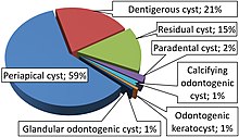
A dentigerous cyst, also known as a follicular cyst, is an epithelial-lined developmental cyst formed by accumulation of fluid between the reduced enamel epithelium and the crown of an unerupted tooth. It is formed when there is an alteration in the reduced enamel epithelium and encloses the crown of an unerupted tooth at the cemento-enamel junction. Fluid is accumulated between reduced enamel epithelium and the crown of an unerupted tooth.

<span class="mw-page-title-main">Stafne defect</span>

The Stafne defect is a depression of the mandible, most commonly located on the lingual surface. The Stafne defect is thought to be a normal anatomical variant, as the depression is created by ectopic salivary gland tissue associated with the submandibular gland and does not represent a pathologic lesion as such. This cavity is commonly observed on panoramic radiograph.

<span class="mw-page-title-main">Central giant-cell granuloma</span> Medical condition

Central giant-cell granuloma (CGCG) is a localised benign condition of the jaws. It is twice as common in females and is more likely to occur before age 30. Central giant-cell granulomas are more common in the anterior mandible, often crossing the midline and causing painless swellings.

<span class="mw-page-title-main">Periapical cyst</span> Medical condition

Commonly known as a dental cyst, the periapical cyst is the most common odontogenic cyst. It may develop rapidly from a periapical granuloma, as a consequence of untreated chronic periapical periodontitis.

<span class="mw-page-title-main">Odontogenic keratocyst</span> Medical condition

An odontogenic keratocyst is a rare and benign but locally aggressive developmental cyst. It most often affects the posterior mandible and most commonly presents in the third decade of life. Odontogenic keratocysts make up around 19% of jaw cysts.

Lateral periodontal cysts (LPCs) are defined as non-keratinised and non-inflammatory developmental cysts located adjacent or lateral to the root of a vital tooth.” LPCs are a rare form of jaw cysts, with the same histopathological characteristics as gingival cysts of adults (GCA). Hence LPCs are regarded as the intraosseous form of the extraosseous GCA. They are commonly found along the lateral periodontium or within the bone between the roots of vital teeth, around mandibular canines and premolars. Standish and Shafer reported the first well-documented case of LPCs in 1958, followed by Holder and Kunkel in the same year although it was called a periodontal cyst. Since then, there has been more than 270 well-documented cases of LPCs in literature.

<span class="mw-page-title-main">Glandular odontogenic cyst</span> Human jaw cyst

A glandular odontogenic cyst (GOC) is a rare and usually benign odontogenic cyst developed at the odontogenic epithelium of the mandible or maxilla. Originally, the cyst was labeled as "sialo-odontogenic cyst" in 1987. However, the World Health Organization (WHO) decided to adopt the medical expression "glandular odontogenic cyst". Following the initial classification, only 60 medically documented cases were present in the population by 2003. GOC was established as its own biological growth after differentiation from other jaw cysts such as the "central mucoepidermoid carcinoma (MEC)", a popular type of neoplasm at the salivary glands. GOC is usually misdiagnosed with other lesions developed at the glandular and salivary gland due to the shared clinical signs. The presence of osteodentin supports the concept of an odontogenic pathway. This odontogenic cyst is commonly described to be a slow and aggressive development. The inclination of GOC to be large and multilocular is associated with a greater chance of remission. GOC is an infrequent manifestation with a 0.2% diagnosis in jaw lesion cases. Reported cases show that GOC mainly impacts the mandible and male individuals. The presentation of GOC at the maxilla has a very low rate of incidence. The GOC development is more common in adults in their fifth and sixth decades.

<span class="mw-page-title-main">Adenomatoid odontogenic tumor</span> Medical condition

The adenomatoid odontogenic tumor is an odontogenic tumor arising from the enamel organ or dental lamina.

Squamous odontogenic tumors (SOTs) are very rare benign locally infiltrative odontogenic neoplasms of epithelial origin. Only some 50 cases have been documented. They occur mostly from 20-40 and are more common in males. Treatment is by simple enucleation and local curettage, and recurrence is rare.

An ameloblastic fibroma is a fibroma of the ameloblastic tissue, that is, an odontogenic tumor arising from the enamel organ or dental lamina. It may be either truly neoplastic or merely hamartomatous. In neoplastic cases, it may be labeled an ameloblastic fibrosarcoma in accord with the terminological distinction that reserves the word fibroma for benign tumors and assigns the word fibrosarcoma to malignant ones. It is more common in the first and second decades of life, when odontogenesis is ongoing, than in later decades. In 50% of cases an unerupted tooth is involved.

<span class="mw-page-title-main">Odontoma</span> Benign tumour of dental tissue

An odontoma, also known as an odontome, is a benign tumour linked to tooth development. Specifically, it is a dental hamartoma, meaning that it is composed of normal dental tissue that has grown in an irregular way. It includes both odontogenic hard and soft tissues. As with normal tooth development, odontomas stop growing once mature which makes them benign.

The odontogenic myxoma is an uncommon benign odontogenic tumor arising from embryonic connective tissue associated with tooth formation. As a myxoma, this tumor consists mainly of spindle shaped cells and scattered collagen fibers distributed through a loose, mucoid material.

The calcifying epithelial odontogenic tumor (CEOT), also known as a Pindborg tumor, is an odontogenic tumor first recognized by the Danish pathologist Jens Jørgen Pindborg in 1955. It was previously described as an adenoid adamantoblastoma, unusual ameloblastoma and a cystic odontoma. Like other odontogenic neoplasms, it is thought to arise from the epithelial element of the enamel origin. It is a typically benign and slow growing, but invasive neoplasm.

Odontogenic cyst are a group of jaw cysts that are formed from tissues involved in odontogenesis. Odontogenic cysts are closed sacs, and have a distinct membrane derived from rests of odontogenic epithelium. It may contain air, fluids, or semi-solid material. Intra-bony cysts are most common in the jaws, because the mandible and maxilla are the only bones with epithelial components. That odontogenic epithelium is critical in normal tooth development. However, epithelial rests may be the origin for the cyst lining later. Not all oral cysts are odontogenic cyst. For example, mucous cyst of the oral mucosa and nasolabial duct cyst are not of odontogenic origin.

A cyst is a pathological epithelial lined cavity that fills with fluid or soft material and usually grows from internal pressure generated by fluid being drawn into the cavity from osmosis. The bones of the jaws, the mandible and maxilla, are the bones with the highest prevalence of cysts in the human body. This is due to the abundant amount of epithelial remnants that can be left in the bones of the jaws. The enamel of teeth is formed from ectoderm, and so remnants of epithelium can be left in the bone during odontogenesis. The bones of the jaws develop from embryologic processes which fuse, and ectodermal tissue may be trapped along the lines of this fusion. This "resting" epithelium is usually dormant or undergoes atrophy, but, when stimulated, may form a cyst. The reasons why resting epithelium may proliferate and undergo cystic transformation are generally unknown, but inflammation is thought to be a major factor. The high prevalence of tooth impactions and dental infections that occur in the bones of the jaws is also significant to explain why cysts are more common at these sites.

A median mandibular cyst is a type of cyst that occurs in the midline of the mandible, thought to be created by proliferation and cystic degeneration of resting epithelial tissue that is left trapped within the substance of the bone during embryologic fusion of the two halves of the mandible, along the plane of fusion later termed the symphysis menti. A true median mandibular cyst would therefore be classified as a non-odontogenic, fissural cyst. The existence of this lesion as a unique clinical entity is controversial, and some reported cases may have represented misdiagnosed odontogenic cysts, which are by far the most common type of intrabony cyst occurring in the jaws. It has also been suggested that the mandible develops as a bilobed proliferation of mesenchyme connected with a central isthmus. Therefore, it is unlikely that epithelial tissue would become trapped as there is no ectoderm separating the lobes in the first instance.

The ameloblastic fibro-odontoma (AFO) is essentially a benign tumor with the features characteristic of ameloblastic fibroma along with enamel and dentin. Though it is generally regarded as benign, there have been cases of its malignant transformation into ameloblastic fibrosarcoma and odontogenic sarcoma. Cahn LR and Blum T, believed in “maturation theory”, which suggested that AFO was an intermediate stage and eventually developed during the period of tooth formation to a complex odontoma thus, being a hamartoma.

References

  1. Kler, Shikha; Palaskar, Sangeeta; Shetty, and, Vishwa Parkash; Bhushan, Anju (2009), "Intraosseous calcifying cystic odontogenic tumor", J Oral Maxillofac Pathol , 13 (1): 27–29, doi:10.4103/0973-029X.48753, PMC   3162852 , PMID   21886994.
  2. 1 2 3 4 5 6 7 8 Zornosa, Ximena; Müller, Susan (2010-07-24). "Calcifying Cystic Odontogenic Tumor". Head and Neck Pathology. 4 (4): 292–294. doi:10.1007/s12105-010-0197-z. ISSN   1936-055X. PMC   2996493 . PMID   20658217.
  3. Gamoh, Shoko; Akiyama, Hironori; Furukawa, Chisato; Matsushima, Yuki; Iseki, Tomio; Wato, Masahiro; Tanaka, Akio; Morita, Shosuke; Shimizutani, Kimishige (2017-11-01). "Calcifying cystic odontogenic tumor accompanied by a dentigerous cyst: A case report". Oncology Letters. 14 (5): 5785–5790. doi:10.3892/ol.2017.6993. ISSN   1792-1074. PMC   5661555 . PMID   29113208.
  4. Leandro Bezerra Borges; Francisco Vagnaldo Fechine; Mário Rogério Lima Mota; Fabrício Bitu Sousa; Ana Paula Negreiros Nunes Alves (2012). "Odontogenic lesions of the jaw: a clinical-pathological study of 461 cases". Revista Gaúcha de Odontologia. 60 (1).{{cite journal}}: CS1 maint: multiple names: authors list (link)
  5. 1 2 3 4 Utumi, Estevam Rubens; Pedron, Irineu Gregnanin; Silva, Leopoldo Penteado Nucci da; Machado, Gustavo Grothe; Rocha, André Caroli (September 2012). "Distintas manifestações do tumor odontogênico cístico calcificante". Einstein (São Paulo). 10 (3): 366–370. doi: 10.1590/S1679-45082012000300019 . ISSN   1679-4508. PMID   23386019.
  6. 1 2 Regezi JA, Sciubba J, Jordan RCK (2012-04-15). Oral Pathology: Clinical Pathologic Correlations. Elsevier Health Sciences. p. 259. ISBN   978-1-4557-0269-5.
  7. 1 2 3 4 5 6 7 8 9 Oral and maxillofacial pathology. Neville, Brad W. (3rd ed.). St. Louis, Mo.: Saunders/Elsevier. 2009. ISBN   978-1-4377-2197-3. OCLC   834142726.{{cite book}}: CS1 maint: others (link)
  8. 1 2 3 4 Kler, Shikha; Palaskar, Sangeeta; Shetty, Vishwa Parkash; Bhushan, Anju (2009). "Intraosseous calcifying cystic odontogenic tumor". Journal of Oral and Maxillofacial Pathology. 13 (1): 27–29. doi:10.4103/0973-029X.48753. ISSN   0973-029X. PMC   3162852 . PMID   21886994.
  9. Mervyn Shear; Paul Speight (2008-04-15). Cysts of the Oral and Maxillofacial Regions. John Wiley & Sons. p. 107. ISBN   978-0-470-75972-1.
  10. 1 2 3 4 5 6 Sonone, Archana; Sabane, V. S.; Desai, Rajeev (2011). "Calcifying Ghost Cell Odontogenic Cyst: Report of a Case and Review of Literature". Case Reports in Dentistry. 2011: 328743. doi: 10.1155/2011/328743 . ISSN   2090-6447. PMC   3335591 . PMID   22567434.
  11. 1 2 3 4 5 Arruda, José-Alcides; Silva, Leni-Verônica; Silva, Leorik; Monteiro, João-Luiz; Álvares, Pamella; Silveira, Marcia; Sobral, Ana-Paula (2018-06-01). "Calcifying odontogenic cyst: A 26-year retrospective clinicopathological analysis and immunohistochemical study". Journal of Clinical and Experimental Dentistry. 10 (6): e542–e547. doi:10.4317/jced.54528. ISSN   1989-5488. PMC   6005085 . PMID   29930772.