
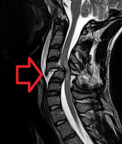
Transverse myelitis (TM) is a rare neurological condition in which the spinal cord is inflamed. Transverse implies that the inflammation extends across the entire width of the spinal cord. Partial transverse myelitis and partial myelitis are terms used to define inflammation of the spinal cord that affects part of the width of the spinal cord. TM is characterized by weakness and numbness of the limbs, deficits in sensation and motor skills, dysfunctional urethral and anal sphincter activities, and dysfunction of the autonomic nervous system that can lead to episodes of high blood pressure. Signs and symptoms vary according to the affected level of the spinal cord. The underlying cause of TM is unknown. The spinal cord inflammation seen in TM has been associated with various infections, immune system disorders, or damage to nerve fibers, by loss of myelin. Decreased electrical conductivity in the nervous system can result.
Tetraplegia, also known as quadriplegia, is paralysis caused by illness or injury that results in the partial or total loss of use of all four limbs and torso; paraplegia is similar but does not affect the arms. The loss is usually sensory and motor, which means that both sensation and control are lost. Tetraparesis or quadriparesis, on the other hand, means muscle weakness affecting all four limbs. It may be flaccid or spastic.
Myelitis is inflammation of the spinal cord which can disrupt the normal responses from the brain to the rest of the body, and from the rest of the body to the brain. Inflammation in the spinal cord, can cause the myelin and axon to be damaged resulting in symptoms such as paralysis and sensory loss. Myelitis is classified to several categories depending on the area or the cause of the lesion; however, any inflammatory attack on the spinal cord is often referred to as transverse myelitis.

The spinothalamic tract is a sensory pathway from the skin to the thalamus. From the ventral posterolateral nucleus in the thalamus, sensory information is relayed upward to the somatosensory cortex of the postcentral gyrus.

The dorsal column–medial lemniscus pathway (DCML) is a sensory pathway of the central nervous system that conveys sensations of fine touch, vibration, two-point discrimination, and proprioception (position) from the skin and joints. It transmits information from the body to the primary somatosensory cortex in the postcentral gyrus of the parietal lobe of the brain. The pathway receives information from sensory receptors throughout the body, and carries this in nerve tracts in the white matter of the dorsal columns of the spinal cord, to the medulla where it is continued in the medial lemniscus, on to the thalamus and relayed from there through the internal capsule and transmitted to the somatosensory cortex. The name dorsal-column medial lemniscus comes from the two structures that carry the sensory information: the dorsal columns of the spinal cord, and the medial lemniscus in the brainstem.

The patellar reflex or knee-jerk (myotatic) (monosynaptic) is a stretch reflex which tests the L2, L3, and L4 segments of the spinal cord.
A spinal cord injury (SCI) is damage to the spinal cord that causes temporary or permanent changes in its function. Symptoms may include loss of muscle function, sensation, or autonomic function in the parts of the body served by the spinal cord below the level of the injury. Injury can occur at any level of the spinal cord and can be complete injury, with a total loss of sensation and muscle function, or incomplete, meaning some nervous signals are able to travel past the injured area of the cord. Depending on the location and severity of damage, the symptoms vary, from numbness to paralysis to incontinence. Long term outcomes also ranges widely, from full recovery to permanent tetraplegia or paraplegia. Complications can include muscle atrophy, pressure sores, infections, and breathing problems.
Myelopathy describes any neurologic deficit related to the spinal cord. When due to trauma, it is known as (acute) spinal cord injury. When inflammatory, it is known as myelitis. Disease that is vascular in nature is known as vascular myelopathy. The most common form of myelopathy in human, cervical spondylotic myelopathy (CSM), is caused by arthritic changes (spondylosis) of the cervical spine, which result in narrowing of the spinal canal ultimately causing compression of the spinal cord. In Asian populations, spinal cord compression often occurs due to a different, inflammatory process affecting the posterior longitudinal ligament.
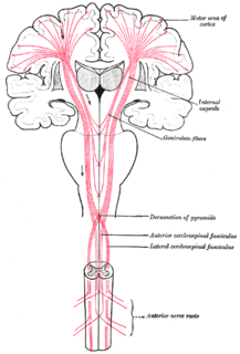
An upper motor neuron lesion occurs in the neural pathway above the anterior horn cell of the spinal cord or motor nuclei of the cranial nerves. Conversely, a lower motor neuron lesion affects nerve fibers traveling from the anterior horn of the spinal cord or the cranial motor nuclei to the relevant muscle(s).
Spinal shock was first defined by Whytt in 1750 as a loss of sensation accompanied by motor paralysis with initial loss but gradual recovery of reflexes, following a spinal cord injury (SCI) – most often a complete transection. Reflexes in the spinal cord below the level of injury are depressed (hyporeflexia) or absent (areflexia), while those above the level of the injury remain unaffected. The 'shock' in spinal shock does not refer to circulatory collapse, and should not be confused with neurogenic shock, which is life-threatening.

The conus medullaris or conus terminalis is the tapered, lower end of the spinal cord. It occurs near lumbar vertebral levels 1 (L1) and 2 (L2), occasionally lower. The upper end of the conus medullaris is usually not well defined, however, its corresponding spinal cord segments are usually S1-S5.

Spinal cord compression develops when the spinal cord is compressed by bone fragments from a vertebral fracture, a tumor, abscess, ruptured intervertebral disc or other lesion. It is regarded as a medical emergency independent of its cause, and requires swift diagnosis and treatment to prevent long-term disability due to irreversible spinal cord injury.
Autonomic dysreflexia (AD), also previously known as mass reflex, is a potential medical emergency classically characterized by uncontrolled hypertension and bradycardia, although tachycardia is known to commonly occur. AD occurs most often in individuals with spinal cord injuries with lesions at or above the T6 spinal cord level, although it has been reported in patients with lesions as low as T10. Guillain–Barré syndrome may also cause Autonomic Dysreflexia.
Tethered cord syndrome (TCS) refers to a group of neurological disorders that relate to malformations of the spinal cord. Various forms include tight filum terminale, lipomeningomyelocele, split cord malformations (diastematomyelia), dermal sinus tracts, and dermoids. All forms involve the pulling of the spinal cord at the base of the spinal canal, literally a tethered cord. The spinal cord normally hangs loose in the canal, free to move up and down with growth, and with bending and stretching. A tethered cord, however, is held taut at the end or at some point in the spinal canal. In children, a tethered cord can force the spinal cord to stretch as they grow. In adults the spinal cord stretches in the course of normal activity, usually leading to progressive spinal cord damage if untreated. TCS is often associated with the closure of a spina bifida. It can be congenital, such as in tight filum terminale, or the result of injury later in life.

Anterior spinal artery syndrome is syndrome caused by ischemia of the anterior spinal artery, resulting in loss of function of the anterior two-thirds of the spinal cord. The region affected includes the descending corticospinal tract, ascending spinothalamic tract, and autonomic fibers. It is characterized by a corresponding loss of motor function, loss of pain and temperature sensation, and hypotension.
Brown-Séquard syndrome is caused by damage to one half of the spinal cord, i.e. hemisection of the spinal cord resulting in paralysis and loss of proprioception on the same side as the injury or lesion, and loss of pain and temperature sensation on the opposite side as the lesion. It is named after physiologist Charles-Édouard Brown-Séquard, who first described the condition in 1850.
Diastematomyelia is a congenital disorder in which a part of the spinal cord is split, usually at the level of the upper lumbar vertebra.

Posterior cord syndrome (PCS), also known as posterior spinal artery syndrome (PSA), is a type of incomplete spinal cord injury. PCS is the least commonly occurring of the six clinical spinal cord injury syndromes, with an incidence rate of less than 1%.
Cobb syndrome is a rare congenital disorder characterized by visible skin lesions with underlying spinal angiomas or arteriovenous malformations (AVMs). The skin lesions of Cobb syndrome typically are present as port wine stains or angiomas, but reports exist of angiokeratomas, angiolipomas, and lymphangioma circumscriptum. The intraspinal lesions may be angiomas or AVMs and occur at levels of the spinal cord corresponding to the affected skin dermatomes. They may in turn produce spinal cord dysfunction and weakness or paralysis.
Congenital dermal sinus is an uncommon form of cranial or spinal dysraphism. It occurs in 1 in 2500 live births. It occurs as a dermal indentation, found along the midline of the neuraxis and often presents alongside infection and neurological deficit. Congenital dermal sinus form due to a focal failure of dysjunction between the cutaneous ectoderm and neuroectoderm during the third to eight week of gestation.Typically observed in the lumbar and lumbosacral region, congenital dermal sinus can occur from the nasion and occiput region down.

